


Abstract
Background: Liver fibrosis represents a critical pathological juncture in the progression of chronic liver disease toward irreversible cirrhosis and hepatocellular carcinoma; however, early diagnosis and effective therapeutic intervention remain significant clinical challenges. Methods: Herein, we report a biomimetic cascade nanozyme platform (termed iSOD) featuring a core–shell nanostructure for the accurate diagnosis and therapy of liver fibrosis. Synthesized via a green, one-pot coprecipitation method utilizing natural superoxide dismutase (SOD) as a structural template, the iSOD comprises an ultrasmall Fe3O4 core (approximately 4.04 nm in diameter) seamlessly enveloped by an active SOD protein shell. Results: This unique architecture provides the SOD-mediated superoxide-scavenging activity and catalase (CAT)-like activity from the ultrasmall iron oxide core. This sequential dual-enzyme cascade can eliminate deleterious reactive oxygen species (ROS) by dismutating superoxide anions and decomposing intermediate hydrogen peroxide (H2O2) into water and oxygen. In vitro evaluations demonstrate that iSOD significantly diminishes intracellular ROS in HSC-T6 cells and protects them against oxidative stress-induced injury. Relaxivity measurements revealed longitudinal (r1) and transverse (r2) relaxivities of 2.874 and 13.878 mM−1 s−1, respectively, indicating the ability of iSOD to influence both T1 and T2 relaxation. In vivo, T1-weighted MRI revealed changes in hepatic signal intensity and spatial distribution 30 min after iSOD administration, with distinct signal patterns observed among healthy, moderately fibrotic, and severely fibrotic rat livers. Histological analyses further demonstrated reduced collagen deposition and improved hepatic tissue morphology following iSOD treatment. Short-term biocompatibility evaluations revealed no obvious adverse effects under the investigated conditions. Conclusions: Collectively, these findings highlight the potential of iSOD as a multifunctional nanoplatform integrating antioxidant therapy with MRI-based assessment of liver fibrosis.
1. Introduction
Hepatic fibrosis is a common pathological repair response to various chronic liver insults, including viral hepatitis, alcoholic liver disease, and non-alcoholic fatty liver disease [1,2]. Pathologically, it is characterized by the excessive deposition of extracellular matrix (ECM) components [3], predominantly collagen fibers [4]. If left unchecked, progressive fibrosis inevitably degenerates into irreversible cirrhosis or hepatocellular carcinoma, posing a serious threat to global health [5]. Currently, liver biopsy remains the clinical gold standard for hepatic fibrosis staging. Nevertheless, its invasiveness, susceptibility to sampling bias, and risk of serious complications greatly limit broad clinical application [6]. While non-invasive modalities such as ultrasound and magnetic resonance elastography offer structural insights [7], they critically lack the molecular imaging probes required to simultaneously gauge disease severity and monitor the underlying pathological microenvironment. Moreover, pharmacological treatment options for liver fibrosis remain very limited, and no anti-fibrotic drug has been approved to date [8]. Therefore, there is an urgent clinical mandate to develop advanced theranostic nanoplatforms that seamlessly integrate non-invasive staging with therapeutic interventions.
The persistent accumulation of reactive oxygen species (ROS) and subsequent oxidative stress act as pivotal drivers in the initiation and exacerbation of liver fibrosis [9,10]. Within the fibrotic hepatic microenvironment, an overabundance of ROS triggers the continuous activation of hepatic stellate cells (HSCs). Activated HSCs transdifferentiate into myofibroblast-like cells, relentlessly secreting type I collagen and other ECM proteins, which ultimately disrupt normal hepatic architecture and function [11]. Therefore, the efficient and exhaustive scavenging of ROS emerges as a highly promising strategy to arrest or reverse fibrotic progression [12,13]. Natural superoxide dismutase (SOD) is a master biological antioxidant that specifically catalyzes the dismutation of superoxide anions (⋅O2−) into hydrogen peroxide (H2O2) and oxygen [14,15,16]. Nevertheless, its direct therapeutic application is severely hampered by inherent protein vulnerabilities, including poor physicochemical stability, a brief circulatory half-life, and formidable physiological barriers [17,18]. More importantly, the unilateral action of SOD leads to the unavoidable accumulation of H2O2, which can induce secondary oxidative damage if not promptly neutralized.
Concurrently, ultrasmall iron oxide nanoparticles (<5 nm) have garnered immense attention as premier T1-weighted magnetic resonance imaging (MRI) contrast agents, distinguished by their favorable magnetic properties and good biosafety profiles [19,20]. Beyond their imaging prowess, specific iron oxide nanomaterials harbor intrinsic catalase (CAT)-like nanozyme activity, facilitating the rapid decomposition of H2O2 into water and oxygen [21,22,23]. We hypothesized that structurally integrating natural SOD with ultrasmall iron oxide nanoparticles could construct a complementary, cascade antioxidant system, effectively resolving the limitations of single-enzyme therapies.
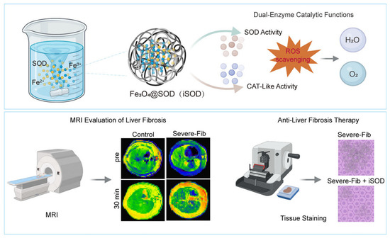
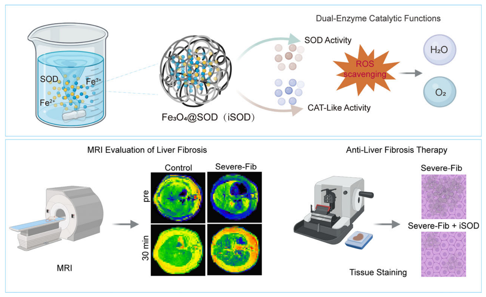
In this study, we engineered an ultrasmall iron oxide nanoplatform (designated as iSOD) utilizing natural SOD as a biological template via a facile, one-step coprecipitation strategy (Scheme 1). This biomimetic design was formulated to achieve four objectives: (1) utilize the functional groups of SOD to facilitate the formation and stabilization of ultrasmall Fe3O4 nanoparticles; (2) integrate SOD-mediated superoxide-scavenging activity and Fe3O4-mediated CAT-like activity to enable a potential sequential pathway for ROS elimination; (3) exploit the magnetic relaxation properties of ultrasmall Fe3O4 for non-invasive MRI assessment of liver fibrosis; and (4) alleviate oxidative stress-associated cellular injury and reduce collagen deposition in fibrotic liver tissue. Through systematic evaluations of its physicochemical properties, cascade enzymatic kinetics, in vitro cytoprotection, in vivo MRI performance, and anti-fibrotic therapeutic outcomes, we present iSOD as a promising translational nanoplatform for the diagnosis and treatment of liver fibrosis.
Scheme 1.
Overview of the biomimetic iSOD nanoplatform for liver fibrosis therapy and MRI-assisted staging. (Top) One-pot green synthesis of Fe3O4@SOD (iSOD) nanoparticles and their sequential dual-enzyme cascade catalysis for ROS scavenging. (Bottom) Translational applications showing non-invasive T1-weighted MRI for distinguishing normal and severe fibrotic livers, alongside histological validation of tissue repair and anti-fibrotic efficacy following iSOD treatment.
2. Materials and Methods
2.1. Materials
Superoxide dismutase (SOD) was obtained from Roche Applied Science, Basel, Switzerland. Ferric chloride hexahydrate (FeCl3·6H2O), ferrous chloride tetrahydrate (FeCl2·4H2O), thioacetamide (TAA), sodium hydroxide (NaOH) and other conventional chemical reagents were purchased from Sigma-Aldrich, St. Louis, MO, USA. Phosphate-buffered saline (PBS), cell culture medium and trypsin–EDTA solution were purchased from Gibco (Thermo Fisher Scientific, Waltham, MA, USA).
2.2. Preparation and Characterization of iSOD Nanoparticles
iSOD nanoparticles were synthesized via a one-step coprecipitation method using SOD as a biomolecular template. In brief, 30 mg of SOD was fully dissolved in 30 mL of deionized water and stirred in a water bath at 37 °C for 10 min. Subsequently, 1 mL of FeCl2·4H2O solution (13.2 mg/mL) and 1 mL of FeCl3·6H2O solution (9.9 mg/mL) were sequentially added. Thereafter, 0.5 mL of 1 M NaOH solution was introduced dropwise to alkalinize the reaction system, and the mixture was vigorously stirred for 30 min. The resulting solution was dialyzed using a dialysis bag (molecular weight cutoff: 14 kDa) to eliminate unreacted ions and free impurities, yielding purified iSOD nanoparticles.
2.3. Enzyme Activity Assays
The SOD-mimetic activity of iSOD was quantified using the Total Superoxide Dismutase Assay Kit (Servicebio, Wuhan, China, Cat. No. G4306) based on the WST-1 colorimetric method. Briefly, the kit detects the decrease in formazan absorbance at 450 nm resulting from the scavenging of superoxide anion radicals (·O2−) after incubation at 37 °C for 20 min. Enzymatic activity was calculated according to the measured inhibition rate, where one unit (U) was defined as the amount of iSOD capable of achieving 50% inhibition of superoxide generation. The ·O2− scavenging capacity of iSOD was further validated by electron spin resonance (ESR) spectroscopy with 5,5-dimethyl-1-pyrroline-N-oxide (DMPO) as the spin-trapping reagent.
The CAT-like activity of iSOD was assessed by monitoring dissolved oxygen production upon H2O2 decomposition (50 μL, 1 mol/L). Briefly, 100 μL, 200 μL and 400 μL of iSOD solution (Fe concentration: 2.6 mM) were rapidly injected into 6 mL deoxygenated PBS, followed by immediate sealing. A dissolved oxygen meter (INESA Scientific Instrument Co., Ltd., Shanghai, China) was used to record the real-time dissolved oxygen concentration of the mixture, and oxygen evolution curves were plotted [24].
2.4. Cell Experiments
HSC-T6 cells were cultured in DMEM supplemented with 10% fetal bovine serum and 1% penicillin–streptomycin in a 37 °C humidified incubator with 5% CO2. For intracellular ROS detection, HSC-T6 cells were pretreated with iSOD at an Fe concentration of 200 μg/mL and then stimulated with H2O2 to induce cellular oxidative stress. The intracellular ROS level was detected using the 2′,7′-dichlorodihydrofluorescein diacetate (DCFH-DA) fluorescent probe and observed under a fluorescence microscope. Cell viability and cytotoxicity were assessed via Calcein acetoxymethyl ester (Calcein-AM)/propidium iodide (PI) double staining and a Cell Counting Kit-8 (CCK-8; Hubei Purmei Biological Technology Co., Ltd., Wuhan, China) assay to evaluate the protective effect of iSOD against oxidative stress-induced cell injury.
2.5. Establishment of Rat Liver Fibrosis Model
Male Sprague-Dawley rats (aged 6–8 weeks, weighing 180–220 g) were obtained from Shanghai SLAC Laboratory Animal Co., Ltd. (Shanghai, China). A rat liver fibrosis model was established using TAA as the inducer according to a previously reported method [25]. TAA was dissolved in sterile physiological saline and administered via intraperitoneal injection. Starting from the 4th week of modeling, rats were sacrificed in batches every week for histopathological examination to identify the optimal time point of moderate liver fibrosis. Sampling and assessment were performed weekly from the 8th week to determine the corresponding time point of severe liver fibrosis. Based on the METAVIR staging system, the 6-week and 10-week time points were selected to represent moderate fibrosis (METAVIR stage ≥ F2 and <F3) and severe fibrosis (METAVIR stage ≥ F3 and <F4), respectively [26].
2.6. Magnetic Resonance Imaging
The longitudinal (T1) and transverse (T2) relaxation times of iSOD solutions with different Fe concentrations were measured using a Bruker minispec mq60 nuclear magnetic resonance analyzer (Bruker BioSpin, Rheinstetten, Germany), and longitudinal relaxivity (r1) and transverse relaxivity (r2) were calculated from the slope of plots of 1/T1 and 1/T2 versus Fe concentration.
In vivo T1-weighted MRI was performed on normal rats, as well as rats with moderate and severe liver fibrosis (n = 3 for each subgroup), using a 3.0 T clinical MRI scanner (Ingenia; Philips, Best, The Netherlands) with an animal coil. Images were acquired before and 30 min after intravenous injection of iSOD via the tail vein (Fe dose: 0.10 mmol/kg) to evaluate the imaging performance and diagnostic potential of iSOD for liver fibrosis. The scanning parameters were set as follows: repetition time = 600 ms; echo time = 20 ms; flip angle = 90°; field of view = 79 × 79 mm; slice thickness = 2.5 mm.
2.7. Drug Intervention in the Established Liver Fibrosis Model
After successful modeling, the dosage of TAA was moderately reduced to maintain a stable fibrotic condition during the experiment. The experimental and model control groups each included moderate-fibrosis and severe-fibrosis rat subgroups (n = 3 per subgroup). Rats in the experimental groups received corresponding drug intervention via tail vein injection (Fe dose: 0.10 mmol/kg), and administration was performed three times weekly for a total treatment period of 2 weeks. The model control group was injected with an equal volume of sterile physiological saline via the same route. The normal control group was injected with saline throughout the experiment (n = 3). At the end of the 2-week treatment cycle, all rats were sacrificed, and liver tissues were harvested for subsequent histopathological analysis.
2.8. Histopathological Examination
Rat liver tissues were fixed in 4% paraformaldehyde, embedded in paraffin and cut into 4 μm thick sections; the sections were stained with hematoxylin and eosin (H&E), Masson’s trichrome and Sirius Red to evaluate hepatic tissue injury, inflammatory infiltration, collagen deposition and the severity of liver fibrosis.
For quantification of collagen deposition, representative fields were randomly selected from each histological section. The collagen-positive area fraction in Masson’s trichrome- and Sirius Red-stained sections was measured using ImageJ (version 1.54g, National Institutes of Health, Bethesda, MD, USA).
2.9. Biosafety and Biocompatibility Evaluation
Fresh red blood cells (RBCs) from healthy mice were washed with PBS and resuspended to a 2% suspension. iSOD solutions at Fe concentrations of 25, 50, 100 and 200 μg/mL were co-incubated with the RBC suspension at 37 °C for 3 h. PBS and deionized water served as the negative and positive controls, respectively. The absorbance of the supernatants at 545 nm was measured to calculate the hemolysis rate (n = 3).
At 1 day and 7 days after iSOD injection (Fe dose: 0.10 mmol/kg), blood samples were collected from healthy mice (n = 3) for serum biochemistry and routine blood examination. Meanwhile, major organs including the heart, liver, spleen, lung and kidney were harvested and subjected to H&E staining to observe histopathological changes, so as to evaluate the in vivo biosafety of iSOD.
2.10. Statistical Analysis
All experimental data were analyzed using SPSS 26.0 software. Quantitative data are presented as mean ± standard deviation. Comparisons among multiple groups were performed using one-way analysis of variance (ANOVA) followed by Tukey’s post hoc test. A p-value < 0.05 was considered statistically significant.
3. Results
3.1. Synthesis and Structural Characterization of iSOD Nanozymes
To construct a biomimetic nanoplatform, ultrasmall iron oxide nanoparticles (iSOD) were synthesized via a facile, one-pot coprecipitation method utilizing natural SOD as a structural template. The physicochemical properties and structural integrity of the as-synthesized iSOD were systematically characterized (Figure 1).
Figure 1.
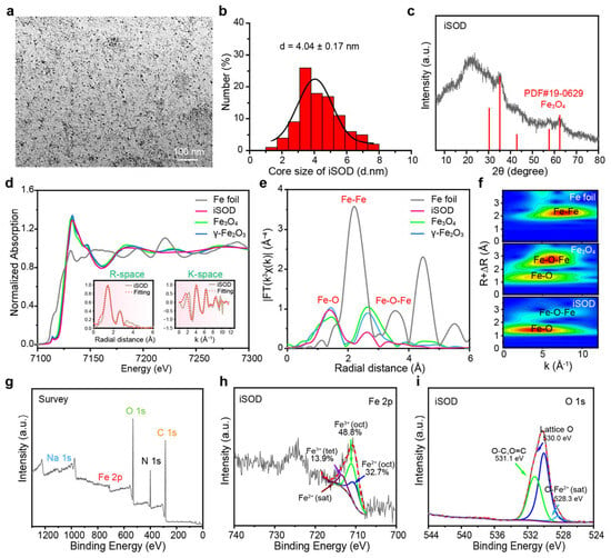
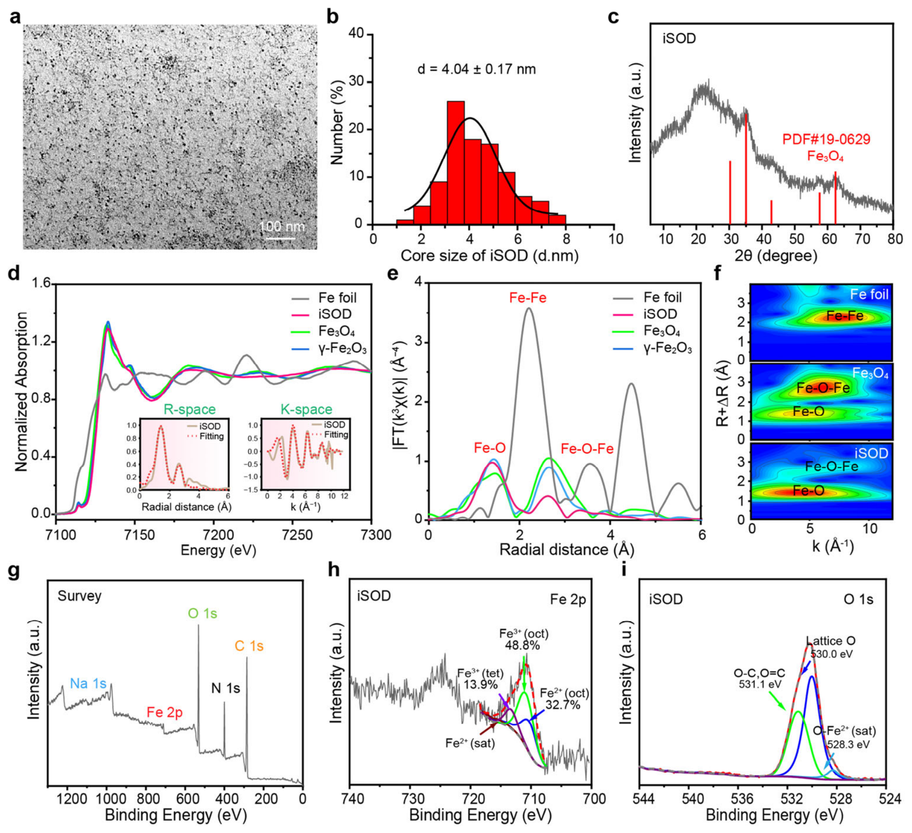
Comprehensive structural and compositional characterization of iSOD nanozymes. (a) Representative TEM image showing the electron-dense Fe3O4 of iSOD. (b) Size distribution histogram derived from TEM analysis, indicating an average diameter of 4.04 nm. (c) XRD pattern of iSOD, matching the standard magnetite Fe3O4 reference (PDF#19-0629). (d–f) Synchrotron radiation XAFS characterizations detailing the local atomic coordination environments (Fe-O and Fe-Fe bonds) and electronic structures of the iron core. (g–i) XPS analyses: (g) wide-scan survey spectrum, (h) high-resolution Fe 2p spectrum highlighting the mixed Fe2+ and Fe3+ valence states, and (i) high-resolution O1s spectrum of iSOD.
Transmission electron microscopy (TEM) revealed that the iSOD nanozymes exhibit a uniformly dispersed, distinctively spherical morphology (Figure 1a). Statistical analysis derived from TEM images confirmed an ultrasmall inorganic core diameter of 4.04 ± 0.17 nm (Figure 1b). This ultrasmall dimension is highly favorable for deep tissue penetration and optimizing catalytic efficiency. Furthermore, dynamic light scattering (DLS) measurements (Figure S1) demonstrated an average hydrodynamic diameter of approximately 28.3 nm. This significant increase in size compared to free SOD, alongside a monodisperse profile, not only corroborates the successful encapsulation of the iron oxide core by the SOD template but also indicates good colloidal stability in aqueous environments. The colloidal stability of iSOD under physiological conditions was further assessed. As shown in Figure S2, iSOD retained uniform size distribution in saline, PBS, 10% FBS and DMEM supplemented with 10% FBS after 24 h incubation at 37 °C, showing favorable stability for intravenous administration.
The crystalline nature of the synthesized nanoparticles was evaluated using X-ray diffraction (XRD). The distinct diffraction peaks present in the iSOD pattern are consistent with the standard reference card for magnetite Fe3O4 (JCPDS No. 19-0629), confirming the precise crystalline phase of the iron oxide core (Figure 1c). To gain deeper, atomic-level insights into the coordination environment and electronic structure, synchrotron radiation-based X-ray absorption fine structure spectroscopy (XAFS) was employed (Figure 1d–f). These high-resolution spectra provided direct evidence of the local Fe-O and Fe-Fe coordination spheres, validating the fine structural integrity of the magnetite core.
Successful nanocomposite formation was further substantiated through comprehensive spectroscopic analyses. Compared to free SOD, the ultraviolet–visible (UV-Vis) spectroscopy of iSOD (Figure S3) exhibited a broader and substantially more intense absorption band spanning 200–600 nm. Concurrently, Fourier-transform infrared (FTIR) spectroscopy (Figure S4) revealed a notably attenuated -OH stretching band at ~3400 cm−1. Together, these findings indicate that the spatial stabilization of the iron oxide is primarily mediated through strong interfacial hydroxyl binding interactions with the SOD protein shell. X-ray photoelectron spectroscopy (XPS) was utilized to probe the elemental composition and chemical valence states (Figure 1g–i). The wide-scan survey confirmed the expected elemental constituents, while the high-resolution Fe 2p spectrum prominently displayed characteristic peaks assigned to both Fe2+ and Fe3+ (Figure 1h). This crucial mixed-valence state serves as the active catalytic foundation for the core’s intrinsic nanozyme functionality.
Collectively, these multifaceted characterizations support the successful fabrication of SOD-templated ultrasmall Fe3O4 nanoparticles. The resultant iSOD nanoplatform boasts uniform dimensions, good dispersibility, and a stable protein corona anchored via stable hydroxyl interactions—thereby laying a solid material foundation for its downstream application as a sensitive MRI contrast agent and cascade antioxidant.
3.2. Dual-Enzyme Cascade Catalysis and Antioxidant Capabilities of iSOD
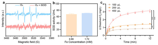
To validate the integrated dual-enzyme cascade functionality of iSOD, we systematically assessed its ⋅O2− scavenging capacity, native SOD activity, and iron oxide-mediated CAT-like kinetics. The results (Figure 2) elucidate the biomimetic catalytic cycle.
Figure 2.
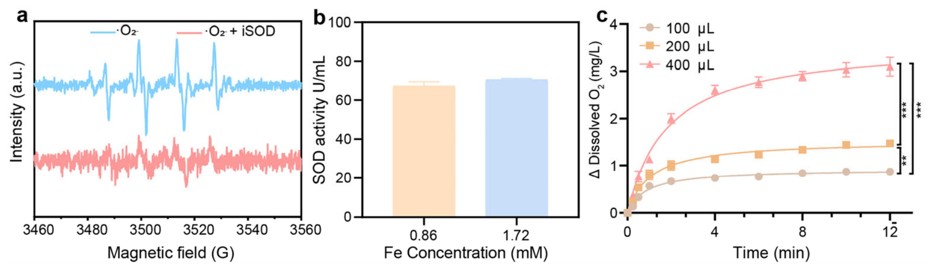
In vitro evaluation of the dual catalytic activities of iSOD. (a) Electron spin resonance (ESR) spectra of DMPO-trapped superoxide radical adducts in the absence and presence of iSOD, demonstrating its superoxide-scavenging activity. (b) Quantitative evaluation of the retained SOD activity of iSOD at different Fe concentrations (0.86 and 1.72 mM). (c) Time-dependent changes in dissolved oxygen concentration following H2O2 decomposition at different iSOD addition volumes (100, 200, and 400 μL), indicating CAT-like catalytic activity. Quantitative data are presented as mean ± standard deviation (SD, n = 3 independent experiments). Statistical comparisons in (c) were performed at the 12 min endpoint using one-way ANOVA followed by Tukey’s post hoc test. ** p < 0.01, *** p < 0.001.
First, ESR spectroscopy coupled with a DMPO spin trap was employed to monitor the real-time scavenging of ⋅O2− (Figure 2a). Upon generation, ⋅O2− forms a stable DMPO/⋅O2− adduct, yielding a prominent and characteristic ESR signal. Upon the introduction of iSOD, this characteristic signal was markedly suppressed. This effective quenching confirms that the iSOD nanoplatform successfully retains the native biological function of the SOD shell, actively dismutating superoxide anions. To further quantify this biological preservation, a standardized colorimetric assay was utilized to measure specific SOD activity (Figure 2b). The quantitative results revealed that iSOD maintains considerable SOD activity (approximately 70 U/mL) across different iron concentrations (0.86 and 1.72 mmol/L). These results indicate that structural templating and hydroxyl-mediated iron anchoring did not eliminate the SOD activity of iSOD under the tested conditions.
A pivotal advantage of this nanoplatform is the resolution of the intrinsic limitation of free SOD—the accumulation of toxic H2O2. We hypothesized that the ultrasmall Fe3O4 core would act as a CAT-like nanozyme to consume this byproduct. Using a dissolved oxygen sensor, we continuously monitored the generation of O2 following the addition of H2O2 to the iSOD solution (Figure 2c). As anticipated, iSOD rapidly catalyzed the decomposition of H2O2, evidenced by a sharp, time-dependent surge in dissolved oxygen levels (O2). Furthermore, this catalytic kinetic profile exhibited a strict dose-dependent relationship; increasing the volume of iSOD (from 100 to 400 μL) significantly accelerated oxygen evolution. Statistical analysis of dissolved oxygen concentration at the 12 min endpoint revealed significant differences. All pairwise comparisons were statistically significant (100 μL vs. 200 μL, p = 0.003; 100 μL vs. 400 μL, p < 0.001; 200 μL vs. 400 μL, p < 0.001), confirming the dose-dependent CAT-mimicking activity of iSOD.
Collectively, these assays confirm that the as-prepared iSOD integrates intrinsic SOD activity and CAT-like activity within a single nanostructure, enabling sequential scavenging of superoxide radicals and decomposition of the generated H2O2 intermediate for ROS elimination. This cascade catalytic property provides a functional basis for subsequent antioxidant and anti-fibrotic evaluation.
3.3. In Vitro Cytoprotective Effects and ROS Scavenging of iSOD Against Oxidative Stress
Having confirmed the dual-enzyme cascade activities of iSOD, we further evaluated its cellular-level cytoprotective efficacy against oxidative stress using HSC-T6 cells (Figure 3).
Figure 3.
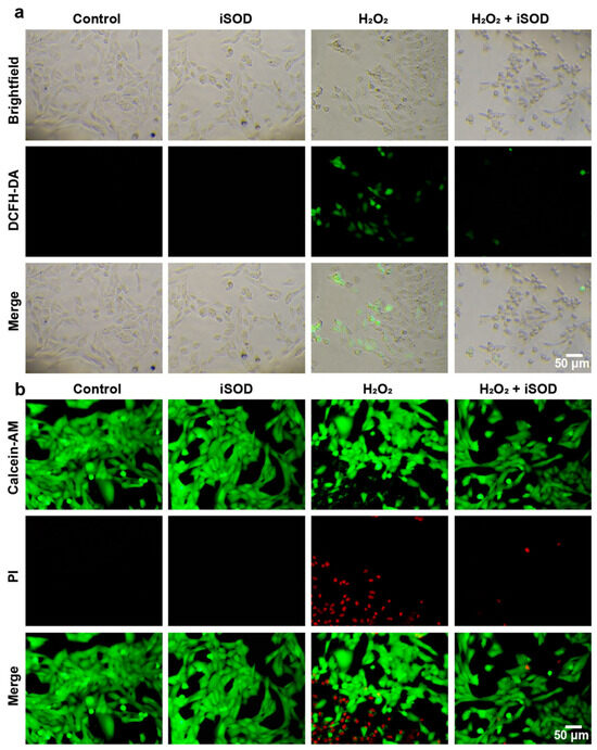
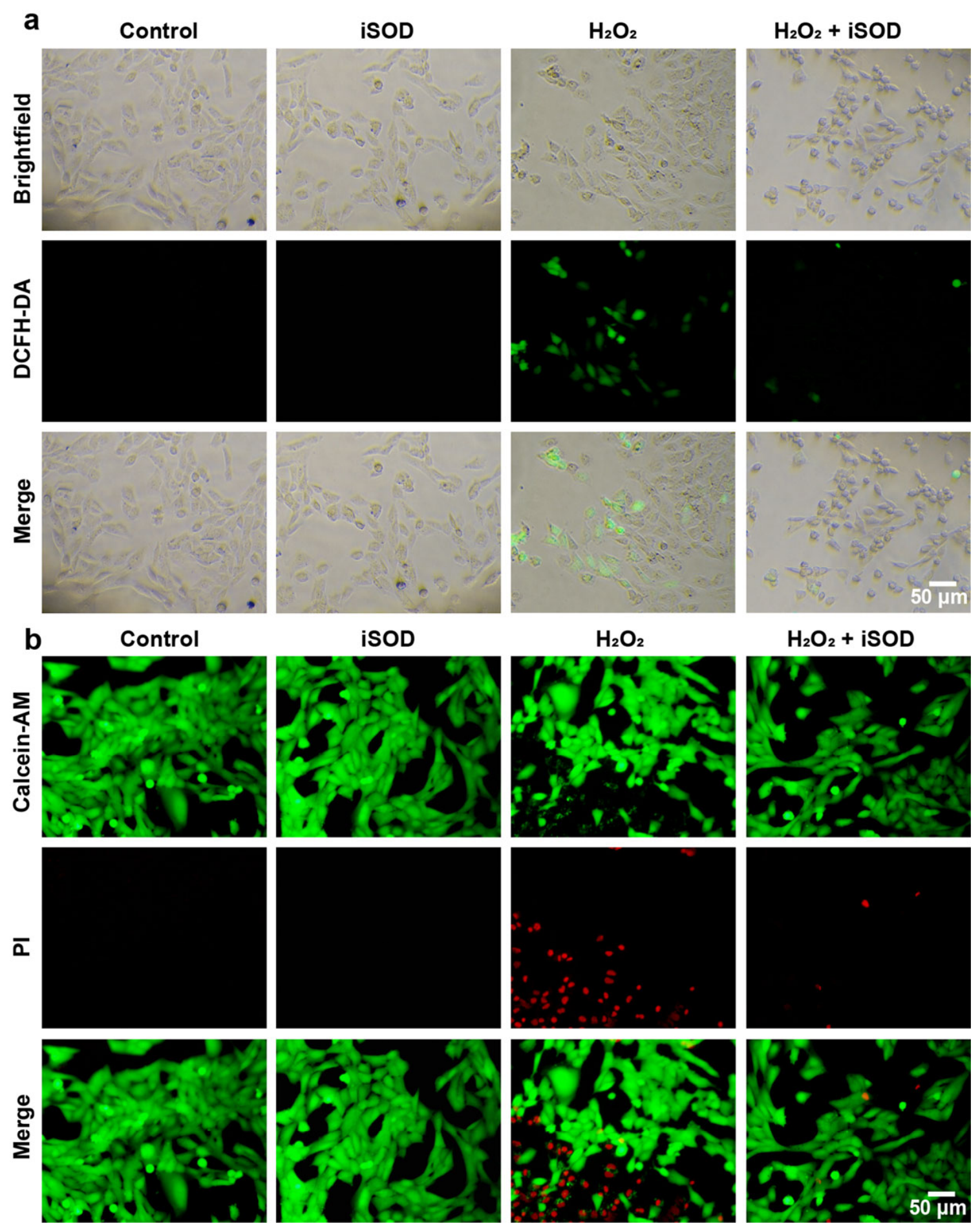
In vitro ROS scavenging and cytoprotective evaluation of iSOD in HSC-T6 cells. (a) Fluorescence microscopic images of HSC-T6 cells stained with DCFH-DA probe after various treatments (Control, iSOD, H2O2, and H2O2 + iSOD), displaying the intracellular ROS elimination capability of iSOD (Scale bar: 50 μm). (b) Live/dead co-staining assays using Calcein-AM (green, live cells) and propidium iodide (PI, red, dead cells) to evaluate cell viability under oxidative stress, verifying the cytoprotective efficacy conferred by iSOD treatment (Scale bar: 50 μm).
Intracellular ROS levels were detected using DCFH-DA as a fluorescent probe, which is oxidized by intracellular ROS into a highly green-fluorescent product. As depicted in Figure 3a (brightfield and fluorescence overlay), cells in the untreated control group and the iSOD-only group exhibited negligible green fluorescence, indicating low baseline ROS levels and confirming the good biosafety and non-toxicity of the iSOD nanozymes. Conversely, exposure to exogenous hydrogen peroxide (H2O2) induced severe oxidative stress, resulting in an intense green fluorescence signal. However, upon pretreatment with iSOD followed by H2O2 challenge, the green fluorescence was attenuated, demonstrating that iSOD can scavenge excessive intracellular ROS and alleviate oxidative stress in HSC-T6 cells.
To further assess the protective capability of iSOD against oxidative injury, Calcein-AM/PI double staining (live/dead assay) was performed (Figure 3b). Live cells are labeled with green fluorescence via intracellular esterase cleavage of Calcein-AM, whereas dead cells with compromised membranes are stained red by PI. The H2O2-treated model group displayed widespread red fluorescence coupled with a drastic reduction in green fluorescence, signifying extensive cell death caused by severe oxidative damage. In stark contrast, the iSOD-pretreated group displayed strong green fluorescence with only sparse red-fluorescent nuclei, and its cell viability was comparable to that of the control group.
Collectively, these in vitro cellular assays show that iSOD can effectively scavenge intracellular ROS, protect HSC-T6 cells from oxidative stress-induced injury, and provide a basis for subsequent in vivo anti-fibrotic treatments.
3.4. In Vitro Relaxivity and In Vivo MRI Staging of Liver Fibrosis
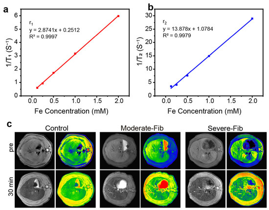
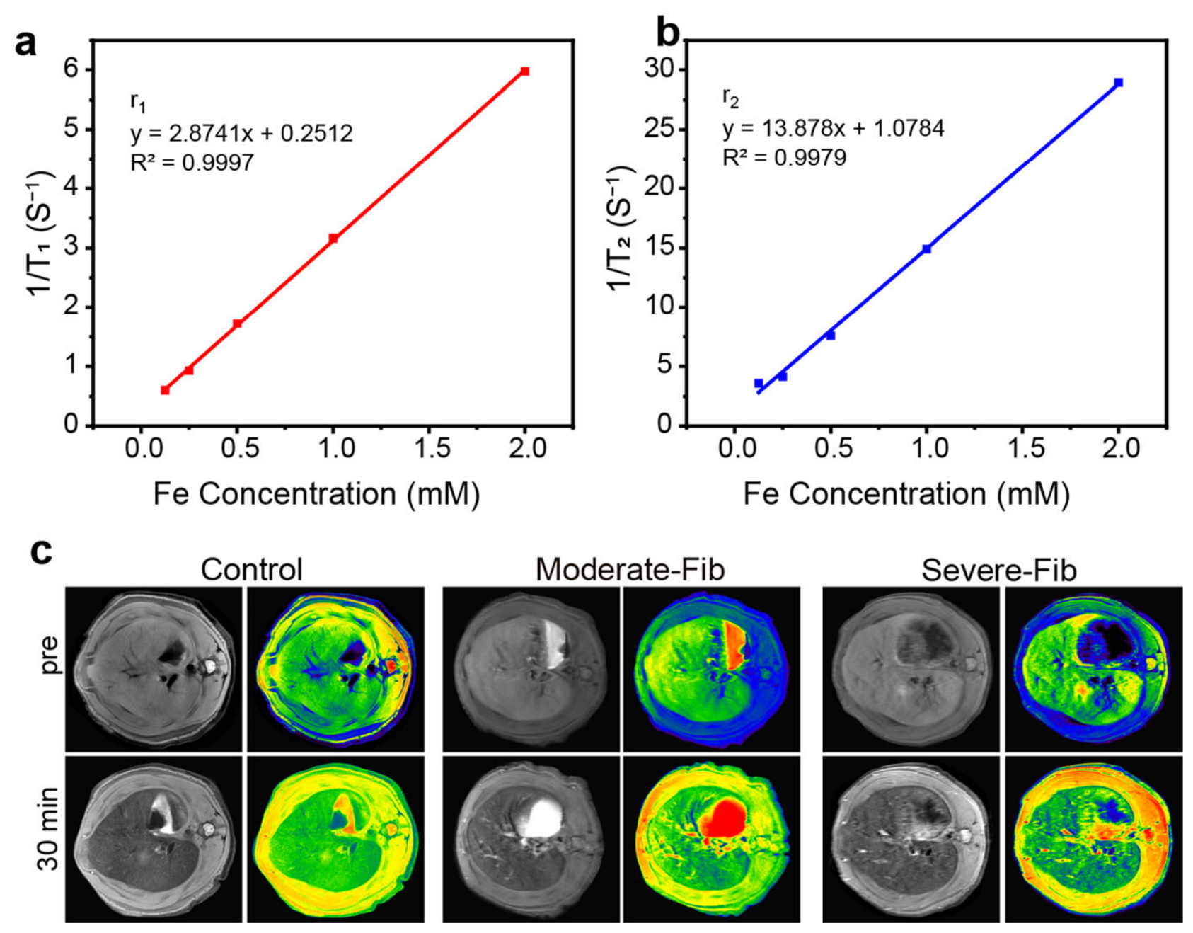
The ultrasmall Fe3O4 core of iSOD provides magnetic relaxation properties, suggesting its potential application as an MRI contrast agent. The in vitro relaxivity and in vivo imaging performance of iSOD were evaluated, as shown in Figure 4.
Figure 4.
In vitro relaxivity characterization and in vivo MRI evaluation of liver fibrosis using iSOD. (a) Linear relationship between the longitudinal relaxation rate (1/T1) and Fe concentration (mM), used to determine the longitudinal relaxivity (r1). (b) Linear relationship between the transverse relaxation rate (1/T2) and Fe concentration (mM), used to determine the transverse relaxivity (r2). (c) Representative in vivo T1-weighted MR images (grayscale and pseudocolor) of rat livers from the healthy control, moderate fibrosis (Moderate-Fib), and severe fibrosis (Severe-Fib) groups, acquired before (pre) and 30 min after intravenous administration of iSOD. Differences in hepatic signal intensity and spatial distribution were observed among the groups following iSOD administration, suggesting potential utility for MRI assessment of liver fibrosis.
The T1 and T2 relaxation times of iSOD aqueous dispersions at different Fe concentrations were measured using a Bruker minispec mq60 NMR analyzer (60 MHz, 1.41 T). As shown in Figure 4a,b, the longitudinal (r1) and transverse (r2) relaxivities were calculated to be 2.874 and 13.878 mM−1 s−1, respectively, yielding an r2/r1 ratio of approximately 4.83. These results demonstrate that iSOD can shorten both T1 and T2 relaxation times, indicating its potential as an MRI contrast agent.
The in vivo MRI performance of iSOD was subsequently investigated in rat liver fibrosis models, including healthy controls, moderate fibrosis (Moderate-Fib), and severe fibrosis (Severe-Fib). As shown in Figure 4c, MR images were acquired before and 30 min after intravenous administration of iSOD. Compared with the pre-injection images, changes in hepatic signal intensity and spatial distribution were observed following iSOD administration. The post-injection images exhibited different signal patterns among the control, Moderate-Fib, and Severe-Fib groups, with heterogeneous signal distributions observed in fibrotic liver tissues (Figure S5).
These findings suggest that iSOD can induce detectable MRI signal changes in liver tissue and may provide imaging information associated with different degrees of liver fibrosis. However, further quantitative MRI analysis is required to establish its accuracy in distinguishing fibrosis stages.
3.5. In Vivo Anti-Fibrotic Efficacy and Histological Remodeling
Prior to in vivo therapeutic evaluation, the in vitro cytotoxicity of iSOD toward HSC-T6 cells was examined using a CCK-8 assay (Figure S6). After 24 h incubation with iSOD at Fe concentrations ranging from 0 to 200 μg/mL, cell viability remained above 90%, with no significant difference from the untreated control group. The favorable cytocompatibility of iSOD laid the foundation for subsequent in vivo therapeutic assessment. To evaluate the in vivo anti-fibrotic efficacy of the iSOD nanoplatform, rat models with different stages of liver fibrosis were established. Rats were stratified into five well-defined cohorts: healthy control (Control), saline-treated moderate fibrosis (Moderate-Fib), iSOD-treated moderate fibrosis (Moderate-Fib + iSOD), saline-treated severe fibrosis (Severe-Fib), and iSOD-treated severe fibrosis (Severe-Fib + iSOD). Following the 2-week treatment, liver tissues were collected for histopathological analysis.
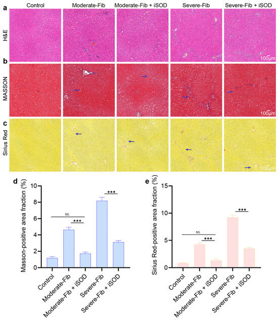
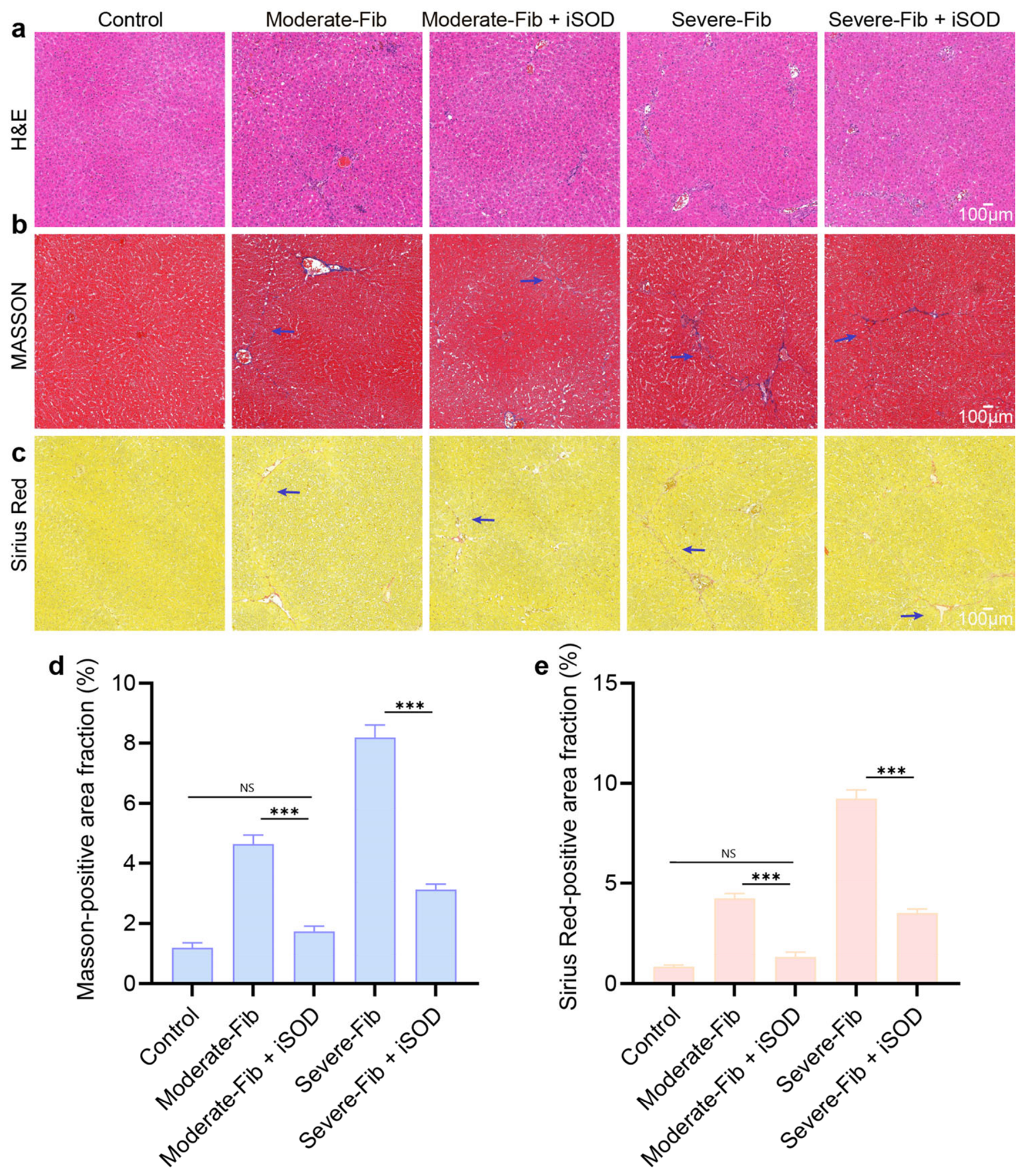
H&E staining was initially performed to assess overall hepatic architecture and inflammatory infiltration (Figure 5a). The Control group displayed healthy, intact liver parenchyma with well-organized hepatic cords and no signs of necrosis or inflammation. In contrast, untreated Moderate-Fib and Severe-Fib groups exhibited classic pathological hallmarks of liver fibrosis, characterized by pronounced inflammatory cell infiltration within the portal tracts and the prominent formation of fibrotic septa that disrupted the normal lobular architecture. Compared with their corresponding saline-treated fibrotic cohorts, the structural integrity of the liver was largely preserved in both the Moderate-Fib + iSOD and Severe-Fib + iSOD cohorts following iSOD administration. The inflammatory infiltration was markedly alleviated, and tissue structural damage was substantially reduced, indicating that iSOD could block fibrosis-related tissue destruction.
Figure 5.
Histopathological evaluation of the antifibrotic effects of iSOD in rat liver fibrosis models. Representative liver tissue sections from five experimental groups (Control, Moderate-Fib, Moderate-Fib + iSOD, Severe-Fib, and Severe-Fib + iSOD) stained with (a) hematoxylin and eosin (H&E), (b) Masson’s trichrome, and (c) Sirius Red to evaluate hepatic histopathological changes and collagen deposition (arrows). Quantitative analysis of collagen-positive areas determined from (d) Masson’s trichrome and (e) Sirius Red staining using ImageJ software. Data are presented as mean ± standard deviation (SD, n = 3 rats per group), with each animal considered an independent biological replicate. Statistical comparisons were performed using one-way ANOVA followed by Tukey’s post hoc test. *** p < 0.001; ns, not significant. Scale bars: 100 μm.
The hallmark of liver fibrosis is the progressive accumulation of ECM components, predominantly collagen fibers. Masson’s trichrome and Sirius Red staining were therefore employed to visualize and evaluate collagen deposition: collagen fibers are visualized in blue in Masson’s trichrome staining (Figure 5b), while they appear red under Sirius Red staining (Figure 5c). Negligible collagen deposition was observed in the Control group. The Moderate-Fib group displayed obvious blue-stained fibrous septa, while thicker, bridging and incomplete septa were observed in the Severe-Fib group. Therapeutic intervention with iSOD attenuated these pathological fibrous networks, with both treated cohorts displaying reduced fibrous septa. This anti-fibrotic effect was further corroborated by Sirius Red staining, in which extensive collagen-positive regions abundant in untreated Moderate-Fib and Severe-Fib cohorts were markedly diminished in their corresponding iSOD-treated groups. Quantitative analysis of collagen-positive area fraction further confirmed that iSOD administration significantly lowered collagen accumulation in both moderate-fibrosis (p < 0.001 vs. Moderate-Fib) and severe-fibrosis (p < 0.001 vs. Severe-Fib) models. Notably, the collagen-positive area of Moderate-Fib + iSOD was not statistically different from the healthy control group (Masson: p = 0.065; Sirius Red: p = 0.075) (Figure 5d,e).
Taken together, these histological results show that iSOD treatment ameliorates hepatic injury and restrains excessive collagen deposition. By remodeling the fibrotic microenvironment, iSOD exhibits potential as a therapeutic modality for attenuating liver fibrosis.
3.6. Biosafety and Biocompatibility Evaluation of iSOD
While confirming the therapeutic efficacy of iSOD against liver fibrosis, comprehensive evaluation of its in vivo biosafety and systemic biocompatibility is a prerequisite for future clinical translation.
In vitro hemocompatibility was first assessed. As presented in Figure S7, iSOD induced negligible hemolysis across all tested concentrations (25–200 μg/mL) after 3 h incubation, with hemolysis rates well below the 5% biocompatibility threshold, confirming excellent hemocompatibility.
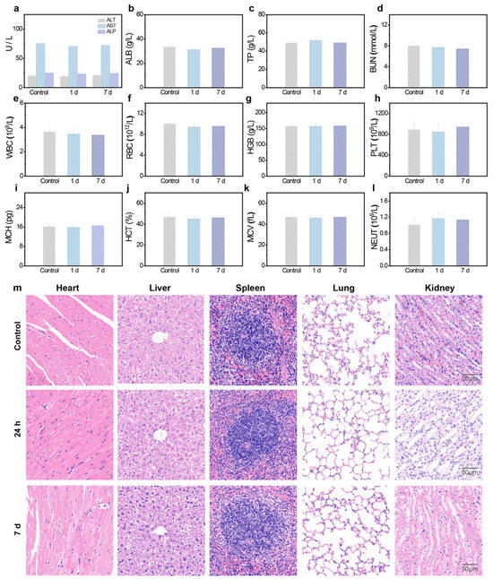
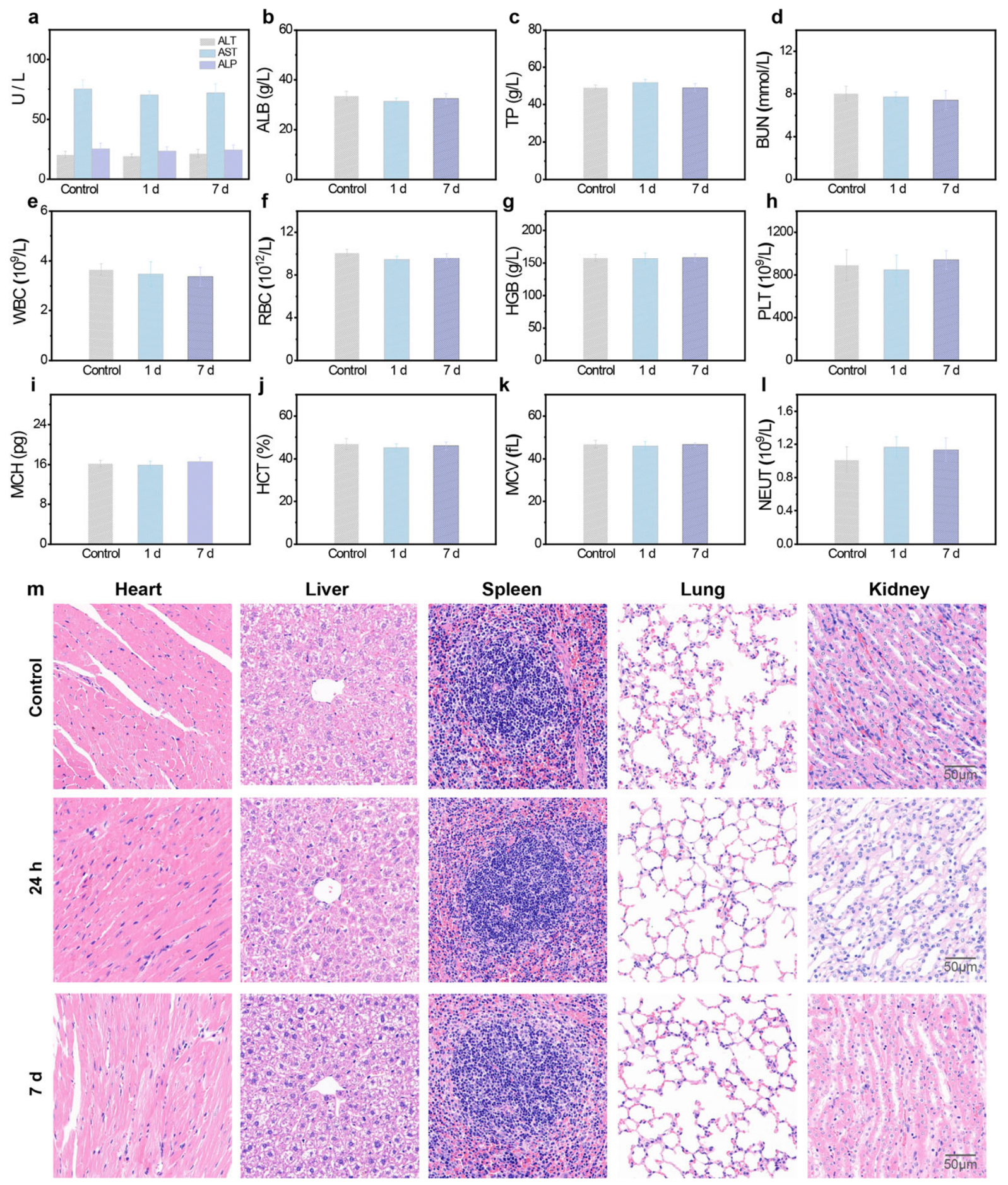
The systematic biocompatibility results are presented in Figure 6. Healthy mice were intravenously administered iSOD, and serum biochemical parameters, complete blood counts, together with histopathological examinations of major organs, were evaluated on day 1 and day 7 post-injection.
Figure 6.
Comprehensive in vivo biosafety and biocompatibility assessment of iSOD. Serum biochemical panels for hepatic and renal function measured at day 1 and day 7 following iSOD injection: (a) ALT, AST and ALP; (b) ALB; (c) TP; (d) BUN. Complete blood count parameters: (e) WBC; (f) RBC; (g) HGB; (h) PLT; (i) MCH; (j) HCT; (k) MCV; (l) NEUT. (m) Representative H&E staining of major organs (heart, liver, spleen, lung, and kidney) from control and iSOD-treated mice. No evident tissue damage or pathological lesions were observed. Scale bars: 100 μm.
To evaluate potential systemic toxicity toward metabolic organs, we measured serum biochemical markers related to liver and renal function (Figure 6a–d). Hepatic indicators included alanine aminotransferase (ALT), aspartate aminotransferase (AST), alkaline phosphatase (ALP), albumin (ALB), and total protein (TP); blood urea nitrogen (BUN) was detected to reflect renal status. Compared with the healthy control group, none of these hepatorenal-related parameters displayed obvious fluctuations in iSOD-treated mice at day 1 or day 7. These findings indicate that systemic iSOD administration does not induce acute or subacute hepatorenal impairment.
Complete blood count parameters were further analyzed to screen for possible systemic inflammation, hematopoietic suppression, or hematological abnormalities (Figure 6e–l). The detected hematological indices covered white blood cell count (WBC), RBC, hemoglobin (HGB), platelet count (PLT), mean corpuscular hemoglobin (MCH), hematocrit (HCT), mean corpuscular volume (MCV), and neutrophil count (NEUT). All parameters stayed within physiological normal ranges, and no obvious deviations were found between the control group and iSOD-treated groups at the two sampling time points. These results rule out sustained systemic inflammation or hematological toxicity after iSOD treatment.
Systemic safety was further validated by H&E staining of major organs (heart, liver, spleen, lung, and kidney) (Figure 6m). At day 1 and day 7 post-injection, tissues from iSOD-treated mice showed well-preserved tissue architecture. No obvious pathological lesions, inflammatory infiltration or fibrotic alterations were detected in comparison with control tissues. Prussian blue staining (Figure S8) was conducted to trace the in vivo metabolic clearance of the iron-based nanozymes. At day 1 following injection, sparse positive blue staining was observed within reticuloendothelial system-enriched organs, including the liver and spleen. Notably, the blue staining signals diminished markedly or were barely detectable by day 7, indicating that iSOD exhibits a favorable clearance profile and can be gradually cleared from the body.
Collectively, these comprehensive biocompatibility evaluations demonstrate that the iSOD nanoplatform exhibits favorable in vivo biosafety, without inducing obvious hepatorenal toxicity, hematological disturbance, or structural damage to major organs, which meets the basic safety criteria for future translational therapeutic applications.
4. Discussion
Herein, we constructed an ultrasmall iron oxide-based theranostic nanoplatform (termed iSOD) through a one-pot green biomineralization route, where SOD was employed as a biological template. The abundant surface hydroxyl, amino, and carboxyl groups of SOD coordinate Fe2+/Fe3+ ions and provide nucleation sites. Under alkaline conditions, these iron precursors undergo in situ coprecipitation to form ultrasmall Fe3O4 crystalline cores, while SOD spontaneously covers the crystal surface through interfacial hydroxyl interactions, forming an inorganic core–protein shell (Fe3O4@SOD) architecture [27]. Conventional TEM mainly visualizes the electron-dense Fe3O4 core, while the presence of the SOD shell is supported by DLS, FTIR, UV–Vis, and XPS analyses.
Compared with conventional enzyme immobilization strategies that rely on physical adsorption or random chemical cross-linking [27], the present synthetic approach takes advantage of the abundant surface hydroxyl moieties of SOD protein to trigger in situ nucleation and stabilization of Fe3O4 cores under mild aqueous conditions without any organic solvents. Such biomineralization-driven synthetic strategy greatly mitigates protein denaturation, a commonly encountered issue in multi-step covalent modification protocols, and enables the straightforward fabrication of compact core–shell nanostructures without additional purification steps. Physicochemical characterizations reveal that the as-prepared iSOD possesses an inorganic core size of 4.04 nm and a hydrodynamic diameter of ~28.3 nm. The SOD provides steric stabilization, rendering iSOD with good colloidal stability in physiologically relevant ionic media and effectively preventing irreversible particle aggregation, which remains a critical bottleneck for bare ultrasmall iron oxide nanoparticles [28].
The iSOD architecture enables a dual-enzyme cascade catalytic reaction. Conventional iron oxide nanozymes merely exhibit catalase-mimetic activity yet lack intrinsic capacity for direct superoxide anion elimination [29]. In contrast, free exogenous SOD is vulnerable to rapid proteolytic degradation and suffers from limited interstitial penetration within fibrotic microenvironments [30]. Simply mixing free SOD with inorganic nanozymes may not provide the same structural organization because the catalytic moieties are physically segregated [31]. Each iSOD nanoparticle comprises an ultrasmall Fe3O4 core with SOD molecules anchored on its surface. The SOD shell converts superoxide anions into hydrogen peroxide, which can be decomposed into water and oxygen by the CAT-like Fe3O4 core. This dual-enzyme cascade suppresses the buildup of intermediate H2O2 and achieves persistent ROS scavenging. iSOD treatment was associated with reduced oxidative stress-driven hepatic stellate cell activation and excessive extracellular matrix deposition [32], thereby slowing the pathological progression of hepatic fibrosis.
Clinically, the management of liver fibrosis faces two major unmet challenges: accurate noninvasive fibrosis staging and effective anti-fibrotic therapeutic interventions [33]. Commercially available liver MRI contrast agents mostly only produce morphological parenchymal enhancement and fail to discriminate different fibrotic grades [34]. The distinct MRI performance of iSOD originates from its ultrasmall dimension and intrinsic magnetic relaxation features. Owing to its ultrasmall Fe3O4 core, iSOD exhibits prominent T1 shortening behaviour with an r1 of 2.874 mM−1s−1. Its moderate r2/r1 ratio (4.83) yields unique imaging signatures critically dependent on its spatial distribution, different from canonical T1 positive contrast agents. Before iSOD administration, T1-weighted MR scans display negligible intrinsic parenchymal signal heterogeneity across healthy, moderate fibrosis, and severe fibrosis animal groups. After intravenous delivery, iSOD is predominantly internalized by hepatic Kupffer cells [35]. In healthy liver tissues, uptake of iSOD by Kupffer cells induces local magnetic susceptibility effects from densely packed particles. Such susceptibility effects dominate over the intrinsic T1-shortening property, resulting in a net homogeneous attenuation of T1-weighted background signals throughout the whole liver [36]. In fibrotic livers, compromised Kupffer-cell function and distorted tissue microarchitecture disrupt this uniform signal reduction and give rise to clear parenchymal signal heterogeneity. The degree of this post contrast signal heterogeneity scales with fibrotic severity, which permits differentiation between moderate and severe liver fibrosis. These observations indicate that iSOD can serve as an imaging-assisted readout for noninvasive liver fibrosis staging.
The diagnostic capability of iSOD is intrinsically coupled with its therapeutic performance. Once accumulated within fibrotic foci, iSOD scavenges surplus local ROS and attenuates oxidative stress propelled fibrotic remodeling. Histological examinations including H&E, Masson’s trichrome, and Sirius Red staining demonstrate that iSOD treatment relieves inflammatory infiltration and curtails excessive collagen accumulation in both moderate and severe fibrosis animal models. The anti-fibrotic performance of iSOD arises from its ROS eliminating capability, which protects hepatic stellate cells against oxidative injury and reduces fibrotic pathological remodeling. Hence, iSOD combines imaging and anti-fibrotic therapy in one platform, where imaging assessment can be implemented simultaneously with anti-fibrotic therapy, offering potential advantages over therapy-only nanomedicines.
Systematic in vivo biocompatibility assessments demonstrate that no apparent hepatorenal damage, hematological anomalies, or pathological lesions in major organs are detected in experimental animals within the 7-day observation window following intravenous iSOD injection. In contrast to conventional inorganic nanozymes that suffer from slow clearance and prolonged organ sequestration [37], iSOD undergoes efficient metabolic clearance attributed to its ultrasmall particle dimension. Meanwhile, the native SOD protein shell forms a good biocompatible outer surface and lowers the risk of long-term toxic responses. Prussian blue staining further corroborates its favorable metabolic clearance behaviour. Taken together, these results validate the suitability of iSOD for in vivo theranostic implementation. Most existing ROS scavenging nanotherapeutics lack real time imaging capacity [32], whereas clinical MRI contrast agents possess negligible anti- fibrotic performance [34]; iSOD combines both functionalities and partially bridges this translational gap.
Admittedly, the present study still bears several limitations. Conventional histological staining and pathological observation constitute our primary evaluation metrics for therapeutic responses. Follow-up investigations will introduce biochemical collagen quantification and other analytical assays to quantitatively decipher iSOD triggered extracellular matrix remodeling. Since only short-term acute biosafety has been evaluated, further efforts are required to elucidate the long-term biosafety profile and comprehensive in vivo biodistribution of iSOD [38]. Future work will aim at improving nanoparticle monodispersity and developing rationally designed targeting modification strategies to enhance lesion-oriented accumulation and in vivo theranostic specificity [39].
5. Conclusions
In summary, we report a facile-to-fabricate, biocompatible multifunctional biomimetic nanoplatform that combines noninvasive MRI and cascade-driven antioxidant therapy for liver fibrosis. As a valuable tool for fibrosis staging and therapeutic intervention, iSOD offers promising translational potential in nanomedicine and advanced biomaterial research.
Supplementary Materials
The following supporting information can be downloaded at https://www.mdpi.com/article/10.3390/pharmaceutics18111387/s1.
Author Contributions
Conceptualization, X.S., F.S. (Fengxiang Song) and Y.S.; methodology, Y.Y.; software, F.S. (Fengxiang Song); validation, X.S., W.G., N.S., Y.S. and W.Y.; formal analysis, X.X.; investigation, Y.Y. and F.S. (Fei Shan); resources, F.S. (Fei Shan); data curation, N.S.; writing—original draft preparation, X.X.; writing—review and editing, X.S. and W.Y.; visualization, X.X., Y.Y., W.G. and F.S. (Fei Shan); supervision, F.S. (Fei Shan) and Y.S.; project administration, W.Y.; funding acquisition, X.S. and W.Y. All authors have read and agreed to the published version of the manuscript.
Funding
This research was sponsored by National Natural Science Foundation of China (82302265), Shanghai Sailing Program (21YF1436600), and the Start-up Fund from Shanghai Public Health Clinical Center (RCJJ2025-07).
Institutional Review Board Statement
All animal experiments were performed according to the protocols approved by the Institutional Animal Care and Use Committee of Shanghai Public Health Clinical Center (Approval No.: 2025-A066-01; approval date: 31 October 2025).
Informed Consent Statement
Not applicable.
Data Availability Statement
The data presented in this work can be obtained from the corresponding authors.
Conflicts of Interest
The authors declare no conflicts of interest.
Abbreviations
The following abbreviations are used in this manuscript:
| ALB | albumin |
| ALP | alkaline phosphatase |
| ALT | alanine aminotransferase |
| AST | aspartate aminotransferase |
| BUN | blood urea nitrogen |
| Calcein-AM | Calcein acetoxymethyl ester |
| CAT | catalase |
| CCK-8 | Cell Counting Kit-8 |
| DCFH-DA | 2′,7′-dichlorodihydrofluorescein diacetate |
| DLS | dynamic light scattering |
| DMPO | 5,5-dimethyl-1-pyrroline-N-oxide |
| ECM | extracellular matrix |
| ESR | electron spin resonance |
| H&E | hematoxylin and eosin |
| HCT | hematocrit |
| HGB | hemoglobin |
| HSCs | hepatic stellate cells |
| MRI | magnetic resonance imaging |
| NEUT | neutrophil count |
| PBS | phosphate-buffered saline |
| PI | propidium iodide |
| PLT | platelet count |
| r1 | longitudinal relaxivity |
| r2 | transverse relaxivity |
| RBC | red blood cell |
| ROS | reactive oxygen species |
| SOD | superoxide dismutase |
| T1 | longitudinal relaxation time |
| T2 | transverse relaxation time |
| TAA | thioacetamide |
| TEM | transmission electron microscopy |
| TP | total protein |
| UV-Vis | ultraviolet–visible spectroscopy |
| WBC | white blood cell count |
| XAFS | X-ray absorption fine structure spectroscopy |
| XPS | X-ray photoelectron spectroscopy |
| XRD | X-ray diffraction |
References
- Taru, V.; Szabo, G.; Mehal, W.; Reiberger, T. Inflammasomes in chronic liver disease: Hepatic injury, fibrosis progression and systemic inflammation. J. Hepatol. 2024, 81, 895–910. [Google Scholar] [CrossRef] [Scilit] [PubMed]
- Ginès, P.; Thiele, M.; Graupera, I.; Serra-Burriel, M.; de Knegt, R.J.; Lammert, F.; Castera, L.; Korenjak, M.; Kamath, P.S.; Grgurević, I.; et al. Screening for fibrosis to diagnose liver diseases early: The LIVERSCREEN project. Nat. Med. 2023, 29, 774–775. [Google Scholar] [CrossRef] [Scilit] [PubMed]
- Foglia, B.; Novo, E.; Protopapa, F.; Maggiora, M.; Bocca, C.; Cannito, S.; Parola, M. Hypoxia, Hypoxia-Inducible Factors and Liver Fibrosis. Cells 2021, 10, 1764. [Google Scholar] [CrossRef] [Scilit] [PubMed]
- Zhang, D.; Zhang, Y.; Sun, B. The Molecular Mechanisms of Liver Fibrosis and Its Potential Therapy in Application. Int. J. Mol. Sci. 2022, 23, 12572. [Google Scholar] [CrossRef] [Scilit] [PubMed]
- Benavides, C.; Kecheva, P.; Solano, F.; Martínez-Arroyo, O.; Esplugues, J.V.; Blas-García, A.; Apostolova, N. The Matrix Reloaded: The Hepatic Matrisome as a Therapeutic Opportunity to Fight Liver Fibrosis. Biomolecules 2026, 16, 884. [Google Scholar] [CrossRef] [Scilit] [PubMed]
- Chowdhury, A.B.; Mehta, K.J. Liver biopsy for assessment of chronic liver diseases: A synopsis. Clin. Exp. Med. 2023, 23, 273–285. [Google Scholar] [CrossRef] [Scilit] [PubMed]
- Huang, W.; Peng, Y.; Kang, L. Advancements of non-invasive imaging technologies for the diagnosis and staging of liver fibrosis: Present and future. View 2024, 5, 20240010. [Google Scholar] [CrossRef] [Scilit]
- Zhao, M.; Wang, L.; Wang, M.; Zhou, S.; Lu, Y.; Cui, H.; Racanelli, A.C.; Zhang, L.; Ye, T.; Ding, B.; et al. Targeting fibrosis, mechanisms and clinical trials. Signal Transduct. Target Ther. 2022, 7, 206. [Google Scholar] [CrossRef] [Scilit] [PubMed]
- Banerjee, P.; Gaddam, N.; Chandler, V.; Chakraborty, S. Oxidative Stress-Induced Liver Damage and Remodeling of the Liver Vasculature. Am. J. Pathol. 2023, 193, 1400–1414. [Google Scholar] [CrossRef] [Scilit] [PubMed]
- Zhang, J.; Zhang, T.; Zhang, Z.; Jia, Q.; Chen, J.; Li, J.; Song, H.; Li, J.; Xiong, Y.; Mo, L.; et al. The Dual-Target Nanoparticles with ROS Sensitivity Inhibit the Hedgehog Signaling Pathway and Decrease Oxidative Stress in Activated Hepatic Stellate Cells to Alleviate Liver Fibrosis. Adv. Funct. Mater. 2024, 34, 2404658. [Google Scholar] [CrossRef] [Scilit]
- Kisseleva, T.; Brenner, D. Molecular and cellular mechanisms of liver fibrosis and its regression. Nat. Rev. Gastroenterol. Hepatol. 2021, 18, 151–166. [Google Scholar] [CrossRef] [Scilit] [PubMed]
- Yang, H.; Wang, W.; Xiao, J.; Yang, R.; Feng, L.; Xu, H.; Xu, L.; Xing, Y. ROS-responsive injectable hydrogels loaded with exosomes carrying miR-4500 reverse liver fibrosis. Biomaterials 2025, 314, 122887. [Google Scholar] [CrossRef] [Scilit] [PubMed]
- Ma, Z.; Tian, X.; Yu, S.; Shu, W.; Zhang, C.; Zhang, L.; Wang, F. Liver Fibrosis Amelioration by Macrophage-Biomimetic Polydopamine Nanoparticles via Synergistically Alleviating Inflammation and Scavenging ROS. Mol. Pharm. 2024, 21, 3040–3052. [Google Scholar] [CrossRef] [Scilit] [PubMed]
- Wang, Z.; Zhang, R.; Hu, Y.; Song, Q.; Fu, X.; Wang, Y.; Chen, M.; Fan, K.; Fan, C.; Sun, D. Chiral metal nanozyme-enabled SOD2 activation and siRNA delivery cooperatively mitigating chronic heart failure. Bioact. Mater. 2025, 58, 203–220. [Google Scholar] [CrossRef] [Scilit] [PubMed]
- Bhagat, S.; Kushwaha, S.; Singh, S. Targeted Gut Delivery of Zn, Cu, and Mn Nanominerals Alleviates Oxidative Stress by Activating Endogenous SOD Enzymes. Adv. Healthc. Mater. 2026, 15, e71209. [Google Scholar] [CrossRef] [Scilit] [PubMed]
- Yang, X.; Tan, M.; Guo, J.; Xiang, J.; Yin, F.; Deng, J.; Luo, J.; Xiao, S.; Mo, M.; Wang, H.; et al. PdZn/CoSA-NC Nanozymes with Highly Efficient SOD/CAT Activities for Treatment of Osteoarthritis via Regulating Immune Microenvironment. Adv. Funct. Mater. 2024, 34, 2401963. [Google Scholar] [CrossRef] [Scilit]
- Li, S.; Li, H.; Xu, X.; Saw, P.E.; Zhang, L. Nanocarrier-mediated antioxidant delivery for liver diseases. Theranostics 2020, 10, 1262–1280. [Google Scholar] [CrossRef] [Scilit] [PubMed]
- Rosa, A.C.; Bruni, N.; Meineri, G.; Corsi, D.; Cavi, N.; Gastaldi, D.; Dosio, F. Strategies to expand the therapeutic potential of superoxide dismutase by exploiting delivery approaches. Int. J. Biol. Macromol. 2021, 168, 846–865. [Google Scholar] [CrossRef] [Scilit] [PubMed]
- Zhang, D.; Zhang, J.; Bian, X.; Zhang, P.; Wu, W.; Zuo, X. Iron Oxide Nanoparticle-Based T1 Contrast Agents for Magnetic Resonance Imaging: A Review. Nanomaterials 2024, 15, 33. [Google Scholar] [CrossRef] [Scilit] [PubMed]
- Gao, Y.; Shi, X.; Shen, M. Intelligent Design of Ultrasmall Iron Oxide Nanoparticle-Based Theranostics. ACS Appl. Mater. Interfaces 2021, 13, 45119–45129. [Google Scholar] [CrossRef] [Scilit] [PubMed]
- Xue, X.; Cui, T.; Liu, D.; Qiu, H.; Wang, H.; Yun, Y.; Qiu, Y.; Yang, W.; Zhang, B. Injury-Retention Strategy for Discriminative Magnetic Resonance Imaging and Enhanced Antioxidant Therapy of Drug-Induced Liver Injury. Nano Lett. 2026, 26, 3846–3856. [Google Scholar] [CrossRef] [Scilit] [PubMed]
- Li, Z.; Wu, B.; Li, W.; Marsili, E.; Tang, K.; Sun, K.; Cao, Z.; Zhang, Y.; Huang, H.; Li, X.; et al. Unlocking an Efficient SOD-to-CAT Catalytic Relay in a MOF Nanozyme via Dopamine-Driven Interfacial Nanoreconstruction for Osteoarthritis Therapy. Adv. Mater. 2026, 38, e72747. [Google Scholar] [CrossRef] [Scilit] [PubMed]
- Roshanghias, H.; Ganjavinia, N.; Zarei-Shokat, S.; Maleki, A. An overview on the multifunctional nanozyme activities of iron oxide magnetic nanoparticles for nanomedicine. Mater. Adv. 2026, 7, 6971–6995. [Google Scholar] [CrossRef] [Scilit]
- Shi, X.; Yang, W.; Ma, Q.; Lu, Y.; Xu, Y.; Bian, K.; Liu, F.; Shi, C.; Wang, H.; Shi, Y.; et al. Hemoglobin-mediated biomimetic synthesis of paramagnetic O2-evolving theranostic nanoprobes for MR imaging-guided enhanced photodynamic therapy of tumor. Theranostics 2020, 10, 11607–11621. [Google Scholar] [CrossRef] [Scilit] [PubMed]
- Wallace, M.; Hamesch, K.; Lunova, M.; Kim, Y.; Weiskirchen, R.; Strnad, P.; Friedman, S. Standard operating procedures in experimental liver research: Thioacetamide model in mice and rats. Lab. Anim. 2015, 49, 21–29. [Google Scholar] [CrossRef] [Scilit] [PubMed]
- Liguori, A.; Zoncapè, M.; Casazza, G.; Easterbrook, P.; Tsochatzis, E.A. Staging liver fibrosis and cirrhosis using non-invasive tests in people with chronic hepatitis B to inform WHO 2024 guidelines: A systematic review and meta-analysis. Lancet Gastroenterol. Hepatol. 2025, 10, 332–349. [Google Scholar] [CrossRef] [Scilit] [PubMed]
- Zhang, D.-G.; Pan, Y.-J.; Chen, B.-Q.; Lu, X.-C.; Xu, Q.-X.; Wang, P.; Kankala, R.K.; Jiang, N.-N.; Wang, S.-B.; Chen, A.-Z. Protein-guided biomimetic nanomaterials: A versatile theranostic nanoplatform for biomedical applications. Nanoscale 2024, 16, 1633–1649. [Google Scholar] [CrossRef] [Scilit] [PubMed]
- Jeon, M.; Halbert, M.V.; Stephen, Z.R.; Zhang, M. Iron Oxide Nanoparticles as T1 Contrast Agents for Magnetic Resonance Imaging: Fundamentals, Challenges, Applications, and Prospectives. Adv. Mater. 2021, 33, e1906539. [Google Scholar] [CrossRef] [Scilit] [PubMed]
- Liang, M.; Yan, X. Nanozymes: From New Concepts, Mechanisms, and Standards to Applications. Acc. Chem. Res. 2019, 52, 2190–2200. [Google Scholar] [CrossRef] [Scilit] [PubMed]
- Zamanian Dastmalchi, H.; Dashtestani, F.; Ghourchian, H. Superoxide dismutase-mimetic nanozymes: A promising alternative to natural superoxide dismutases for biomedical and industrial applications. Colloids Surf. B Biointerfaces 2026, 257, 115138. [Google Scholar] [CrossRef] [Scilit] [PubMed]
- Zhu, Y.; Wang, W.; Cheng, J.; Qu, Y.; Dai, Y.; Liu, M.; Yu, J.; Wang, C.; Wang, H.; Wang, S.; et al. Stimuli-Responsive Manganese Single-Atom Nanozyme for Tumor Therapy via Integrated Cascade Reactions. Angew. Chem. Int. Ed. Engl. 2021, 60, 9480–9488. [Google Scholar] [CrossRef] [Scilit] [PubMed]
- Jing, H.; Ren, Y.; Zhou, Y.; Xu, M.; Krizkova, S.; Heger, Z.; Lu, Q.; Wang, S.; Liang, X.; Adam, V.; et al. Remodeling of the liver fibrosis microenvironment based on nilotinib-loaded multicatalytic nanozymes with boosted antifibrogenic activity. Acta Pharm. Sin. B 2023, 13, 5030–5047. [Google Scholar] [CrossRef] [Scilit] [PubMed]
- Xie, D.; Xie, L.; Fang, C.; Du, Z.; Cao, Z.; Su, C.; Huo, Y. New advances of nanozymes for the diagnosis and treatment of digestive system diseases (Review). Int. J. Mol. Med. 2025, 56, 176. [Google Scholar] [CrossRef] [Scilit] [PubMed]
- Hu, X.; Xu, M.; Xiao, X.; Wang, N.; Zhang, Q.; Ge, J.; Zeng, J. Molecular Imaging Probes for Early Detection and Staging of Liver Fibrosis. Chem. Biomed. Imaging 2025, 3, 702–714. [Google Scholar] [CrossRef] [Scilit] [PubMed]
- Kiessling, F.; Mertens, M.E.; Grimm, J.; Lammers, T. Nanoparticles for imaging: Top or flop? Radiology 2014, 273, 10–28. [Google Scholar] [CrossRef] [Scilit] [PubMed]
- Daldrup-Link, H.E. Ten Things You Might Not Know about Iron Oxide Nanoparticles. Radiology 2017, 284, 616–629. [Google Scholar] [CrossRef] [Scilit] [PubMed]
- Xu, K.; Liu, Y.; Chen, C. Future prospects in clinical translation of inorganic nanoparticles. Acta Pharm. Sin. B 2024, 14, 5082–5084. [Google Scholar] [CrossRef] [Scilit] [PubMed]
- Meng, X.; Zhu, G.; Sun, S.; Yao, W.; Zhang, Y.; Yang, Y.; Sun, T. Nanozymes for Liver Disease Therapy: Advances in Catalytic Activity, Targeting Strategies, and Clinical Translation. Adv. Sci. 2026, 13, e74590. [Google Scholar] [CrossRef] [Scilit] [PubMed]
- Mao, Y.; Gong, Y.; Bai, X. The Dual-Faceted Role of Metal-Based Nanomaterials in Hepatic Fibrosis Therapy. Int. J. Nanomed. 2026, 21, 574077. [Google Scholar] [CrossRef] [Scilit] [PubMed]
Disclaimer/Publisher’s Note: The statements, opinions and data contained in all publications are solely those of the individual author(s) and contributor(s) and not of MDPI and/or the editor(s). MDPI and/or the editor(s) disclaim responsibility for any injury to people or property resulting from any ideas, methods, instructions or products referred to in the content. |
© 2026 by the authors. Licensee MDPI, Basel, Switzerland. This article is an open access article distributed under the terms and conditions of the Creative Commons Attribution (CC BY) license.