


Abstract
The accumulation of senescent cells in tissues serves as a fundamental driver of aging and age-related pathologies, including chronic inflammation, tissue dysfunction, and organ degeneration. Conventional anti-aging interventions often suffer from limited specificity and off-target toxicity. Currently, developing therapeutic strategies capable of precisely targeting senescent cells is urgent. This review focuses on recent advances in delivery strategies designed to achieve either the selective elimination or functional modulation of senescent cells, with the goal of transitioning from broad-spectrum interventions to precision senotherapy. We systematically discuss the latest developments in senescence-targeting delivery platforms. To realize in vivo senescent-cell clearance, nanocarrier-based systems which are responsive to the senescent microenvironment and immune cell-based approaches are engineered. In parallel, we also reviewed emerging strategies for functional remodeling. Advanced delivery vehicles including lipid nanoparticles, extracellular vesicles, and engineered cell therapies are used to realize metabolic reprogramming and epigenetic modulation. The critical mechanisms of delivery systems in overcoming diverse barriers to improve specificity, bioavailability, and safety are highlighted. In this review, we aim to provide a preliminary framework that could serve as a useful reference for guiding the clinical translation of emerging senotherapies.
1. Introduction
Cellular senescence is a state of irreversible cell-cycle arrest triggered by DNA damage, telomere attrition and oncogenic stress [1,2,3,4]. It is characterized by elevated senescence-associated β-galactosidase (SA-β-gal) activity and the senescence-associated secretory phenotype (SASP) [5,6]. SASP is a complex mixture of pro-inflammatory cytokines, chemokines, growth factors and matrix-remodeling enzymes [7,8]. Therefore, senescent cells drive chronic inflammation, disrupt tissue architecture and impair stem cell function [9,10,11]. Results from both preclinical and clinical studies firmly reveal that the accumulation of senescent cells causally contributes to aging and diverse pathologies including atherosclerosis, osteoarthritis, neurodegeneration, pulmonary fibrosis and metabolic dysfunction, positioning these cells as a prime therapeutic target [12,13,14,15,16,17].
Currently, senotherapies are broadly classified as senolytics, which selectively eliminate senescent cells, and senomorphics, which modify the senescent phenotype or suppress SASP without necessarily killing these cells [18]. Senolytics require high selectivity over non-senescent cells and controlled apoptosis induction, whereas senomorphics favor sustained local exposure while avoiding broad immunosuppression and preserving beneficial senescence. Their biomarkers and safety considerations are also different. Senolytic efficacy requires evidence of senescent-cell clearance, not merely marker reduction, while senomorphic efficacy is assessed by reduced SASP and improved tissue function despite persistent senescent cells. Given these differences, platforms with dual-action are emerging [12,19]. The senolytic approach, pioneered by agents like dasatinib and quercetin, has shown preclinical efficacy in reducing senescent burden across multiple disease models [20,21,22]. Generally, ABT263 (navitoclax), quercetin, and fisetin are primarily classified as senolytics, as they selectively induce apoptosis in senescent cells by targeting anti-apoptotic pathways. However, clinical translation is hampered by insufficient selectivity, off-target toxicity (e.g., thrombocytopenia with ABT263) [23], and poor bioavailability [24]. Recently, antibody–drug conjugates have gained great clinical success [25,26,27,28,29]. Moreover, nanotechnology also provides great potential to amplify the efficacy [30,31,32,33]. Therefore, recent efforts to overcome these limitations include antibody–drug conjugates targeting uPAR [34,35] and nanoparticle systems exploiting senescence-associated metabolic features such as lipofuscin accumulation or elevated β-galactosidase activity [36,37,38]. These advances underscore the pivotal role of delivery systems in enhancing specificity and safety [39,40,41,42].
Senomorphic strategies offer a complementary route [12,20,43]. These strategies aim to modulate the SASP, restore homeostasis or reverse senescence phenotypes via metabolic or epigenetic reprogramming [18,44,45]. Rapamycin and dexamethasone are more appropriately classified as senomorphics since their principal effect is to suppress the SASP or modulate the senescent phenotype without directly eliminating senescent cells. Senomorphic approaches might be advantageous when senescent cells play beneficial roles, such as in wound healing or development [12,46,47]. Nevertheless, senomorphic interventions face several hurdles: (1) SASP heterogeneity across cell types and tissues complicates target identification [20,48]; (2) sustained administration raises long-term safety and compliance concerns [49,50,51]; (3) systemic administration of senomorphic agents might affect non-senescent cells or interfere with essential physiological processes [9,20,52]. Recent progress in delivery technologies hold great potential to reduce systemic toxicity and surmount these barriers [53,54]. For instance, glucose-modified hybrid nanocarriers encapsulating NAD+ precursors have enabled dynamic, temporally controlled targeting of distinct cell populations during tissue repair [5,55]. Engineered mesenchymal progenitor cells resistant to senescence (SRCs) function as living delivery vehicles, persistently secreting rejuvenating factors and reversing multi-organ aging in non-human primates [56]. In addition, extracellular vesicles are emerging as promising carriers for drug delivery [57,58,59,60]. Extracellular vesicle platforms loaded with α-ketoglutarate or derived from antler blastema progenitors have shown efficacy in ameliorating skin aging and bone loss in murine and primate models [61,62,63]. These examples highlight how advanced delivery systems can achieve the specificity, durability and safety essential for effective senotherapies [64,65,66].
In the following sections, we review recent advances in delivery platforms for both senolytic elimination and senomorphic remodeling of senescent cells (Figure 1). The design principles, mechanistic insights and translational potential were discussed. Recent microenvironment-responsive nanocarriers, living cell-based therapies, stem cell-derived therapeutics and organelle delivery approaches against senescence are reviewed. We aim to provide a framework that guides the rational development of next-generation senotherapies.
Figure 1.
Schematic illustration of mechanisms for senescence-targeting delivery strategies. Schematic illustration is created in BioRender. Zefan Liu (2026) https://BioRender.com/foyo4is.
2. Strategies for Precise Elimination of Senescent Cells
2.1. Microenvironment-Responsive Targeted Delivery Based on Prodrug or Nanoparticles
Currently, the hallmarks of aging contain multiple regulatory pathways including genomic instability, telomere attrition, epigenetic alterations, loss of proteostasis, and mitochondrial dysfunction [67,68,69,70]. Complex molecular events derived from the interplay of these signaling pathways eventually converge on senescent cells, which function as a crucial constituent [1,71]. The microscopic molecular damage and pathway imbalances persistently drive normal cells into senescence, leading to the accumulation and dysfunction of senescent cells [1,72,73]. Therefore, the clearance of senescent cells represents a promising therapeutic approach [67].
In recent years, anti-aging treatments employ non-specific systemic drug administration [5]. The core strategy is to directly intervene in the aging process using identified senolytic compounds [74]. Results from current studies have confirmed the anti-aging potential of molecules such as quercetin, fisetin, ABT263 (Navitoclax), rapamycin, and dexamethasone [19,20,34,75,76,77,78,79]. Although these agents all exhibit anti-aging potential, their mechanisms of action are not identical. Quercetin, fisetin and ABT263 (Navitoclax) are primarily classified as senolytics. Rapamycin and dexamethasone are considered to be senomorphics, delaying aging via senolytic-independent mechanisms [80,81,82,83,84]. For example, oral systemic administration of ABT263 to naturally aged mice and irradiation-induced premature aging mice effectively eliminates various senescent cells by inhibiting the anti-apoptotic proteins BCL-2/BCL-xL [85]. The ABT263 administration significantly ameliorates the aging phenotypes of the hematopoietic system and skeletal muscle [85,86].
However, this non-specific systemic administration approach has unavoidable limitations [87,88,89]. Firstly, after administration, the active ingredient is hindered by low bioavailability and rapid metabolization by the liver, making it difficult to reach the effective therapeutic concentration in specific lesion tissues [90,91]. For example, the oral bioavailability of the natural flavonoid quercetin is less than 1% [92,93,94]. Moreover, the penetration efficiency of these free drugs is extremely low against physiological barriers such as the blood–brain barrier, dense cartilage matrix, and the nucleus pulposus of the intervertebral disc [95,96,97,98]. This is a key reason why the systemic use of quercetin alone is ineffective in intervening in the aging of bones, joints and intervertebral discs [99,100]. Quercetin failed to function on nucleus pulposus cells and senescent chondrocytes due to the insufficient drug accumulation [101]. Secondly, current drugs for senescent-cell clearance lack specificity, causing off-target toxicity against normal proliferating cells [102,103]. ABT263 serves as a representative example of how senolytic agents affect normal cells [104]. ABT263 eliminates senescent cells by inhibiting BCL family proteins but this pathway is also present in platelets and bone marrow stem cells [105]. After systemic administration, ABT263 disrupts platelet homeostasis and induces hematological toxicities such as thrombocytopenia [106]. The proliferative function of normal stem cells is also impaired [85,107].
In this context, nanocarrier-based targeted delivery has emerged as a promising direction for senescent-cell clearance [108]. These systems exhibit unique physicochemical properties, tissue penetration and controlled release profiles [109,110]. These features further increase drug accumulation at senescent lesions and enable sustained release [111] (Table 1).
Lorenza Garau Paganella et al. developed a PNP supramolecular hydrogel loaded with navitoclax nanoparticles (PNP-Navitoclax) by blending navitoclax with an injectable hydrogel. The in vitro experiments showed that PNP-Navitoclax gradually eroded, with 70% total weight loss after 96 h. This enables a sustained release of navitoclax. The IC50 of PNP-Navitoclax was 4 μM against senescent endothelial cells but 16 μM against healthy endothelial cells. This result indicated that PNP-Navitoclax exhibited a wider therapeutic window, indicating relative selectivity at lower concentrations. Thus, the corresponding therapeutic window is substantially broadened and side effects might be reduced [112] (Figure 2). Increasing research attention has been directed toward hydrogel-based delivery systems [113].
Figure 2.
Representative microenvironment-responsive hydrogel nanoplatforms for local clearance of senescent cells [112]. Reprint with permission from [112] Copyright {2025} American Chemical Society (ACS). ** p < 0.01, *** p < 0.001.
While direct intra-lesional injection can increase drug concentration locally, these formulations rely solely on passive erosion by bodily fluids and fail to achieve site-specific drug release [114,115,116]. Normal tissues exposed to the gel might also trigger slow drug leakage over time [117,118,119]. Moreover, during local injection of hydrogel-based nanomaterials, the shear force from syringe plunging might disrupt the superficial gel network, causing minor initial burst release [112,120,121]. To further address these issues, senescence microenvironment-responsive nanocarriers have been explored [122]. By exploiting the physicochemical and biological differences between degenerated senescent tissues and normal tissues, these intelligent systems can specifically recognize and respond to local signals of the senescent niche [122]. This design enables prolonged drug retention at the lesion site while achieving precise accumulation near the target cells for senescent-cell clearance [113].
Reactive oxygen species (ROS) are important responsive targets within the senescent microenvironment [123,124]. ROS are routine byproducts of cellular metabolism and are normally maintained at balanced levels under physiological conditions [125,126]. In aging tissues, however, the antioxidant defense system declines, leading to progressive ROS accumulation and a characteristic hyperoxidative microenvironment [127]. ROS not only drive cellular senescence and tissue damage but also serve as highly identifiable pathological signals at senescent lesions [1,128].
Taking advantage of this feature, ROS-responsive nanocarriers for precise drug release at senescent sites have been developed. Zhongke Wang et al. used fisetin which antagonizes BCL proteins to overcome the apoptosis resistance of senescent cells. Firstly, they prepared fisetin-loaded liposomes (F-Lip) via a thin-film dispersion method. Separately, they synthesized GelMA (methacrylated gelatin) from a gelatin backbone and permanently grafted 3-acrylamidophenylboronic acid (APBA) onto the GelMA polymer via free radical copolymerization. This process yielded a boronic acid-functionalized GelMA hydrogel loaded with fisetin liposomes and MnO2 nanoenzymes, referred to as FMAG. Compared with unmodified GelMA controls, the FMAG hydrogel relied on dynamic boronate ester crosslinking to achieve notable ROS-responsive release. Under normal physiological conditions (pH 7.4), only 9.2% of fisetin and 8.5% of MnO2 were released within 48 h, indicating minimal drug leakage. In contrast, under simulated periodontal high-ROS conditions (pH 6.6 with H2O2), both components exceeded 70% release within 48 h, with markedly enlarged swelling pores. This allowed synchronized release of senolytic F-Lip and antioxidant MnO2, enabling on-demand drug release at lesions while minimizing off-target effects in healthy tissues. In vivo, after 28 days of treatment, the FMAG group showed only 6.8% p16-positive area, far lower than the periodontitis model group (47.6%) and monotherapy groups. The expression levels of senescence markers p16 and p21 in the FMAG group approached those of healthy controls. Moreover, FMAG significantly enhanced macrophage efferocytosis and promoted M2 macrophage polarization. These results demonstrated that FMAG effectively eliminated senescent gingival fibroblasts and interrupted the vicious cycle of SASP-driven inflammation at its source [113].
Although ROS-responsive release systems based on boronate ester bonds (such as APBA) can achieve passive drug release at the tissue level, they have inherent limitations in practice [129,130,131]. Inflammation leads to non-specific oxidation stimulation [132,133]. The various substances in bodily fluids including weak acids and polyols which can disrupt the crosslinking bonds caused drug leakage into healthy tissues [134,135]. Moreover, the modified hydrogels remain vulnerable to mechanical forces and suffer from short release durations. Therefore, achieving active and precise targeting at the cellular level represents the next research priority [113,136,137].
Dipeptidyl Peptidase 4 (DPP4) is localized on the membrane of senescent chondrocytes but is barely detectable on normal chondrocytes [138,139]. Upon oxidative stress or injury, senescent chondrocytes significantly upregulate membrane DPP4 expression [140]. In the context of age-related disease, DPP4 competitively binds to MYH9 (myosin heavy chain 9), thereby blocking its degradation by E3 ubiquitin ligase [141]. This leads to MYH9 accumulation, which in turn triggers excessive mitochondrial fission and dysfunction. As a result, oxidative stress surges and drives permanent chondrocyte cell-cycle arrest, further aggravating senescence [142,143]. DPP4 might be considered a relative rather than absolute marker of senescent chondrocytes. It is markedly upregulated on senescent chondrocytes under oxidative stress or injury, while barely detectable on normal chondrocytes. However, DPP4 is not exclusive to senescent cells [144]. Local intra-articular delivery might provide sufficient selectivity, whereas systemic senotherapy would likely require combination with additional senescence-associated markers or microenvironment-responsive approaches.
Peng et al. developed a nanocarrier system that uses an anti-DPP4 antibody to deliver sabutoclax specifically to senescent cells for the treatment of osteoarthritis. This system termed BS@MD consists of a core formed by co-assembly of bortezomib and sabutoclax. Then, it is coated with macrophage membrane vesicles derived from LPS-pretreated RAW264.7 cells and functionalized with DPP4 antibody. The macrophage membrane coating serves two functions. Firstly, cytokine receptors on the membrane broadly neutralize senescence-associated secretory phenotype (SASP) factors. Secondly, the membrane carries CD47 protein, which engages the CD47–SIRPα pathway to achieve immune evasion and phagocytic clearance. To enable senescent-cell targeting, anti-DPP4 antibodies were conjugated onto the membrane surface. BS@MD showed a positive uptake portion of 58.3% in senescent chondrocytes, compared with only 12.0% in normal chondrocytes in vitro. When the DPP4 binding sites on senescent chondrocytes were pre-blocked with anti-DPP4 antibody, BS@MD uptake dropped by 92%, confirming the targeting specificity. p16 immunohistochemistry results revealed that BS@MD treatment significantly reduced the number of p16-positive senescent chondrocytes compared with controls in vivo. In 20-month-old aged mice with spontaneous osteoarthritis, BS@MD treatment preserved cartilage integrity and resulted in smoother joint surfaces. Quantitative analysis showed that the OARSI score and cartilage damage score were reduced by approximately 70% relative to controls. Immunohistochemistry further confirmed that BS@MD markedly decreased the expression of p16, IL-6, and TNF-α in aged articular cartilage [142] (Figure 3A). Notably, passive accumulation in aged or diseased tissues is different from actual senescent-cell targeting. Passive or microenvironment-responsive systems can provide partial selectivity. For example, PNP-Navitoclax showed an IC50 of 4 μM in senescent endothelial cells versus 16 μM in healthy endothelial cells. FMAG released only 9.2% fisetin and 8.5% MnO2 under normal pH 7.4 but more than 70% of both under high-ROS conditions. These results indicate that stimulus-responsive systems can exploit senescence-associated microenvironmental signals to achieve lesion-level selective release, with advantages in local retention, on-demand release, and reduced exposure of normal tissues. However, such systems mainly rely on local erosion, stimulus uniformity, and passive diffusion, and their cellular-level targeting precision may be affected by microenvironmental heterogeneity and background signals. Active targeting strategies, in contrast, recognize senescence-associated surface features and provide more direct molecular recognition at the cellular level. For example, BS@MD showed an uptake rate of 58.3% in senescent chondrocytes versus 12.0% in normal chondrocytes. It should be noted that release selectivity and cellular uptake selectivity represent different dimensions of evaluation and cannot be directly compared. Although active targeting is conventionally considered to have an advantage in discriminating senescent from non-senescent cells, stimulus-responsive systems also possess irreplaceable value at the tissue level. Therefore, a more reasonable interpretation is that these strategies respectively provide selectivity at different levels and are complementary rather than simply substitutive. All three are administered locally, but their targeting fidelity increases from passive accumulation (PNP-Navitoclax) to microenvironment-responsive release (FMAG) and finally to active ligand–receptor recognition (BS@MD). Nevertheless, all remain at the preclinical stage, and their efficacy has been demonstrated only in rodent models of periodontal disease or osteoarthritis, without human clinical evidence.
Figure 3.
Representative biomimetic membrane-coated nanocarriers for targeted elimination of senescent cells [142,145]. (A) Reprint with permission from [142]. Copyright {2026} Elsevier. (B) Reprint with permission from [145]. Copyright {2025} Elsevier.
Although this review is structured around the senolytic and senomorphic categories, this binary should not be regarded as absolute. Indeed, there are few platforms designed to engage both mechanisms, simultaneously eliminating senescent cells and suppressing their deleterious secretory ability. FMAG illustrates this dual-action concept: fisetin-loaded liposomes promote apoptosis of senescent cells by antagonizing BCL family proteins, while the co-delivered MnO2 nanozyme scavenges excessive ROS, and preserves mitochondrial homeostasis. It can attenuate SASP, thereby remodeling the senescent microenvironment. Similarly, BS@MD combines sabutoclax-mediated senolysis with bortezomib-dependent inhibition of inflammatory signaling and macrophage membrane-mediated neutralization of SASP factors. These examples suggest that the senolytic/senomorphic distinction is not a strict dichotomy. Because senescent cells and their secretory program are mutually reinforcing, dual-action platforms may offer greater therapeutic durability and broader efficacy than single-mechanism approaches. Future delivery design may therefore benefit from synergistically integrating senolytic and senomorphic activities.
Although conjugating anti-DPP4 antibody to macrophage membranes enables cell-level targeting of nanocarriers, the antibody itself lacks ER targeting and transmembrane anchor sequences [142]. Therefore, it cannot be autonomously transported and integrated into the membrane. An alternative approach is to transfect macrophages with a fusion plasmid encoding an anti-DPP4 single-chain variable fragment (scFv) that carries a signal peptide and a transmembrane domain [146]. The transfected cells can then synthesize and display the targeting moiety on the membrane via the secretory pathway [146]. However, there is no study reporting such genetically engineered macrophages expressing anti-DPP4 on their membranes.
Chemical conjugation, which links anti-DPP4 antibodies to nanomedicines, serves as a substitute strategy [25,142]. However, this method may randomly attach to antibody amino groups, potentially blocking the Fab binding region and thereby reducing targeting efficiency [26,147]. Moreover, exogenous IgG carries immunogenicity risks; repeated administration may elicit anti-antibody responses and shorten in vivo retention [147]. To achieve active and precise targeting at the cellular level, it is desirable to identify natural transmembrane targeting elements that could offer lower immunogenicity [142].
NKG2D (c) is a transmembrane innate immune receptor [148]. The ectodomain specifically binds to NKG2D ligands, including Rae1, Mult1, and H60a [149]. They are broadly upregulated on senescent cells but barely detectable on healthy cells [150]. This differential expression enables precise targeting of senescent cells [150]. Importantly, NKG2D carries intrinsic membrane localization elements, including an N-terminal ER sorting signal peptide and a hydrophobic transmembrane helix in its middle region [151]. Following translation, the protein spontaneously integrates into the phospholipid bilayer of the cell membrane [152]. By transfecting chondrocytes with a full-length NKG2D-encoding plasmid under a strong promoter, cells continuously synthesize intact NKG2D protein, which is stably displayed on the outer membrane via its self-localizing sequences [153]. This approach avoids the need for chemical conjugation of targeting ligands in vitro. Consequently, engineered cell membrane vesicles with senescence-recognition capability can be directly prepared, simplifying the construction of targeting carriers [145,153].
Wang et al. prepared ABT263-loaded liposomes and mixed them with NKG2D-overexpressing chondrocyte membrane vesicles (NCM) at a 1:1 mass ratio to obtain ANCM nanoparticles (ABT263-liposome coated with NKG2D-overexpressing chondrocyte membrane). These nanoparticles were then encapsulated within SHM hydrogel microspheres (methacrylated hyaluronic acid injectable microspheres co-loaded with ANCM and SDF-1α) to yield the final formulation ANCM@SHM. At senescent cartilage lesions, NKG2D on ANCM specifically binds to NKG2DL(NKG2D ligand) on senescent chondrocytes, enabling targeted accumulation. The released ABT263 inhibits Bcl-2 anti-apoptotic proteins and activates the BAX/caspase pathway, selectively inducing apoptosis and clearing senescent cells. Flow cytometry results showed that senescent chondrocytes internalized ANCM at a rate 2.8-fold higher than that of normal chondrocyte membrane-coated nanoparticles. Internalization by erythrocyte membrane-coated nanoparticles was only 35% of that of ANCM, demonstrating a strong preference of ANCM for senescent chondrocytes. In NKG2D blocking experiments, receptor blockade reduced cellular uptake by 62%, confirming that the targeting effect relies entirely on NKG2D–NKG2DL pairing. In a mouse model of osteoarthritis, the ANCM@SHM group showed reduced expression of senescence markers p16 and γ-H2AX compared with controls, confirming successful senescent-cell clearance. In addition, compared with controls, COL2A1 (a cartilage matrix synthesis marker) in the ANCM@SHM group increased by 2.6-fold, while MMP13 (a degrading enzyme) decreased by 72%. The inflammatory marker iNOS decreased by 66%, and synovial hyperplasia was markedly alleviated. Osteophyte volume was reduced by 69%. The cartilage erosion and fibrosis were greatly attenuated. These results demonstrated that genetically engineered ANCM biomimetic nanoparticles, with high surface expression of NKG2D, possess potent active targeting ability toward senescent chondrocytes, substantially increasing drug efficacy at the disease site [145] (Figure 3B). Cellular senescence is a multifactorial state and no single marker is sufficiently specific for the definitive identification [114,154]. SA-β-gal activity reflects increased lysosomal content but can be influenced by pH and metabolic stress. p16 and p21 indicate cell-cycle arrest but may also be induced in non-senescent cells under various stresses. γH2AX reports DNA damage but does not necessarily prove irreversible arrest and SASP is highly heterogeneous across cell types and tissues. Therefore, multiparametric confirmation is essential. This principle is supported by the studies reviewed here. FMAG reduced the p16-positive area from 47.6% to 6.8% and restored p16/p21 levels toward healthy controls, while also promoting M2 macrophage polarization. ANCM@SHM decreased p16 and γH2AX and increased COL2A1 by 2.6-fold while reducing MMP13 by 72%, demonstrating both senescent-cell clearance and functional tissue repair. For strategies designed to clear senescent cells, complementary observations of cell-cycle arrest, DNA damage, SASP, apoptosis and normal-cell sparing are needed. Accordingly, the marker reduction is necessary but not sufficient to establish selective senolysis. Rapamycin is a classic senomorphic agent, reducing markers such as p16 and p21, but it is not used to directly eliminate senescent cells.
NKG2DL is induced in senescence triggered by oxidative stress or DNA damage [155], but its surface density is relatively low in terminally differentiated cells, including chondrocytes [156]. In addition, proteases including ADAMs and MMPs cleave the extracellular domain of NKG2DL, releasing soluble sNKG2DL that competes for NKG2D binding and impairs target recognition [157]. Therefore, NKG2D/NKG2DL targeting is applicable primarily to NKG2DL-positive senescent cell subpopulations and is best-suited to local delivery settings, where ligand density and inflammatory conditions can be more favorable. This limitation can be partially overcome by engineering carrier membranes with high NKG2D surface density, as demonstrated by ANCM@SHM. However, for systemic senotherapy, NKG2D/NKG2DL targeting might require combination with additional senescence markers or microenvironment-responsive strategies to ensure sufficient selectivity [145,158].
Lipofuscin is a macromolecular deposit within lysosomes, composed of non-degradable crosslinked oxidized proteins, lipids and metals [159]. Senescent cells are frequently characterized by mitochondrial dysfunction, including impaired electron transport chain (ETC) activity and increased electron leakage, which together promote ROS generation. Excessive ROS continuously oxidize intracellular lipids and proteins, generating aldehydes and carbonylated macromolecules that serve as precursors for lipofuscin [160,161]. As a non-degradable aggregate, lipofuscin accumulates extensively in senescent cells induced by replicative senescence or oxidative stress [162]. It is neither cleaved by proteases to release soluble ligands nor regulated by inflammatory cytokines [163]. Notably, lipofuscin specifically binds to lipophilic dyes such as Sudan Black B (SBB) [36,164].
Magkouta et al. developed a novel nanomedicine based on a multi-signal cascade-responsive system, termed mGL392. This formulation encapsulates the GL392 compound in a micelle nanocarrier. The outermost layer consists of poly(ethylene oxide)-block-poly(ε-caprolactone) (PEO-b-PCL). Upon intravenous administration, the PEO shell helps mGL392 evade clearance by the mononuclear phagocyte system, thereby prolonging its circulation time. Both normal and senescent cells can internalize PEO-b-PCL-coated nanoparticles. However, senescent cells undergo metabolic reprogramming, which acidifies their cytoplasm Under this acidic environment, the PCL core rapidly disassembles and releases the encapsulated GL392. In contrast, normal cells maintain a neutral intracellular pH, where drug release is minimal, and most intact nanoparticles are directly expelled via exocytosis [36].
Following acidic release in senescent cells, the GL9 domain of GL392, which is a lipofuscin-binding moiety derived from Sudan Black B, specifically binds to lipofuscin accumulated in senescent lysosomes. In normal cells, lipofuscin is nearly absent, and unbound GL392 is rapidly cleared. Subsequently, high intracellular esterase activity in senescent cells cleaves the succinate ester linker of GL392, releasing dasatinib to exert its senolytic effect. The in vitro experiments showed that treatment of 10 nM mGL392 for 4 days reduced SA-β-gal-positive senescent cells by approximately 40%. The selectivity index (SI) of mGL392 exceeded 8.3, whereas free dasatinib had an SI of only 0.83. This result indicated that at equivalent concentrations, free dasatinib caused substantial killing of normal proliferating cells, whereas mGL392 selectively cleared senescent cells with minimal effects on normal cells. In 3D human lung organoids, 500 nM mGL392 specifically eliminated senescent cells without damaging healthy organoids. After 4 days, mGL392 reduced GLF16-labeled senescent cells by approximately 38%. The MTT assays showed that healthy non-senescent organoid viability remained above 94%, comparable to the vehicle control (96%). In contrast, free dasatinib reduced senescent cells by about 36% but caused normal organoid viability to drop to 52%. The dasatinib plus quercetin combination resulted in only 58% viability, also showing marked off-target toxicity. Importantly, reduction in senescence-associated markers should not be equated with selective senescent-cell clearance. Decreased SA-β-gal activity, p16, p21, ROS or SASP factors might reflect suppression of the senescent phenotype or attenuation of inflammation, rather than actual elimination of senescent cells. Selective senolysis requires additional evidence, including a reduction in senescent-cell number, induction of apoptosis within senescent cells and relative sparing of non-senescent cells. Among the studies reviewed here, mGL392 provides stronger support for selective clearance, as it achieved a selectivity index above 8.3 and increased cleaved caspase-3 positivity, indicating apoptosis of senescent cells.
In palbociclib-induced senescent B16 melanoma mouse models, five groups were compared: saline control, palbociclib alone, palbociclib plus free dasatinib, palbociclib plus dasatinib/quercetin (D+Q), and palbociclib plus mGL392. After 9 days, palbociclib alone induced tumor senescence but reduced tumor volume by only 12% and decreased intratumoral SA-β-gal-positive cells by 21%. Palbociclib combined with free dasatinib or D+Q reduced tumor volume by 31% and 34%, with senescent cell reductions of 42% and 45%, respectively. In contrast, palbociclib plus mGL392 achieved a 62% reduction in tumor volume and a 76% clearance rate of SA-β-gal-positive cells, with significantly higher cleaved caspase-3 positivity than all control groups [36] (Figure 4).
Figure 4.
Representative lipofuscin-targeted multi-signal cascade-responsive micelle senolytic platform mGL392 for systemic clearance of senescent cells [36]. (A,B) Reprint with permission from [36]. Copyright {2025} Springer Nature. **** p < 0.0001.
The development of agents such as mGL392, which target lipofuscin, provides a new delivery paradigm for systemic intervention in senescence-related diseases. Although mGL392 showed promising potential, it relies critically on senescent microenvironmental features including an acidified cytoplasm and elevated esterase activity. The efficacy of mGL392 is limited in senescent cells that have accumulated sufficient lipofuscin. The early senescence or non-lipofuscin-accumulating subtypes might be excluded. ANCM@SHM and mGL392 represent two contrasting strategies for improving senescent-cell selectivity. ANCM@SHM uses NKG2D-overexpressing chondrocyte membrane vesicles to actively target NKG2DL on senescent chondrocytes, achieving 2.8-fold higher uptake than normal chondrocyte membrane-coated nanoparticles. It is delivered locally via injectable microspheres for osteoarthritis. mGL392, by contrast, is a systemically administered micelle that exploits a multi-signal cascade. The acidic cytoplasmic pH triggers PEO-b-PCL disassembly and the GL9 moiety binds to lipofuscin. Then, the elevated esterase activity releases dasatinib. Its selectivity index exceeds 8.3, compared with 0.83 for free dasatinib. Thus, ANCM@SHM achieves high local targeting fidelity through active ligand recognition, whereas mGL392 provides broad, systemic targeting via senescence-associated metabolic features. Both ANCM@SHM and mGL392 remain preclinical. Although mGL392 offers a more scalable synthetic route and broader tissue applicability, the targeting ability of mGL392 depends on sufficient lipofuscin accumulation and acidic intracellular pH. Overall, this approach offers a low-toxicity, universal and translationally feasible senolytic strategy for a wide range of systemic aging-related diseases, including pulmonary fibrosis, degenerative joint disorders, and therapy-induced tumor recurrence.
2.2. Targeted Delivery Based on Living Cells
Targeted anti-aging therapy based on living cells is an emerging field with limited studies (Figure 5). Here, we focus primarily on CAR-T-related therapeutic strategies. Chimeric antigen receptor T cell (CAR-T) therapy involves genetically engineering a patient’s own T cells to express chimeric receptors that recognize specific target antigens [165,166]. (Table 2) This strategy enables highly efficient and specific killing of target cells [167]. This technology has achieved breakthrough success in tumor immunotherapy [168]. Consequently, researchers have explored the use of CAR-T cells for targeted clearance of senescent cells [35,169,170].
Figure 5.
Schematic illustration of targeted delivery strategies based on living cells. Schematic illustration is created in BioRender. Zefan Liu (2026) https://BioRender.com/foyo4is.
To identify suitable target antigens, the team led by Corina Amor firstly used multi-omics approaches to screen and identify uPAR. This is a membrane protein specifically overexpressed on the surface of senescent cells. Based on this target, they constructed chimeric antigen receptor T cells targeting uPAR (uPAR CAR-T). In a mouse model of liver fibrosis induced by carbon tetrachloride (CCl4), intravenous infusion of uPAR CAR-T cells led to specific infiltration into the liver and clearance of accumulated senescent hepatic stellate cells. This treatment significantly reduced the number of senescence-associated β-galactosidase (SA-β-Gal)-positive cells [35].
Compared with the untreated model group (UT) and the negative CAR-T control group, uPAR CAR-T treatment eliminated more than 85% of senescent cells in the liver. The proportions of SA-β-Gal-positive cells were approximately 14.5% in the UT group and 12.5% in the negative control group, respectively, but only 1.8% in the treatment group. These results confirmed that uPAR CAR-T efficiently removes pathological senescent cells and restores tissue senescence burden to near-physiological levels. Moreover, uPAR CAR-T significantly reduced liver fibrosis area, alleviating over 80% of collagen deposition compared with both control groups. Quantification of Sirius red-stained fibrotic areas showed values of 4.6% in the UT group, 5.0% in the negative control group and only 0.9% in the treatment group. Compared with the UT group, the treatment group showed an 80.4% reduction. Compared with the negative control, the reduction was 82.0%. These results further confirmed that uPAR CAR-T alleviates fibrotic lesions [35].
In addition, uPAR CAR-T reduced circulating suPAR levels by 26% to 36% and decreased liver transaminase levels by 64% to 78% compared with controls. Relative suPAR expression in the uPAR CAR-T group was reduced by 36.4% versus the UT group and by 26.3% versus the negative CAR-T group. This study extended CAR-T therapy from oncology to aging-related diseases, demonstrating that the immunologically engineered “senolytic CAR-T” strategy can precisely eliminate pathological senescent cells, reverse tissue fibrosis and restore organ function. It provides a novel and universal therapeutic platform for treating a range of chronic aging-related diseases, including liver fibrosis, non-alcoholic steatohepatitis and atherosclerosis [35].
Subsequently, Corina Amor et al. further expanded the application of uPAR CAR-T therapy by investigating the long-term preventive effects of a single infusion in physiological aging models, aiming to advance clinical translation. Two aged mouse models were established. The first consisted of 18- to 20-month-old C57BL/6 naturally aged mice. In this model, senescent cells spontaneously accumulated with age, along with typical geriatric phenotypes including glucose homeostasis imbalance and reduced exercise endurance. The second model was a metabolic syndrome model induced by long-term high-fat diet, characterized by obesity, insulin resistance and impaired glucose tolerance [171].
All experimental groups received a single low-dose intravenous infusion of m.uPAR-m.28z CAR-T cells (0.5 × 10⁶ cells). Multidimensional assessments revealed that a single low-dose infusion achieved safe and long-lasting anti-aging effects. In aged mice with established senescence-related damage, uPAR CAR-T treatment effectively reversed metabolic disorders including glucose dysregulation and declined exercise endurance. In young mice, prophylactic infusion significantly delayed metabolic dysfunction associated with physiological aging. The protective effects can last over 12 months. In the high-fat diet model, the benefits of prophylactic uPAR CAR-T administration persisted for at least 5.5 months. No significant adverse effects were observed [171].
Quantitative histological staining further corroborated the therapeutic effects of CAR-T treatment in three key sites including liver, gonadal adipose tissue and pancreas where senescent cells tend to accumulate. uPAR CAR-T significantly reduced the number of SA-β-Gal-positive cells and markedly alleviated the senescence burden. These findings confirmed that a single low-dose infusion of uPAR CAR-T reverses established chronic metabolic damage by specifically eliminating senescent cells. At the same time, it blocks further accumulation of senescent cells, thereby delaying physiological aging. This strategy thus exerts both therapeutic and preventive anti-aging effects [171] (Figure 6A).
Figure 6.
Therapeutic performance of uPAR-targeted senolytic CAR-T cells against age-related metabolic dysfunction and tumor stromal senescence [171,172]. (A) Reprint with permission from [171]. Copyright {2024} Springer Nature. (B) Reprint with permission from [172]. Copyright {2026} Elsevier.
Recent studies have shown that age-related decline in immune function, which is known as immunosenescence, is a major contributor to the high incidence of cancer and infection [173,174,175]. Clearing senescent stromal cells including fibroblasts and myeloid cells can break the physical and immune barriers [176]. This might facilitate the infiltration of therapeutic T cells into diseased tissues [176]. In the context of anti-aging, this also suggests that eliminating senescent cells may alleviate the aged or dysfunctional tissue microenvironment and restore the normal immune surveillance and regenerative capacity [169]. In a study by Zeda Zhang’s team, they found that uPAR+ epithelial progenitor cells and the surrounding uPAR+ stromal cells (fibroblasts and macrophages) form a niche that promotes tumor progression [172].
Researchers designed a humanized CAR T cell targeting the uPAR D2-D3 domain. In a mouse xenograft tumor model, they separately administered two variants: h.uPAR CAR-T which specifically kills tumor cells without affecting mouse stromal cells and m.uPAR CAR-T which specifically targets mouse stromal cells without acting on human tumor cells [172].
Experimental data showed that the combination of h.uPAR CAR-T and m.uPAR CAR-T achieved the best tumor control and longest survival among all groups, significantly outperforming single-targeting regimens against either tumor or stroma alone. In the combination group, fibrotic areas within tumors were markedly reduced. Quantitative analysis revealed that fibrosis area decreased by over 76% compared with the blank control. Compared with the single tumor-targeting group which had approximately 40% fibrosis area, the combination regimen reduced fibrosis by 70%. In addition, the infiltration of h.uPAR CAR-T cells into tumors was substantially increased in the combination group. Compared with the single tumor-targeting group (about 4% infiltration), the combination group showed a 275% increase in CAR-T infiltration. In the single stroma-targeting group (about 9% infiltration), the combination regimen still achieved a 67% increase. Overall, the combination strategy greatly enhanced CAR-T cell enrichment within the lesions [172].
Dynamic bioluminescence tracking over a 54-day period revealed distinct tumor growth patterns among the groups. In the single tumor-targeting group, tumor growth was only transiently suppressed for 28 days, followed by recurrence and visible lesions at the endpoint. In the single stroma-targeting group, bioluminescence signal continuously increased from day 0 to day 21, peaking on day 21. In contrast, the combination group showed a sharp decline in signal by day 28, with nearly no recurrence from day 28 to the end of observation at day 54, indicating sustained and stable tumor control. In summary, these findings revealed that targeting senescent stromal cells remodels the tumor microenvironment and reduces physical barriers (fibrosis), enabling tumor-specific CAR T cells (h.uPAR CAR-T) to infiltrate and eliminate tumor cells [172] (Figure 6B).
Among living-cell-based delivery systems, uPAR CAR-T and NKG2D-CAR T cells represent two antigen-directed strategies with distinct targeting mechanisms. uPAR CAR-T cells recognize uPAR on senescent cells and, upon intravenous infusion, eliminate more than 85% of senescent cells in liver fibrosis. NKG2D-CAR T cells instead target NKG2DL and have been shown to clear senescent cells in aged mice and non-human primates. Both are administered intravenously and both achieve high targeting fidelity through chimeric antigen receptor recognition. But uPAR CAR-T has demonstrated long-lasting preventive effects, with a single low-dose infusion delaying metabolic dysfunction for over 12 months in aged mice. NKG2D-CAR T, however, might be limited by weak NKG2DL expression in terminally differentiated cells and by soluble NKG2DL acting as a competitive inhibitor. Both remain at the preclinical stage, and neither has entered clinical trials for senescence-targeting indications.
Autologous CAR-T therapy is produced for each patient individually, requiring small-scale, decentralized manufacturing, whereas allogeneic therapy is manufactured as larger batches intended for multiple recipients [177,178]. This difference fundamentally affects scalability, cost, and quality control. Autologous therapy functions as a service, with one batch per patient, while allogeneic therapy operates as a product, with one batch serving many patients. Labor represents nearly half of the cost of goods sold in autologous manufacturing, making it economically challenging for large patient populations. Batch-to-batch consistency is a major challenge. For autologous products, patient-to-patient variability in starting material, driven by disease state, prior treatments and collection method, is extremely hard to standardize. Allogeneic CAR-T therapies offer potential advantages in scalability and cost, as a single donor batch may yield multiple doses [179]. However, allogeneic platforms face their own challenges, including the need for donor screening, the risk of host disease and the requirement for genome editing to reduce immunogenicity. Quality control is also a bottleneck for both modalities, as current CAR-T analysis relies heavily on expensive and time-consuming assays, including flow cytometry and qPCR [180,181].
Therapeutic approaches based on NK cells hold considerable promise for tackling the challenges of aging. A clinical study by Tang et al. provided early human evidence for the senolytic potential of autologous NK cell infusion [182]. In this open-label trial, 32 middle-aged healthy participants received two intravenous infusions of ex vivo expanded NK cells, while five received saline as controls. Within four weeks after infusion, senescent CD4+ and CD8+ T cell subsets, including CD28−, CD57+, CD28−CD57+ and CD28−KLRG1+ populations, were significantly reduced and remained lower than baseline. Exhausted T cells expressing PD-1 or TIM-3 also decreased. In parallel, plasma levels of SASP-related factors such as IL-6, IL-8, IL-1α and MIP-1α were markedly lowered, whereas IFN-γ remained unchanged. In vitro co-culture assays confirmed that expanded NK cells selectively killed senescent CD28−CD57+CD4+ T cells but spared normal CD28+CD4+ T cells in a dose-dependent manner. No serious adverse events were observed. This study supports living-cell-based delivery as a feasible senotherapeutic strategy by demonstrating that adoptively transferred NK cells can clear senescent T cells and alleviate SASP in humans.
Bai et al. investigated whether combining adoptive NK cell transfer with Acein, a peptide that stimulates endogenous dopamine release, could enhance senescent-cell clearance in aged mice [183]. In a human cohort of 26 volunteers, NK cell infusion reduced the proportion of SA-β-gal-positive CD3+ T cells and downregulated p16, p21, and PAI1 mRNA, while MCP-1 and IL-6 remained unchanged. When human NK cells were co-cultured with senescent adipose tissue, perforin and CD69 expression increased, and the tissue showed decreased p16 and p21 levels together with reduced secretion of SASP-related factors. In aged mice, NK cell infusion alone lowered senescence markers in multiple tissues. The further combination with Acein further decreased SA-β-gal-positive cells and p16/p21 expression in adipose tissue. NK cell infusion and Acein together increased Ki67+BrdU+ cells, reducing SASP factors in the liver, fat and eye. Notably, Acein alone had no senolytic effect. Mechanistically, dopamine enhanced NK cell cytotoxicity against senescent cells through D1-like receptors. This study illustrates that pairing living-cell delivery with a pharmacological activator of NK cell function can potentiate senescent cell elimination and alleviate SASP in vivo.
Engineered immune cells, particularly senolytic CAR-T and NK cell-based therapies, represent a promising strategy for eliminating senescent cells. For example, uPAR CAR-T cells eliminated more than 85% of senescent cells in liver fibrosis and reduced collagen deposition by over 80%. A single low-dose infusion reversed metabolic dysfunction and delayed aging for over 12 months in aged mice. NKG2D-CAR T cells similarly cleared senescent cells in aged mice and non-human primates. However, their clinical translation is restricted by several risks. Persistent immune activation and cytokine release are inherent concerns for CAR-T and NK cell-based therapies, which might cause systemic toxicity, including cytokine release syndrome and neurotoxicity. Due to the fact that senescent cells are widely distributed, the surface markers such as uPAR and NKG2DL are not exclusively expressed on pathological senescent cells [170]. Excessive or sustained clearance might damage tissues that depend on beneficial senescence, such as during wound healing and tumor surveillance. The therapeutic potential of adoptively transferred T cells can be curtailed by declining effector function over time. In addition, their ability to reach fibrotic or immunologically protected lesions is often insufficient, which may diminish treatment responses and call for repeated administration [184,185]. Therefore, although engineered immune cells hold considerable anti-aging potential, precise control of activation, dosing and tissue targeting will be essential to ensure their safety and clinical feasibility.
Senolytic vaccination represents an antigen-specific strategy that harnesses the immune system to eliminate senescent cells. Suda et al. identified glycoprotein nonmetastatic melanoma protein B (GPNMB) as a seno-antigen preferentially expressed on senescent vascular endothelial cells and upregulated in atherosclerotic lesions and aged tissues [186]. Immunization with a GPNMB peptide vaccine induced specific antibodies that mediated antibody-dependent cellular cytotoxicity through NK cells, leading to selective clearance of GPNMB-positive senescent cells. In high-fat diet-fed mice, vaccination reduced SA-β-gal activity and Cdkn2a expression in visceral adipose tissue and improved glucose tolerance and insulin sensitivity. In ApoE knockout mice, it significantly reduced atherosclerotic plaque area. Notably, in Zmpste24 knockout progeroid mice, GPNMB vaccination extended median lifespan in males from 21.7 ± 1.27 to 27.1 ± 1.53 weeks (p < 0.01) and in the combined cohort from 21.1 ± 0.85 to 25.3 ± 1.10 weeks (p < 0.01). Compared with dasatinib plus quercetin and navitoclax, the vaccine provided more sustained benefits at 24 weeks without causing leukopenia, thrombocytopenia, or prolonged bleeding time. These findings support senolytic vaccination as a durable and potentially safer approach for senescent-cell clearance.
Tissue-resident macrophages (TRMs) offer an alternative living-cell strategy for senescent-cell clearance that does not require exogenous cell engineering. TRMs are long-lived, self-renewing phagocytes that constitute the majority of macrophages in major organs and maintain tissue homeostasis through efferocytosis. During aging, however, elevated prostaglandin E2 receptor EP2 signaling suppresses their phagocytic function, leading to the accumulation of senescent neutrophils that exhibit SASP, DNA damage responses, and NETosis, thereby causing paracrine stress and organ dysfunction. Tan et al. demonstrated that genetic deletion or pharmacological inhibition of EP2 restored TRM efferocytic capacity, ameliorated multiple age-related phenotypes including cognitive impairment, frailty, muscle loss, fat accumulation, and cardiac dysfunction, and reduced systemic inflammation [187]. Notably, aged TRMs showed an approximately 10-fold reduction in clearing senescent neutrophils compared with young TRMs, whereas clearance of apoptotic substrates was only 2.5-fold lower, indicating a selective vulnerability toward senescent neutrophils. Pharmacological EP2 inhibition with PF-04418948 also restored senescent neutrophil clearance in liver, spleen, and bone marrow. Elevated TRM EP2 and senescent neutrophils were conserved in aged human tissues. This strategy exhibits a living-cell clearance paradigm distinct from CAR-T. Rather than engineering immune cells to recognize a defined senescence antigen, it restores the endogenous capacity of tissue-resident macrophages, suggesting that senescence-targeted strategies could also aim to rejuvenate the clearance machinery of tissue-resident phagocytes.
3. Strategies for Function Modulation of Senescent Cells
3.1. Strategies of Modulating Function Based on Nanoparticles
The senescence-associated secretory phenotype (SASP) is a characteristic functional feature of senescent cells [188]. It emerges after cells undergo cell-cycle arrest triggered by stimuli such as DNA damage, oxidative stress or oncogene activation [1]. This process is driven by transcriptional reprogramming via signaling pathways including NF-κB, C/EBPβ, and p38 MAPK, ultimately leading to SASP production [188]. In aged tissues, SASP manifests as abundant secretion of soluble factors, including pro-inflammatory cytokines, chemokines, growth factors and matrix-remodeling enzymes [9]. On one hand, SASP can induce neighboring normal cells to enter senescence via paracrine signaling. This process is also known as paracrine senescence [189]. On the other hand, persistent SASP stimulation not only generates low-grade inflammation, but also disrupts tissue microenvironment homeostasis, accelerating organ dysfunction and the progression of various age-related diseases [190,191]. Therefore, modulating SASP expression to suppress its detrimental pro-inflammatory and pro-senescence effects has become an important intervention strategy for delaying aging and improving living quality [9]. In recent years, various nanomaterial-based targeted delivery systems have been developed for this purpose [192]. These include stimuli-responsive systems (e.g., ROS/pH-responsive), ligand–receptor targeted systems and cell membrane-camouflaged biomimetic systems [193,194,195]. Among these strategies for delivering SASP inhibitors, different platforms have their own advantages and disadvantages in terms of targeting precision, delivery efficiency and applicability (Table 3).
Firstly, stimuli-responsive delivery systems responding to ROS or pH are widely studied approaches [196,197,198]. Their core principle relies on exploiting the significant microenvironmental differences between diseased and normal tissues for targeted action [196,199]. In aging- or inflammation-associated lesions, persistent SASP often leads to elevated ROS levels and acidic pH [200]. In contrast, normal physiological conditions maintain low ROS and neutral pH. Based on these differences, researchers conjugate or encapsulate drugs within nanocarriers via responsive chemical bonds, including boronate esters, hydrazones and thioacetals [201], or alternatively, using responsive materials such as diselenide-containing polymers [202]. When the nanoparticles reach the lesion site, the high ROS concentration or low pH triggers bond cleavage, material degradation or conformational changes [203]. This enables the active ingredient to be released in a spatially controlled manner, thereby achieving precise intervention in the diseased region [204].
Bang et al. developed a glucose/ROS dual-responsive targeted delivery platform named CCM@CS-HG. The hydrogel backbone was composed of carboxymethyl chitosan and oxidized sodium alginate, with boronate ester and Schiff base bonds serving as dynamic crosslinks. Nanoparticles co-loaded with fullerenol, carnosine and the mitochondrial-targeting peptide MTP-131 were encapsulated within the three-dimensional network. Under the high-glucose and high-ROS microenvironment at the lesion site, these stimuli continuously cleaved the boronate ester and Schiff base bonds, leading to sustained drug release. The released fullerenol neutralized excessive intracellular ROS, carnosine alleviated glycation damage induced by high glucose and MTP-131 stabilized mitochondrial membrane potential. The in vitro experiments showed that under continuous injury by high glucose and H2O2, CCM@CS-HG treatment reduced total intracellular ROS accumulation and mitochondrial ROS by more than 50%. It also significantly downregulated the senescence markers p16 and p21, thereby inhibiting cellular senescence and local chronic inflammation in wounds [205].
In addition, CCM@CS-HG effectively suppressed SASP factor secretion and reversed high glucose-induced oxidative stress-related fibroblast senescence. The in vivo experiments confirmed that CCM@CS-HG significantly accelerated diabetic wound healing. Quantitative analysis of wound area showed that on day 7, the wound area was reduced by approximately 72.5% compared with the control group. By day 14, the residual wound area in the CCM@CS-HG group was only about 2%, corresponding to a healing rate of nearly 98%, which was significantly better than all other groups. Protein analysis of wound tissues further confirmed that local SASP levels were markedly inhibited. The expression of senescence markers p16 and p21, as well as inflammatory cytokines TNF-α, IL-6 and IL-1β were reduced by more than 50%. In addition, Masson’s trichrome staining and CD31 vascular endothelial staining demonstrated that CCM@CS-HG markedly promoted collagen deposition and increased capillary density. These findings indicate that CCM@CS-HG accelerates chronic skin wound repair through multiple pathways [205].
However, the therapeutic efficacy of ROS/pH-responsive delivery heavily depends on the uniformity of stimulus levels within the lesion [206]. These systems also face challenges such as inconsistent response thresholds and potential leakage risks [207]. The fundamental reason is that drug release from these nanocarriers relies on the absolute concentration and spatial distribution of endogenous microenvironmental signals [208]. ROS concentrations and pH values may vary markedly across individuals, diseases and even different regions of the same lesion. The responsive chemical bonds in the carriers such as boronate esters and hydrazones which typically require a specific threshold for cleavage [209]. When the local stimulus falls below this threshold, drug release efficiency drops sharply, resulting in insufficient treatment. Conversely, exceeding the threshold may trigger burst release [210]. Secondly, lesions often exhibit gradient distributions. For example, ROS levels in the hypoxic core might differ several-fold from those in the inflammatory margin [211]. This leads to inconsistent release kinetics in different areas, making uniform delivery throughout the lesion difficult to achieve [211]. Furthermore, low-level background stimuli—such as physiological oxidative metabolites or mild local inflammation—may exist in the bloodstream or non-target organs [212]. Although far below lesion levels, these may still induce non-specific release from some carriers [212].
Unlike ROS/pH-responsive delivery, which relies on microenvironmental chemical signals, ligand–receptor targeted delivery systems function via different mechanisms. Specific ligands such as peptides, antibodies, or carbohydrates are covalently or non-covalently attached to the nanocarrier surface. This allows the carrier to actively recognize and bind to specific receptors overexpressed on target cell membranes. The dissociation constant of such interactions can be as low as nanomolar or even picomolar levels, endowing the carrier with extremely high selectivity and binding affinity. This significantly reduces non-specific distribution in non-target cells or tissues. More importantly, many target receptors including folate receptor and FGFR1 trigger receptor-mediated endocytosis upon ligand binding. This drives efficient transmembrane entry of nanoparticles into cells, enabling direct cytosolic or lysosomal drug delivery [213].
Wu et al. exploited the high-affinity ligand–receptor interaction between CXCL10 and CXCR3 to achieve active targeting of nanomedicines to senescent cells. In this study, extracellular vesicles (D-EVs) secreted by mesenchymal stem cells under a senescence microenvironment were firstly isolated. These D-EVs were then conjugated to a thermosensitive hydrogel via a linker peptide. Under senescence-associated stimulation, mesenchymal stem cells abundantly express CXCL10, a chemokine and secretory protein. Following expression, CXCL10 can fuse with the cell membrane and be selectively retained on the vesicle membrane surface or encapsulated within the vesicles. When CXCL10-bearing D-EVs entered the lesion site, the CXCL10–CXCR3 receptor–ligand recognition enabled targeted delivery of the encapsulated GPX4 protein directly into CXCR3-expressing senescent cells [214].
This enabled successful delivery of GPX4 protein into senescent cells. Leveraging the intracellular natural antioxidant glutathione, GPX4 converts toxic lipid peroxides into non-toxic lipids, thereby alleviating intracellular oxidative stress and inhibiting ferroptosis. As GPX4 continued to act, the synthesis of senescence-associated proteins (p16 and p21) was gradually suppressed, leading to substantial improvement in SASP. Data showed that D-EVs retained in the intervertebral disc for up to 28 days when delivered via the hydrogel, significantly longer than free D-EVs (28 days vs. 7 days). Further detection of GPX4 (a ferroptosis inhibitory protein) and 4-HNE (a marker of ferroptosis/SASP activation) revealed that the D-EVs@Gelᴿᴼˢ group exhibited markedly elevated GPX4 levels in lesional tissues (20.7% ± 1.0% vs. 7.4% ± 0.9% in controls). In parallel, 4-HNE levels were significantly reduced in the D-EVs@GelROS group (6.4% ± 0.7%) compared with controls (24.1% ± 1.2% in the N-EVs group) [214].
Among nanoparticle-based functional modulation strategies, CCM@CS-HG and D-EVs@GelROS exemplify two distinct approaches to suppressing senescence-associated dysfunction. CCM@CS-HG is a glucose/ROS dual-responsive hydrogel that locally releases fullerene, carnosine and MTP-131, reducing intracellular and mitochondrial ROS by more than 50%. D-EVs@GelROS uses senescence-microenvironment-educated MSC-derived extracellular vesicles bearing CXCL10, which binds CXCR3 on senescent nucleus pulposus cells and delivers GPX4. D-EVs@GelROS increases GPX4 content and reduces 4-HNE. Although CCM@CS-HG and D-EVs@GelROS are both delivered locally and suppress SASP and senescence markers, CCM@CS-HG relies on microenvironmental chemical signals, whereas D-EVs@GelROS employs active ligand–receptor targeting. Neither has progressed beyond preclinical animal models, and their targeting fidelity is limited by the heterogeneity of ROS/glucose levels and CXCR3 expression, respectively.
Although the currently known candidate targets such as CXCR3 and DPP4 are upregulated in senescent cells [215], they are also present on certain normal cells. Moreover, when local inflammation is severe and large numbers of immune cells infiltrate, these immune cells might also express CXCR3, leading to non-specific binding [216]. In addition, receptor expression profiles across different senescence subtypes are highly heterogeneous, making it difficult to identify a universal senescence receptor [217]. Furthermore, ligands especially peptides and antibodies have poor in vivo stability [218]. They are susceptible to degradation by plasma or tissue proteases [219]. They might also elicit immunogenic responses [219]. Moreover, the nanoparticle preparation process might cause conformational inactivation of ligands [220]. The protein corona formed in vivo could also shield the ligand’s recognition function, thereby significantly compromising the precision and efficiency of active targeting [221].
In recent years, cell membrane-camouflaged biomimetic delivery has emerged as a research hotspot [222]. This technology involves coating the surface of nanocarriers with natural cell membranes derived from macrophages, red blood cells or stem cells [223,224]. This endows the nanoparticles with the surface protein profile of the source cells, allowing them to mimic the corresponding biological behavior in vivo [225]. A key advantage of this approach is immune evasion, which is largely attributed to the retention of self-recognition signaling molecules on the membrane, most notably CD47 [226]. CD47 binds to the inhibitory receptor SIRPα on macrophages, significantly suppressing recognition and clearance by the mononuclear phagocyte system [227].
In parallel, chemokine receptors retained on the source cell membrane such as CCR2 and CXCR can specifically recognize and respond to chemokine ligands at the lesion site [228]. This enables the camouflaged nanoparticles to migrate directionally toward tissues rich in senescent cells or inflammation [229]. In addition, macrophage membranes possess intrinsic “inflammatory tropism” [230]. They also retain adhesion molecules such as integrins and selectins, which mediate interactions with activated vascular endothelial cells or the extracellular matrix, thereby promoting anchoring and endocytosis of the drug-loaded nanoparticles within target tissues [231].
For example, Liu et al. firstly prepared PLGA nanoparticles loaded with evolocumab and PLGA nanoparticles loaded with curcumin separately. These two types of nanoparticles were then mixed at a defined ratio to obtain the P@(Evol+Cur) nanosystem. Subsequently, the nanoparticles were coated with RAW264.7 macrophage membranes via co-incubation. Finally, hyaluronic acid (HA) targeting ligands were conjugated onto the particle surface, yielding the HA-M@P@(Evol+Cur) formulation which is a macrophage membrane-biomimetic dual-targeted nano-delivery system [232] (Figure 7), which when taken via the intravenous route leads to choroidal inflammation, macrophage senescence and SASP, which drive plaque instability [233]. This platform targets CD44 on activated macrophages and modulates the JAK2/STAT3 inflammatory pathway, a signaling axis linked to SASP regulation. Although senescent cells were not directly quantified, it illustrates how delivery systems can intervene in senescence-associated inflammation, providing information in future senotherapy.
Figure 7.
Fabrication process and in vivo anti-atherosclerosis efficacy of macrophage membrane-camouflaged dual-drug nanocomplex HA-M@P@(Evol+Cur) [232]. Reprint with permission from [232] . Copyright {2025} BioMed Central (BMC).
This delivery system relies on CD47 protein on the outer macrophage membrane to bind to SIRPα receptors on mononuclear phagocytes, thereby evading clearance by the reticuloendothelial system in vivo. Meanwhile, the macrophage membrane carries inflammation-related chemotactic proteins, enabling directional migration along chemokine gradients at the lesion site. Combined with hyaluronic acid targeting ligands that specifically bind to CD44 highly expressed on activated macrophages in inflamed lesions, this achieves dual-targeted enrichment at atherosclerotic plaques. Within the HA-M@P@(Evol+Cur) system, the two active agents act synergistically. Evolocumab inhibits PCSK9 expression and upregulates ABCA1/ABCG1, thereby promoting cholesterol efflux from macrophages. Curcumin blocks the JAK2/STAT3 inflammatory signaling pathway, alleviating persistent chronic inflammation within plaques. Quantitative analysis of aortic root lesions in ApoE−/− mice showed that the plaque area in the high-fat diet group reached 36.49%. After treatment with the HA-M@P@(Evol+Cur) biomimetic nanoformulation, the plaque area decreased to 14.15%, representing a 61.2% reduction in plaque burden relative to controls. In contrast, free evolocumab, free evolocumab plus curcumin, and the nanoformulation without dual targeting each achieved less than 20% plaque burden reduction. Further measurement of the necrotic core revealed that HA-M@P@(Evol+Cur) significantly reduced the necrotic area and improved plaque stability [232].
Xia Weiyi et al. developed macrophage membrane-camouflaged rapamycin-loaded PLGA nanoparticles (MRaNPs). This system avoids the clinical risks associated with intravitreal injection, such as intraocular hemorrhage and endophthalmitis. It also enables precise targeted delivery to choroidal neovascularization (CNV) lesions via non-invasive intravenous administration. These biomimetic nanoparticles leverage the intact bilayer structure of macrophage membranes to achieve multiple targeting advantages. The CD47 protein on the membrane surface mediates immune evasion, reducing phagocytic clearance by the reticuloendothelial system and prolonging circulation half-life. In addition, the membrane retains two key adhesion molecules, CD11b and CD49d, which specifically recognize ICAM-1 and VCAM-1 overexpressed on inflamed vascular endothelium. This allows the nanoparticles to migrate directionally along chemokine gradients, penetrate the damaged blood–retinal barrier, and accumulate in CNV regions. Consequently, rapamycin is efficiently delivered to vascular endothelial cells and retinal pigment epithelial cells. Through inhibition of the mTOR pathway, the formulation exerts triple therapeutic effects: anti-angiogenesis, anti-inflammation, and restoration of retinal pigment epithelium function [234] (Figure 8A). Choroidal neovascularization in wet age-related macular degeneration is associated with senescence of retinal pigment epithelial and endothelial cells, and mTOR is a central regulator of SASP [234,235]. Rapamycin is a well-established senomorphic agent, but its clinical use is limited by poor targeting. MRaNP uses macrophage membrane camouflage to deliver rapamycin to inflamed endothelium, reducing inflammation and angiogenesis. Although senescent cells were not directly measured, it demonstrates how biomimetic delivery can improve the efficacy of a senomorphic agent.
Figure 8.
Two representative biomimetic delivery systems for senescent-cell functional remodeling: macrophage membrane rapamycin nanoparticles and mitochondria-loaded enucleated microvesicle microneedles [234,236]. (A) Reprint with permission from [234]. Copyright {2022} Elsevier. (B) Reprint with permission from [236]. Copyright {2025} Wiley-VCH. *** p < 0.001, **** p < 0.0001 for PVA-MN@euMVs vs. Control, ## p < 0.01 for ID@euMVs vs. Control, & p < 0.05, && p < 0.01, &&& p < 0.001 for PVA-MN vs. Control.
The in vitro data showed that MRaNP treatment reduced relative cell proliferation to 0.43, representing a 57% inhibition of endothelial cell proliferation compared with the PBS-treated injury model. This effect was significantly superior to that of uncoated RaNP nanoparticles. In an LPS-induced ARPE-19 retinal pigment epithelial inflammation model, ELISA results showed that MRaNP reduced IL-6 secretion by approximately 69.5% and IL-8 secretion by about 46% compared with the LPS-stimulated model group. Overall, pro-inflammatory cytokine release was reduced by more than 50%, indicating marked suppression of the RPE inflammatory response. In a laser-induced CNV mouse model, MRaNP reduced choroidal neovascularization area by 65.7% compared with the saline control. It also downregulated mTOR phosphorylation and pro-inflammatory protein expression in the lesions by more than half. By modulating the mTOR-autophagy pathway, MRaNP improved the inflammatory microenvironment of the fundus, offering a novel non-invasive systemic administration strategy for wet age-related macular degeneration [234]. HA-M@P@(Evol+Cur) and MRaNP illustrate the growing use of macrophage membrane-camouflaged nanoparticles for modulating senescence-associated inflammation. HA-M@P@(Evol+Cur) combines macrophage membrane camouflage with hyaluronic acid targeting of CD44 on activated macrophages, and is administered intravenously to atherosclerotic mice, reducing plaque area from 36.49% to 14.15%. MRaNP similarly uses macrophage membrane camouflage to target ICAM-1 and VCAM-1 on inflamed endothelium, delivering rapamycin intravenously to choroidal neovascularization lesions and reducing neovascular area by 65.7%. They both achieve immune evasion and inflammation-directed accumulation, but neither directly targets senescent cells. They both remain preclinical. Their targeting fidelity depends on adhesion molecule expression in inflamed vasculature rather than on senescence-specific markers. The batch-to-batch variability of cell membrane coating remains a major translational barrier.
Although cell membrane-camouflaged biomimetic delivery addresses the rapid immune clearance of synthetic nanocarriers and harnesses the intrinsic homing ability of cell membranes for precise delivery to complex lesions [237], the clinical translation of cell-membrane-coated platforms continues to face several substantial barriers [238,239]. A major limitation lies in the reproducibility of membrane isolation and coating processes, which vary considerably between laboratories. Established techniques such as extrusion, sonication, and microfluidics frequently result in incomplete membrane coverage and pronounced inter-batch heterogeneity. Moreover, critical parameters including the membrane-to-nanoparticle ratio, homogenization intensity, temperature, and pH are typically determined empirically rather than through standardized protocols, leading to inconsistent coating quality that directly compromises targeting efficacy. A second obstacle concerns the sourcing of GMP-compliant membranes. Although platelets, red blood cells, and immune cells can be procured from blood banks, cell types that require in vitro expansion necessitate bioreactor-based culture systems. Ensuring batch-to-batch consistency for such cells further demands continuous genotyping and the establishment of master cell banks. A third issue relates to the structural integrity of membrane proteins during processing. Operations such as freeze-drying, filtration, and lyophilization can trigger protein denaturation, aggregation, and partial loss of functional targeting ligands, thereby impairing both immune evasion and active targeting. Scalability represents a fourth challenge: although extrusion is widely adopted in laboratory settings owing to its uniformity, it suffers from low throughput and is difficult to adapt for industrial-scale production. Consequently, obtaining sufficient quantities of high-quality, functional cell membranes at an acceptable cost remains a significant hurdle for large-scale manufacturing. Finally, the absence of well-defined product specifications for cell-membrane-coated nanoparticles—covering critical quality attributes such as coating completeness, membrane orientation, marker density, and membrane-to-core ratio—poses additional difficulties for regulatory assessment and batch release. Collectively, these limitations hinder large-scale production and clinical translation of this technology [240].
3.2. Function Modulation Strategies Based on Living Cells or Organelles
Deeper investigations into aging have revealed that the decline in stem cell function and the loss of tissue regenerative capacity are among the causes of tissue aging [241]. However, conclusive evidence on how to systematically reverse this process has long been lacking. To address this challenge, a research team has proposed making stem cells resistant to senescence through gene editing. These live stem cells can then be delivered back to the body, where they exert systemic repair functions in aged hosts [56] (Table 4).
To this end, Lei et al. introduced two mutations (S253A and S315A) into the FOXO3 gene. These mutations prevent FOXO3 protein from being inactivated by AKT phosphorylation, resulting in sustained activation of nuclear FOXO3. As a transcription factor, FOXO3 translocates into the nucleus and regulates thousands of genes. It mediates antioxidant stress defense, DNA damage repair and metabolic regulation to maintain cellular homeostasis. It also protects the viability and self-renewal capacity of adult stem cells, such as neural stem cells. After these gene-enhanced stem cells were directionally differentiated into mesenchymal progenitor cells, they were termed senescent-resistant mesenchymal progenitor cells (SRCs).
In aged cynomolgus monkeys receiving intravenous infusion of SRCs, the treatment group achieved significantly higher accuracy in delayed memory tasks compared with controls. Magnetic resonance imaging revealed that SRC infusion (2 × 10⁸ cells/kg) markedly restored cortical thickness and volume in the frontal and parietal lobes of aged monkeys. Whole-brain quantitative analysis showed recovery of cortical thickness in 17,788 regions and increased brain volume in 9888 regions. Hippocampal function was also improved, as evidenced by enhanced whole-loop functional connectivity and repair of neural fiber connections in seven aging-damaged brain regions. The restoration of signaling efficiency in eight memory-related regions and upregulation of myelin sheath integrity was also observed. In addition, SRC treatment effectively reversed age-related trabecular bone loss. Compared with aged controls (BV/TV ≈ 54%, Tb.N ≈ 0.42 mm−1, Tb.Sp ≈ 2.0 mm, Tb.Th ≈ 0.32 mm), the SRC-treated group showed a BV/TV increase to 64%, Tb.N recovery to 0.64 mm−1, Tb.Sp reduction to 1.3 mm, and Tb.Th improvement to 0.52 mm. These results confirmed that SRC effectively reversed age-related trabecular bone loss and improves bone microarchitecture.
More importantly, comparative analysis of transcriptomic and DNA methylation data from 61 tissues revealed that SRC treatment reversed the transcriptomic age of multiple organs by an average of 3.34 years, compared with 2.80 years for the WTC group (Wild-Type Control human mesenchymal progenitor cells). DNA methylation assessment showed that SRC reduced biological age by 4.98 years in the brain, 4.02 years in skeletal muscle and 2.12 years in the spleen. Notably, SRC exhibited stronger reversal of aging-related gene expression than WTC. Among all gene clusters, SRC downregulated senescence-upregulated genes by 35.9% (vs. 30.4% in WTC) and restored senescence-downregulated genes by 51.8% (vs. 44.4% in WTC). Histological examination further confirmed that SRC treatment significantly reduced senescence markers (SA-β-Gal and p21) by more than 50% and attenuated infiltration of inflammatory cells (CD45+, CD3+, and MPO+) in multiple organs, including the lung, liver, heart, brain, and kidney. Deeper single-nucleus and single-cell transcriptomic analyses showed that SRC exerted pronounced rejuvenating effects on immature neurons and oligodendrocytes in the hippocampus, as well as on oocytes and granulosa cells in the ovary, reversing their age by up to 5–7 years.
The reported SRC study indicated reversal of transcriptomic and epigenetic aging in aged monkeys, but it originates from a single laboratory and a limited number of non-human primates. The results lack independent replication and long-term safety data. Similarly, mGL392 achieved a high selectivity index and efficient clearance of senescent cells. But the selectivity depends on the lipofuscin accumulation and acidic pH. Moreover, the anti-tumor model might not be appropriate to predict efficacy in chronic age-related diseases. In addition, the appropriateness of chosen animal models in current studies remains a critical concern. Many platforms rely on induced senescence or young animals that do not recapitulate natural human aging. More standardized senescence models, natural aging models and validation in human-relevant aged tissues are essential for future clinical translation.
To further elucidate the anti-aging mechanism, researchers discovered that SRC primarily exerts its effects through secreted exosomes (SRC-Exo). In aged mice, SRC-Exo (single tail vein injection of 20 μg total exosomes per mouse, administered once every 7 days for 12 weeks) delayed transcriptomic aging in the liver, lung, kidney, skeletal muscle and brain. The exosomes can reduce senescent cells and inflammatory factors. In five types of human primary cells including neurons, ovarian stromal cells, ovarian endothelial cells, aortic endothelial cells and hepatocytes, the SRC-Exo similarly reduced SA-β-Gal and p21 expression and restored heterochromatin stability, alleviating lipid droplet accumulation and protein aggregation.
Although multiple studies have shown that stem cell delivery can mitigate aging progression, direct administration of live stem cells still carries several risks. For instance, live MSCs have the capacity for proliferation and multi-lineage differentiation [242]. When the in vivo microenvironment is disturbed, they might undergo abnormal proliferation or differentiation, potentially leading to tumor formation or ectopic tissue [242]. In addition, allogeneic MSCs express major histocompatibility complex (MHC) molecules on their surface [243]. Upon entry into the body, they are easily recognized by the immune system, triggering immune rejection and local inflammation. Moreover, after entering a pathological microenvironment characterized by injury, high glucose or hypoxia, the membrane structures and organelles of transplanted MSCs are rapidly damaged, leading to apoptosis and loss of function [244]. To address these issues, some researchers have proposed further processing stem cells to prepare enucleated MSC-derived micron-sized microvesicles (Mito@euMVs) [236].
Dong et al. firstly treated mesenchymal stem cells (MSCs) with cytochalasin B to disrupt the cytoskeleton and loosen the connection between the nucleus and cytoplasm. They then isolated enucleated cytoplasts that retained intact mitochondria and cell membranes through density gradient centrifugation. These cytoplasts were resuspended and extruded repeatedly through a liposome extruder equipped with a 3 μm polycarbonate membrane. Taking advantage of membrane self-assembly, this process generated micron-sized microvesicles with uniform particle size. The vesicles were finally purified by high-speed centrifugation to obtain Mito@euMVs. The Mito@euMVs were then delivered transdermally using polyvinyl alcohol (PVA) dissolvable microneedles [236] (Figure 8B).
The in vitro experiments showed that treatment with Mito@euMVs at 20 or 40 μg/mL significantly reduced the percentage of senescence-associated β-galactosidase (SA-β-gal)-positive fibroblasts and human umbilical vein endothelial cells, compared with the senescent control group. The levels of pro-inflammatory cytokines IL-6 and TNF-α and intracellular ROS were reduced. Specifically, in fibroblasts, the SA-β-gal-positive rate in the high-glucose senescence control group reached approximately 38% on day 5. Free naked mitochondria at 20 or 40 μg/mL showed no significant difference from the senescence control (about 37% and 32%, respectively). In contrast, treatment with 20 μg/mL Mito@euMVs reduced the positive rate to about 23%, while 40 μg/mL Mito@euMVs further reduced it to approximately 11%—a 71% reduction compared with the senescence control. These results demonstrated that the Mito@euMVs delivery system effectively inhibited high glucose-induced fibroblast senescence, whereas free naked mitochondria exhibited no notable anti-aging effect [236].
The in vivo experiments showed that wounds in the control group healed slowly, with massive infiltration of senescent and inflammatory cells. In contrast, in the microneedle-delivered Mito@euMVs group, wounds were completely closed after 21 days of treatment. The wound tissue exhibited markedly increased collagen deposition and neovascularization, along with a reduced proportion of M1 pro-inflammatory macrophages and an elevated proportion of M2 reparative macrophages. Cellular senescence and local inflammation were effectively suppressed [236]. Although EVs hold great promise, the heterogeneity of EV hinders its further application for senescence-targeted delivery [245,246]. EVs are not a uniform entity and their composition varies with source cell type, senescent state, culture conditions and isolation method. Consequently, the differences in size, surface markers and cargo such as proteins, RNAs, and metabolites exist. Cargo loading is also variable. Methods such as passive incubation, electroporation, sonication, extrusion and parent-cell engineering differ in loading efficiency, cargo stability and membrane integrity. Isolation and purification procedures including ultracentrifugation, density gradient centrifugation, size-exclusion chromatography and microfluidics, affect yield, purity and subpopulation distribution. They may also co-isolate non-vesicular particles that confound potency and safety. Potency assays also remain poorly standardized, requiring combined physicochemical characterization, cargo quantification, uptake analysis and functional readouts in senescence models with normal-cell controls. Importantly, EVs might transfer unwanted biological signals, including pro-inflammatory or senescence-promoting cargo, potentially exacerbating rather than resolving senescence. These concerns apply broadly to EV-based senotherapies and must be addressed through standardized isolation, characterization, potency testing and safety assessment to support clinical translation.
Notably, functional remodeling should be distinguished from simple SASP reduction and broader metabolic or epigenetic reprogramming. SASP suppression refers to decreased secretion of pro-inflammatory cytokines, chemokines or matrix-remodeling enzymes. For example, CCM@CS-HG reduced TNF-α, IL-6 and IL-1β by more than 50% and downregulated p16/p21 in diabetic wounds. BS@MD neutralized SASP factors and reduced IL-6 and TNF-α in aged cartilage. These effects indicate the attenuation of the deleterious secretory program but do not necessarily demonstrate reversal of cell-cycle arrest. In contrast, metabolic remodeling involves restoration of mitochondrial function, oxidative phosphorylation, ATP production and redox homeostasis. Mito@euMVs transferred intact mitochondria to senescent fibroblasts and endothelial cells, reducing SA-β-gal positivity from approximately 38% to 11%. They further decreased IL-6, TNF-α and ROS, accelerating wound closure with increased collagen deposition. Epigenetic reprogramming is broader still and is demonstrated by changes in DNA methylation and transcriptomic age. SRC treatment reversed transcriptomic age by an average of 3.34 years and reduced DNA methylation age by 4.98 years in the brain, 4.02 years in skeletal muscle and 2.12 years in the spleen. SRC reduced SA-β-gal and p21 by more than 50%. Therefore, successful remodeling should be demonstrated by combined evidence including reduced SASP factors, decreased senescence markers, improved metabolic function, epigenetic age reversal or functional tissue recovery. However, most current studies lack long-term lineage tracing and serial phenotyping. Whether treated cells fully exit senescence, remain senescent with a suppressed SASP, or acquire a distinct stable phenotype remains unresolved.
In senescent cells, mitochondria shift from being the energy hub to the core driver of dysfunction. Their structure, energy metabolism, quality control and signaling become imbalanced. Mitochondria not only drive senescence but also sustain the senescent state. They further amplify pathological effects such as inflammation and oxidative damage, creating a vicious cycle [247]. For aging-related diseases such as neurodegenerative disorders, optic nerve atrophy and heart failure, conventional drugs are mostly symptomatic treatments, for example, antioxidants or neuroprotective agents. These can only alleviate symptoms but cannot repair or replace damaged mitochondria, making it difficult to reverse disease progression [247]. Mitochondrial transplantation, in contrast, directly supplies intact and functional mitochondria. This approach restores cellular energy metabolism pathways and addresses the problem at its root cause. Moreover, human blood naturally contains abundant free functional mitochondria, which provides an inherent basis for immune tolerance. Therefore, mitochondrial transplantation is far safer than live cell transplantation. In addition, mitochondria are tiny in size. When combined with targeted modification, they can penetrate deep into tissues such as nerves, retina and myocardium, reaching otherwise inaccessible lesions [248].
Studies have shown that tunnel nanotube-assisted mitochondrial transfer from human mesenchymal stem cells (hMSCs) to damaged cells can effectively restore mitochondrial membrane potential, scavenge ROS, return ATP levels to the normal range and reverse respiratory chain damage.
hMSCs (donor cells) were pretreated with MoS2 nanoflowers and then co-cultured with rotenone-damaged recipient cells at a 1:1 ratio. Mitochondrial transfer occurred via spontaneously formed tunneling nanotubes in vitro. MoS2 treatment doubled the total mitochondrial abundance of hMSCs and boosted the efficiency of mitochondrial transfer to damaged cells by 2–4-fold, with high-energy-demanding target cells receiving 3- to 4-fold more mitochondria. As a result, mitochondrial membrane potential was significantly restored, ROS levels decreased by more than 60%, ATP returned to normal physiological levels and respiratory chain damage was reversed. Although the in vivo animal experiments are lacking, this study fully elucidates the mechanism and in vitro efficacy of MoS2 nanoflowers with abundant atomic vacancies in enhancing intercellular mitochondrial transfer. It opens a novel technical pathway for the intervention of mitochondria-related diseases.
Free mitochondria rely solely on weak, non-specific electrostatic adsorption mediated by heparan sulfate (HS) and lack a molecular recognition mechanism. After entering the body, they diffuse randomly and are taken up indiscriminately by both normal and diseased cells, failing to achieve targeted enrichment in lesional cells. Alternative strategies using liposomes, nanoparticles or exosomes as carriers can only achieve crude enrichment at the tissue level. These approaches also suffer from poor targeting, low overall efficiency, intracellular degradation, insufficient stability and difficult-to-control side effects.
Temurkhan Ayupov et al. applied a nano-delivery platform (MitoCatch) for mitochondrial delivery into senescent cells. This nanomaterial employs the (GGGS)2G peptide sequence as a linker scaffold. The scaffold binds to a specific antigen on the outer mitochondrial membrane at one end. The other end binds to an antigen on the senescent cell membrane via a customized antibody. This binding triggers endocytosis, leading to the engulfment of the mitochondria together with the nanocarrier by the cell. MitoCatch stably carries mitochondria by binding to the TOMM20 and TOMM70 proteins. This is because the outer mitochondrial membrane is enriched with TOM complex receptors, whose extracellular binding domains are fully exposed. MitoCatch can bind these receptors stably at nanomolar affinities, and the binding event does not disrupt the integrity of the outer mitochondrial membrane or impair oxidative respiration, while the other side of MitoCatch can be customized to recognize unique surface markers of target cells such as neurons and cardiomyocytes [249]. The data showed that following MitoCatch-mediated mitochondrial delivery into senescent cells in vitro, cellular respiratory function was significantly improved, and the energy metabolism deficits of the senescent cells were reversed. To detect the effect of MitoCatch-mediated mitochondrial delivery, the dGBP1-TagBFP fluorescent reporter probe was deployed in human neurons cells to detect mitochondria delivered into the cytoplasm. The results showed that, compared with the control group, the fluorescence intensity of cytosolic mitochondria under MitoCatch-mediated delivery approximately doubled. Live-cell time-lapse imaging further confirmed that almost no functional mitochondria persisted in control cells, validating that receptor-mediated targeted uptake facilitates endosomal escape and enables mitochondria to exert reparative effects [249] (Figure 9).
Figure 9.
Schematic construction and in vitro cell rescue validation of cell-type-specific mitochondrial delivery platform MitoCatch [249]. Reprint with permission from [249]. Copyright {2026} Springer Nature. ** p < 0.01.
Successful clinical translation is one of the ultimate goals for researchers focusing on senescence-targeting delivery. Nonetheless, the majority of current approaches discussed in this review remain at the in vitro or preclinical animal stage. These include microenvironment-responsive nanocarriers (FMAG, PNP-Navitoclax), biomimetic membrane-coated systems (BS@MD, ANCM@SHM), lipofuscin-targeted micelles (mGL392), organelle-based platforms (Mito@euMVs, MitoCatch), engineered living cells and their derivatives (uPAR CAR-T, NKG2D-CAR T, SRCs, SRC-Exo). Even uPAR CAR-T-based therapy showing promising efficacy in aged mice or non-human primates have not entered clinical trials. In contrast, human clinical evidence for senotherapy is currently limited to a small number of pharmacological agents instead of engineered platforms. The most extensively studied is the senolytic combination of dasatinib plus quercetin (D+Q), which has been evaluated in early-phase trials for conditions including diabetic kidney disease, idiopathic pulmonary fibrosis and Alzheimer’s disease. Importantly, a recent analysis noted that only nine published clinical trials of senolytics exist to date, only two of which included a control group [250]. The initial findings suggest possible biological efficacy with no significant safety concerns but also highlight the heterogeneity of aging mechanisms in humans. Senomorphic agents such as metformin have been evaluated in clinical trials for other indications, but their senescence-specific clinical evidence remains limited.
4. Conclusions
The accumulation of senescent cells is a key driver of aging and age-related diseases, making these cells an attractive target for therapeutic intervention. Senotherapy has evolved along two principal axes: senolytics for selective elimination and senomorphics for functional modulation. The advances in delivery systems exhibited promising potential to facilitate further clinical translation. Drug delivery systems including stimuli-responsive nanocarriers, biomimetic systems, living cell-based therapies and extracellular vesicles have gradually addressed the specificity, safety, and durability challenges that constrained early senotherapeutic agents. Nonetheless, it should be noted that sophisticated delivery platforms alone cannot solve the specificity problem of clinical senotherapy. Senescent cells are heterogeneous and markers such as SA-β-gal, p16 and p21 are not exclusively associated with pathological senescence. Senescence also plays beneficial roles in wound healing and tumor surveillance, so indiscriminate clearance might impair normal physiology. Successful clinical senotherapy will therefore require an integrated approach. Firstly, reliable identification of pathological senescent cells is essential, ideally through multiparametric confirmation using complementary markers and emerging tools. Secondly, selective delivery or target engagement should spare non-senescent cells, and strategies such as combinatorial antigen recognition may improve specificity. Thirdly, preservation of beneficial senescence should also be considered, particularly where transient senescence is physiologically important. Then, controlled dosing is necessary because optimal dose, duration, and scheduling remain undefined. Long-term safety assessment is required, as the chronic effects of senotherapies in humans remain uncertain. Finally, validation in physiologically relevant aged human tissues is indispensable. Generally, delivery platforms are necessary but not sufficient and clinical translation of senotherapy will depend on reliable target identification, selective engagement, preservation of beneficial senescence, controlled dosing, long-term safety and validation in human-relevant aged tissues.
The ethical and societal implications of senescence-targeted therapies warrant explicit consideration. As most delivery platforms remain preclinical, claims of substantial lifespan extension risk overpromising to elderly patients. A more responsible framework should prioritize healthspan—preserving independence, function and quality of life, rather than lifespan alone. Equitable access is also critical, since complex manufacturing and high costs could exacerbate existing gaps. Transparent communication of uncertainty, long-term safety monitoring and preservation of beneficial senescence are essential for responsible clinical translation. While challenges remain in translating these sophisticated platforms to clinical practice, the continued innovation in delivery design holds significant promise. With deeper understanding of senescent cell heterogeneity and the development of adaptive, combinatory strategies, anti-senescence therapies are poised to reshape the therapeutic landscape of age-related diseases and ultimately improve human healthspan.
Table 1.
Representative microenvironment-responsive prodrug/nanoparticle-based platforms for precise senescent cell elimination. Summary of targeting strategies, carriers, payloads, therapeutic effects and references of typical nanocarriers for senolytic clearance of senescent cells.
Table 2.
Living-cell-based targeted delivery systems for senescent-cell clearance. Summary of NK-cell and CAR-T-based senolytic platforms targeting senescent cell.
Table 3.
Nanoparticle-based platforms for functional modulation of senescent cells. Summary of representative senomorphic nanomedicines for SASP regulation and tissue homeostasis restoration.
Table 4.
Living-cell and organelle-based platforms for senescent-cell functional remodeling. Summary of targeting strategies, biological carriers, cargos, efficacy and references of cell-derived and mitochondrial delivery systems for tissue rejuvenation.
Author Contributions
Z.L.: Writing—original draft; Z.H.: Writing—original draft; A.X.: Investigation; Q.W.: Visualization; L.L.: Validation and investigation; M.Z.: Funding acquisition; Y.X.: Conceptualization, Funding acquisition and Writing—review and editing; J.R.: Writing—review and editing. All authors have read and agreed to the published version of the manuscript.
Funding
This work was supported by National Natural Science Foundation of China (Grant No. 82304395) and National Natural Science Foundation of China (grant number 82504700, China).
Institutional Review Board Statement
Not applicable.
Informed Consent Statement
Not applicable.
Data Availability Statement
No new data were created or analyzed in this study. Data sharing is not applicable to this article.
Conflicts of Interest
The authors declare no conflicts of interest.
References
- Bojan, S.; Jovanovic, I.; Milica, D.S.; Stojanovic, B.S.; Vojin, K.; Ivan, R.; Danijela, J.; Marina, M.K.; Nenad, Z.; Arsic, A.A. Oxidative stress-driven cellular senescence: Mechanistic crosstalk and therapeutic horizons. Antioxidants 2025, 14, 987. [Google Scholar] [CrossRef] [Scilit]
- Hussain, A.; Tatar, M.; Gong, R. Molecular mechanisms and recent advances in cellular senescence. Cell Biosci. 2026, 13, 99. [Google Scholar] [CrossRef] [Scilit]
- Zou, L.; Meng, L.; Xu, Y.; Wang, K.A.; Zhang, J.W. Revealing the diagnostic value and immune infiltration of senescence-related genes in endometriosis: A combined single-cell and machine learning analysis. Front. Pharmacol. 2023, 14, 1259467. [Google Scholar] [CrossRef] [Scilit]
- Wu, Q.M.; Liu, J.; Mu, Y.S.; Li, J.; Tian, Y.J.; Wang, Z.; Deng, M.; Qiu, Y.M.; Zhou, S.; Zhang, Z.Y.; et al. UBC9-mediated p53 SUMOylation drives tubular senescence and exacerbates acute kidney injury in mice. Acta Pharmacol. Sin. 2026, 47, 1204–1218. [Google Scholar] [CrossRef] [Scilit]
- Wang, Y.; Liu, M.; Chen, X.; Wang, S.; Li, J.; Sun, Y.; Zheng, X.; Zhang, J.; Li, H. The drug discovery and therapeutic nano-strategies targeting cellular senescence. Mater. Today Bio 2025, 27, 102480. [Google Scholar] [CrossRef] [Scilit]
- Ajoolabady, A.; Pratico, D.; Bahijri, S.; Eldakhakhny, B.; Tuomilehto, J.; Wu, F.; Ren, J. Hallmarks and mechanisms of cellular senescence in aging and disease. Cell Death Discov. 2025, 11, 364. [Google Scholar] [CrossRef] [Scilit]
- Parekh, M.; Adhikari, Y.; Deshpande, N.; Wong, R.; Price, M.O.; Price, F.W.; Jurkunas, U.V. p16-mediated G0/G1 cell cycle arrest leads to SASP and fibrosis in Fuchs endothelial corneal dystrophy. Cell Death Dis. 2026, 17, 197. [Google Scholar] [CrossRef] [Scilit]
- Zou, X.; Yang, Y.X.; Yue, B.J.; Yang, H.Y.; Yang, Y.R.; Li, M.Y.; Du, O.; Qiao, G.; Wu, Y.J.; Du, J.R.; et al. 5-HT7R Deficiency Alleviates ADP-Heptose-Induced Cognitive Impairment via Inhibiting Ferroptosis and Neuroinflammation in Mice. CNS Neurosci. Ther. 2025, 31, e70455. [Google Scholar] [CrossRef] [Scilit]
- Alqahtani, S.; Alqahtani, T.; Venkatesan, K.; Sivadasan, D.; Ahmed, R.; Sirag, N.; Elfadil, H.; Abdullah Mohamed, H.; T.A., H.; Elsayed Ahmed, R.; et al. SASP modulation for cellular rejuvenation and tissue homeostasis: Therapeutic strategies and molecular insights. Cells 2025, 14, 608. [Google Scholar] [CrossRef] [Scilit]
- Saavedra, D.; Añé-Kourí, A.L.; Barzilai, N.; Caruso, C.; Cho, K.-H.; Fontana, L.; Franceschi, C.; Frasca, D.; Ledón, N.; Niedernhofer, L.J.; et al. Aging and chronic inflammation: Highlights from a multidisciplinary workshop. Immun. Ageing 2023, 20, 25. [Google Scholar] [CrossRef] [Scilit]
- Ran, L.Y.; Wei, C.; Wang, X.Y.; Zhao, Y.A.; Li, P.J.; Huang, D.; Gu, W.Y.; Wu, X.; Liang, Z.A.; Wang, X.Y.; et al. SPHK2 inhibition alleviates chronic intermittent hypoxia-induced inflammation in adipose tissue by decreasing endoplasmic reticulum stress. Eur. J. Pharmacol. 2025, 1002, 177841. [Google Scholar] [CrossRef] [Scilit]
- Alum, E.U.; Izah, S.C.; Uti, D.E.; Ugwu, O.P.-C.; Betiang, P.A.; Basajja, M.; Ejemot-Nwadiaro, R.I. Targeting cellular senescence for healthy aging: Advances in senolytics and senomorphics. Drug Des. Dev. Ther. 2025, 19, 8489–8522. [Google Scholar] [CrossRef] [Scilit]
- Molnar, A.A.; Pasztor, D.T.; Tarcza, Z.; Merkely, B. Cells in atherosclerosis: Focus on cellular senescence from basic science to clinical practice. Int. J. Mol. Sci. 2023, 24, 17129. [Google Scholar] [CrossRef] [Scilit]
- Wu, C.-J.; Liu, R.-X.; Huan, S.-W.; Tang, W.; Zeng, Y.-K.; Zhang, J.-C.; Yang, J.; Li, Z.-Y.; Zhou, Y.; Zha, Z.-G. Senescent skeletal cells cross-talk with synovial cells plays a key role in the pathogenesis of osteoarthritis. Arthritis Res. Ther. 2022, 24, 59. [Google Scholar] [CrossRef] [Scilit]
- Wang, Y.; Kuca, K.; You, L.; Nepovimova, E.; Heger, Z.; Valko, M.; Adam, V.; Wu, Q.; Jomova, K. The role of cellular senescence in neurodegenerative diseases. Arch. Toxicol. 2024, 98, 2393–2408. [Google Scholar] [CrossRef] [Scilit]
- Ma, W.; Zhou, T.Y.; Tang, S.L.; Gan, L.; Cao, Y. Advantages and disadvantages of targeting senescent endothelial cells in cardiovascular and cerebrovascular diseases based on small extracellular vesicles. Expert Opin. Ther. Targets 2024, 28, 1001–1015. [Google Scholar] [CrossRef] [Scilit]
- Zhang, R.; Yu, Y.; Xue, J.L.; Zhang, L.; Jiang, J.L. A Meta-Analytical Study Appraising the Effectiveness and Safety of Diclofenac Sodium for Senescent Knee Arthritis Treatment. Indian J. Pharm. Sci. 2024, 86, 182–190. [Google Scholar] [CrossRef] [Scilit]
- Zhang, L.; Pitcher, L.E.; Prahalad, V.; Niedernhofer, L.J.; Robbins, P.D. Targeting cellular senescence with senotherapeutics: Senolytics and senomorphics. FEBS J. 2023, 290, 1362–1383. [Google Scholar] [CrossRef] [Scilit]
- Zheng, L.; He, S.; Wang, H.; Li, J.; Liu, Y.; Liu, S. Targeting cellular senescence in aging and age-related diseases: Challenges, considerations, and the emerging role of senolytic and senomorphic therapies. Aging Dis. 2024, 15, 2554. [Google Scholar] [CrossRef] [Scilit]
- Saliev, T.; Singh, P.B. Targeting senescence: A review of senolytics and senomorphics in anti-aging interventions. Biomolecules 2025, 15, 860. [Google Scholar] [CrossRef] [Scilit]
- Wang, S.; Zhai, J.; Heng, K.; Sha, L.; Song, X.; Zhai, H.; Dai, C.; Li, J.; Teng, F.; Huang, J. Senolytic cocktail dasatinib and quercetin attenuates chronic high altitude hypoxia associated bone loss in mice. Sci. Rep. 2024, 14, 30417. [Google Scholar] [CrossRef] [Scilit]
- Cai, Z.; Wang, H.; Hu, J.; Qu, C.; Chen, S.; Liu, X.; Fan, C. Senolytic Combination of Dasatinib and Quercetin Promotes Tendon-to-Bone Healing by Mitigating Age-Related Senescence in Rats. Am. J. Sports Med. 2025, 53, 3182–3191. [Google Scholar] [CrossRef] [Scilit]
- Adamczyk-Grochala, J.; Lewinska, A. Nano-based theranostic tools for the detection and elimination of senescent cells. Cells 2020, 9, 2659. [Google Scholar] [CrossRef] [Scilit]
- Skwarska, A.; Konopleva, M. BCL-xL targeting to induce apoptosis and to eliminate chemotherapy-induced senescent tumor cells: From navitoclax to platelet-sparing BCL-xL PROTACs. Cancer Res. 2023, 83, 3501–3503. [Google Scholar] [CrossRef] [Scilit]
- Tao, Y.R.; Lu, Y.; Yu, B.; Wang, Y.X. Molecular glue meets antibody: Next-generationantibody-drug conjugates. Trends Pharmacol. Sci. 2025, 46, 520–534. [Google Scholar] [CrossRef] [Scilit]
- Ren, J.W.; Chen, Z.Y.; Bai, Y.J.; Han, P. Efficacy and safety of antibody-drug conjugates in the treatment of urothelial cell carcinoma: A systematic review and meta-analysis of prospective clinical trials. Front. Pharmacol. 2024, 15, 1377924. [Google Scholar] [CrossRef] [Scilit]
- Wang, Y.X.; Guo, C.Y.; Li, W.M. Artificial intelligence in antibody-drug conjugate development. Trends Pharmacol. Sci. 2025, 46, 1209–1223. [Google Scholar] [CrossRef] [Scilit]
- Wang, Z.J.; Li, H.X.; Gou, L.T.; Li, W.; Wang, Y.X. Antibody-drug conjugates: Recent advances in payloads. Acta Pharm. Sin. B 2023, 13, 4025–4059. [Google Scholar] [CrossRef] [Scilit]
- Tao, Y.R.; Lu, Y.; Xue, T.; Lai, Q.H.; Song, H.R.; Chen, X.F.; Guo, C.Y.; Yang, J.L.; Wang, Y.X. Antibody-drug conjugates targeting DDR1 as a novel strategy for treatment of breast cancer. J. Drug Target. 2024, 32, 1295–1304. [Google Scholar] [CrossRef] [Scilit]
- Huang, Y.Z.; Zhan, Q.; Wu, C.Z.; Liao, N.L.; Jiang, Z.; Ding, H.R.; Wang, K.Y.; Li, Y. Trends and Hotspots in Nanoparticles for the Targeted Delivery of Nucleic Acids: A Ten-Year Bibliometric Study. Front. Pharmacol. 2022, 13, 868398. [Google Scholar] [CrossRef] [Scilit]
- Huang, J.X.; Xiao, K. Nanoparticles-Based Strategies to Improve the Delivery of Therapeutic Small Interfering RNA in Precision Oncology. Pharmaceutics 2022, 14, 1586. [Google Scholar] [CrossRef] [Scilit]
- Qin, X.; Zhang, M.; Liang, J.L.; Xu, S.Q.; Fu, X.; Liu, Z.Q.; Tian, T.R.; Song, J.L.; Lin, Y.F. Nanoparticles encapsulating antigenic peptides induce tolerogenic dendritic cells in situ for treating systemic lupus erythematosus. J. Control. Release 2025, 380, 943–956. [Google Scholar] [CrossRef] [Scilit]
- Zeng, M.T.; Hu, C.J.; Chen, T.; Zhao, T.R.; Dai, X.H. Advancements in Cell Membrane-Derived Biomimetic Nanotherapeutics for Breast Cancer. Int. J. Nanomed. 2025, 20, 6059–6083. [Google Scholar] [CrossRef] [Scilit]
- Lelarge, V.; Capelle, R.; Oger, F.; Mathieu, T.; Le Calvé, B. Senolytics: From pharmacological inhibitors to immunotherapies, a promising future for patients’ treatment. npj Aging 2024, 10, 12. [Google Scholar] [CrossRef] [Scilit]
- Amor, C.; Feucht, J.; Leibold, J.; Ho, Y.-J.; Zhu, C.; Alonso-Curbelo, D.; Mansilla-Soto, J.; Boyer, J.A.; Li, X.; Giavridis, T.; et al. Senolytic CAR T cells reverse senescence-associated pathologies. Nature 2020, 583, 127–132. [Google Scholar] [CrossRef] [Scilit]
- Magkouta, S.; Veroutis, D.; Papaspyropoulos, A.; Georgiou, M.; Lougiakis, N.; Pippa, N.; Havaki, S.; Palaiologou, A.; Thanos, D.-F.; Kambas, K.; et al. Generation of a selective senolytic platform using a micelle-encapsulated Sudan Black B conjugated analog. Nat. Aging 2025, 5, 162–175. [Google Scholar] [CrossRef] [Scilit]
- Garcia-Fleitas, J.; Garcia-Fernandez, A.; Marti-Centelles, V.; Sancenon, F.; Bernardos, A.; Martinez-Manez, R. Chemical strategies for the detection and elimination of senescent cells. Acc. Chem. Res. 2024, 57, 1238–1253. [Google Scholar] [CrossRef] [Scilit]
- Pandurangan, S.; Kuppuram, D.; Arunkumar, R.; Nagarajan, N. Targeting cellular senescence: Therapeutic advances in senolytic and senomorphic interventions for age-related pathologies. Curr. Top. Med. Chem. 2026. [Google Scholar] [CrossRef] [Scilit]
- Li, C.; Wang, J.; Wang, Y.; Gao, H.; Wei, G.; Huang, Y.; Yu, H.; Gan, Y.; Wang, Y.; Mei, L.; et al. Recent progress in drug delivery. Acta Pharm. Sin. B 2019, 9, 1145–1162. [Google Scholar] [CrossRef] [Scilit]
- Zhou, Y.; Liu, R.; Shevtsov, M.; Gao, H. When imaging meets size-transformable nanosystems. Adv. Drug Deliv. Rev. 2022, 183, 114176. [Google Scholar] [CrossRef] [Scilit]
- Porro, G.; Basile, M.; Xie, Z.D.; Tuveri, G.M.; Battaglia, G.; Lopes, C.D.F. A new era in brain drug delivery: Integrating multivalency and computational optimisation for blood-brain barrier permeation. Adv. Drug Deliv. Rev. 2025, 224, 115637. [Google Scholar] [CrossRef] [Scilit]
- Liu, W.; Fan, B.; Fang, T.; Li, H.Y.; Zhang, J.; Liu, B.; Liu, Z.Y. Unraveling the meta-hallmarks between senescent and tumor cells: A new perspective for senolytic drug discovery. Acta Pharm. Sin. B 2025, 15, 5071–5098. [Google Scholar] [CrossRef] [Scilit]
- Chen, Q.Q.; Xin, G.; Li, S.Y.; Dong, Y.M.; Yu, X.X.; Wan, C.Y.; Wei, Z.L.; Zhu, Y.D.; Zhang, K.; Wang, Y.L.; et al. Berberine-mediated REDD1 down-regulation ameliorates senescence of retinal pigment epithelium by interrupting the ROS-DDR positive feedback loop. Phytomedicine 2022, 104, 154181. [Google Scholar] [CrossRef] [Scilit]
- Zhao, Y. “Metabolic memory” of aging: Anchoring, transmission, and frontiers of transgenerational intervention. Biogerontology 2026, 27, 60. [Google Scholar] [CrossRef] [Scilit]
- He, S.; Wang, L.; Wu, D.; Tong, F.; Zhao, H.; Li, H.; Gong, T.; Gao, H.; Zhou, Y. Dual-responsive supramolecular photodynamic nanomedicine with activatable immunomodulation for enhanced antitumor therapy. Acta Pharm. Sin. B 2024, 14, 765–780. [Google Scholar] [CrossRef] [Scilit]
- Lai, P.; Liu, Y.X. Angelica sinensis polysaccharides inhibit endothelial progenitor cell senescence through the reduction of oxidative stress and activation of the Akt/hTERT pathway. Pharm. Biol. 2015, 53, 1842–1849. [Google Scholar] [CrossRef] [Scilit]
- Liu, F.; Yuan, L.H.; Li, L.; Yang, J.C.; Liu, J.P.; Chen, Y.A.; Zhang, J.; Lu, Y.R.; Yuan, Y.J.; Cheng, J.Q. S-sulfhydration of SIRT3 combats BMSC senescence and ameliorates osteoporosis via stabilizing heterochromatic and mitochondrial homeostasis. Pharmacol. Res. 2023, 192, 106788. [Google Scholar] [CrossRef] [Scilit]
- Faget, D.V.; Ren, Q.; Stewart, S.A. Unmasking senescence: Context-dependent effects of SASP in cancer. Nat. Rev. Cancer 2019, 19, 439–453. [Google Scholar] [CrossRef] [Scilit]
- Fielder, E.; Wan, T.; Alimohammadiha, G.; Ishaq, A.; Low, E.; Weigand, B.M.; Kelly, G.; Parker, C.; Griffin, B.; Jurk, D. Short senolytic or senostatic interventions rescue progression of radiation-induced frailty and premature ageing in mice. eLife 2021, 11, e75492. [Google Scholar] [CrossRef] [Scilit]
- Zhu, Q.-C.; Zheng, Q.; Zhang, H.; Tu, Y.; Wang, P.; Liu, X.-J. Cellular Senescence and the SASP in HFpEF: Pathogenic Mechanisms and Therapeutic Targeting. Int. J. Mol. Sci. 2026, 27, 5278. [Google Scholar] [CrossRef] [Scilit]
- Chen, W.; Yung, B.C.; Qian, Z.Y.; Chen, X.Y. Improving long-term subcutaneous drug delivery by regulating material-bioenvironment interaction. Adv. Drug Deliv. Rev. 2018, 127, 20–34. [Google Scholar] [CrossRef] [Scilit]
- Chaib, S.; Palmer, A.K.; Wyles, S.P.; Musi, N.; Kirkland, J.L.; Tchkonia, T. Translating cellular senescence research into clinical practice for metabolic disease. Nat. Rev. Endocrinol. 2025, 22, 102–115. [Google Scholar] [CrossRef] [Scilit]
- Liu, Y.K.; Zhao, Z.H.; Li, M. Overcoming the cellular barriers and beyond: Recent progress on cell penetrating peptide modified nanomedicine in combating physiological and pathological barriers. Asian J. Pharm. Sci. 2022, 17, 523–543. [Google Scholar] [CrossRef] [Scilit]
- Wang, X.X.; Fan, R.R.; Wang, G.Y.; Han, B.; Zhou, L.X.; Tong, A.P.; Zou, B.W.; Guo, G. Overcoming resistance in oncolytic virotherapy: Nano-engineered solutions for systemic delivery and efficacy boost. Drug Resist. Updat. 2026, 85, 101345. [Google Scholar] [CrossRef] [Scilit]
- Liang, K.; Zhao, L.; Zhang, S.; Zheng, L.; Zhang, Z.; Wang, S.; Chen, J.; Xu, W.; Wang, W.; Yang, H.; et al. Spatiotemporal-adaptive nanotherapeutics promote post-injury regeneration in ageing through metabolic modulation. Nat. Nanotechnol. 2025, 20, 1700–1712. [Google Scholar] [CrossRef] [Scilit]
- Lei, J.; Xin, Z.; Liu, N.; Ning, T.; Jing, Y.; Qiao, Y.; He, Z.; Jiang, M.; Yang, Y.; Zhang, Z. Senescence-resistant human mesenchymal progenitor cells counter aging in primates. Cell 2025, 188, 5039–5061.e35. [Google Scholar] [CrossRef] [Scilit]
- Yan, Q.Q.; Liu, M.Y.; Mao, J.Y.; Zhao, Z.H.; Wang, B. Extracellular Vesicles in Acute Kidney Injury Mechanisms, Biomarkers, and Therapeutic Potential. Int. J. Nanomed. 2025, 20, 6271–6288. [Google Scholar] [CrossRef] [Scilit]
- Ni, M.J.; Xing, L.Y.; Wang, Y.T.; Liu, X.; Zhang, L.; Li, Y.T.; Li, L.; Huang, Y. Bioengineered milk-derived extracellular vesicles implementing high drug loading and membrane integrity for efficient oral drug delivery. Asian J. Pharm. Sci. 2025, 20, 101093. [Google Scholar] [CrossRef] [Scilit]
- Liu, J.Z.; Dong, J.; Pei, X.B. Apoptotic Extracellular Vesicles Derived from Human Umbilical Vein Endothelial Cells Promote Skin Repair by Enhancing Angiogenesis: From Death to Regeneration. Int. J. Nanomed. 2024, 19, 415–428. [Google Scholar] [CrossRef] [Scilit]
- Zhou, Y.J.; Liu, S.Y.; Zhao, M.; Wang, C.S.; Li, L.; Yuan, Y.J.; Li, L.; Liao, G.N.; Bresette, W.; Zhang, J.; et al. Injectable extracellular vesicle-released self-assembling peptide nanofiber hydrogel as an enhanced cell-free therapy for tissue regeneration. J. Control. Release 2019, 316, 93–104. [Google Scholar] [CrossRef] [Scilit]
- Wu, Z.; Guan, Y.; Chen, Q.; Song, R.; Xie, J.; Zhang, X.; Wang, Y.; Chen, Q.; Chen, X. Essential role of the metabolite α-ketoglutarate in bone tissue and bone-related diseases: Role of α-ketoglutarate in bone tissue and bone-related diseases. Acta Biochim. Et. Biophys. Sin. 2025, 57, 1207–1221. [Google Scholar] [CrossRef] [Scilit]
- Hao, Y.; Yu, B.; Qin, M.; Qin, T.; Wang, J.; Wei, Y.; Zhao, C.; Xing, Y.; Yuan, Y.; Xue, T. Extracellular vesicles from antler blastema progenitor cells reverse bone loss and mitigate aging-related phenotypes in mice and macaques. Nat. Aging 2025, 5, 1790–1809. [Google Scholar] [CrossRef] [Scilit]
- Meng, W.R.; Wang, L.L.; Du, X.Y.; Xie, M.Z.; Yang, F.; Li, F.; Wu, Z.E.; Gan, J.G.; Wei, H.X.; Cao, C.; et al. Engineered mesenchymal stem cell-derived extracellular vesicles constitute a versatile platform for targeted drug delivery. J. Control. Release 2023, 363, 235–252. [Google Scholar] [CrossRef] [Scilit]
- Zhang, Z.; Ma, L.; Luo, J. Chondroitin Sulfate-Modified Liposomes for Targeted Co-Delivery of Doxorubicin and Retinoic Acid to Suppress Breast Cancer Lung Metastasis. Pharmaceutics 2021, 13, 406. [Google Scholar] [CrossRef] [Scilit]
- Li, W.; Zhou, C.; Fu, Y.; Chen, T.; Liu, X.; Zhang, Z.; Gong, T. Targeted delivery of hyaluronic acid nanomicelles to hepatic stellate cells in hepatic fibrosis rats. Acta Pharm. Sin. B 2020, 10, 693–710. [Google Scholar] [CrossRef] [Scilit]
- Qin, X.; Xu, Y.; Zhou, X.; Gong, T.; Zhang, Z.-R.; Fu, Y. An injectable micelle-hydrogel hybrid for localized and prolonged drug delivery in the management of renal fibrosis. Acta Pharm. Sin. B 2021, 11, 835–847. [Google Scholar] [CrossRef] [Scilit]
- López-Otín, C.; Blasco, M.A.; Partridge, L.; Serrano, M.; Kroemer, G. Hallmarks of aging: An expanding universe. Cell 2023, 186, 243–278. [Google Scholar] [CrossRef] [Scilit]
- López-Otín, C.; Blasco, M.A.; Partridge, L.; Serrano, M.; Kroemer, G. The hallmarks of aging. Cell 2013, 153, 1194–1217. [Google Scholar] [CrossRef] [Scilit]
- Maldonado, E.; Morales-Pison, S.; Urbina, F.; Solari, A. Aging hallmarks and the role of oxidative stress. Antioxidants 2023, 12, 651. [Google Scholar] [CrossRef] [Scilit]
- Guo, J.; Huang, X.; Dou, L.; Yan, M.; Shen, T.; Tang, W.; Li, J. Aging and aging-related diseases: From molecular mechanisms to interventions and treatments. Signal Transduct. Target. Ther. 2022, 7, 391. [Google Scholar] [CrossRef] [Scilit]
- Wu, Z.; Qu, J.; Liu, G.-H. Roles of chromatin and genome instability in cellular senescence and their relevance to ageing and related diseases. Nat. Rev. Mol. Cell Biol. 2024, 25, 979–1000. [Google Scholar] [CrossRef] [Scilit]
- Di Micco, R.; Krizhanovsky, V.; Baker, D.; d’Adda di Fagagna, F. Cellular senescence in ageing: From mechanisms to therapeutic opportunities. Nat. Rev. Mol. Cell Biol. 2021, 22, 75–95. [Google Scholar] [CrossRef] [Scilit]
- Chen, Y.; Yim, W.Y.; Li, C.; Wang, Z.; Hou, J.; Peng, Y.; Liu, F.; Liu, Z.; Dong, N.; Wang, Y. T cell senescence induced by DNMT1 deletion mediates cardiac transplant tolerance via altered CDKN1A methylation. Mol. Ther. 2026, 34, 3484–3502. [Google Scholar] [CrossRef] [Scilit]
- Xu, M.; Pirtskhalava, T.; Farr, J.N.; Weigand, B.M.; Palmer, A.K.; Weivoda, M.M.; Inman, C.L.; Ogrodnik, M.B.; Hachfeld, C.M.; Fraser, D.G. Senolytics improve physical function and increase lifespan in old age. Nat. Med. 2018, 24, 1246–1256. [Google Scholar] [CrossRef] [Scilit]
- Henschke, A.; Grześkowiak, B.; Ivashchenko, O.; Sánchez-Cerviño, M.C.; Coy, E.; Moya, S. Targeting Cellular Senescence with Liposome-Encapsulated Fisetin: Evidence of Senomorphic Effect. Int. J. Mol. Sci. 2025, 26, 7489. [Google Scholar] [CrossRef] [Scilit]
- Shin, E.J.; Lee, J.S.; Hong, S.; Lim, T.-G.; Byun, S. Quercetin directly targets JAK2 and PKCδ and prevents UV-induced photoaging in human skin. Int. J. Mol. Sci. 2019, 20, 5262. [Google Scholar] [CrossRef] [Scilit]
- Lee, J.H.; Park, J.; Shin, D.W. The molecular mechanism of polyphenols with anti-aging activity in aged human dermal fibroblasts. Molecules 2022, 27, 4351. [Google Scholar] [CrossRef] [Scilit]
- Lamming, D.W.; Ye, L.; Sabatini, D.M.; Baur, J.A. Rapalogs and mTOR inhibitors as anti-aging therapeutics. J. Clin. Investig. 2013, 123, 980–989. [Google Scholar] [CrossRef] [Scilit]
- Xu, W.T.; Zhao, T.T.; Chen, H.H.; Huang, N.; Gong, H.; Zhang, J.; Yang, Y.; Li, T.P.; Zhang, G.C.; Gong, C.H.; et al. Pan-mTOR inhibitors sensitize the senolytic activity of navitoclax via mTORC2 inhibition-mediated apoptotic signaling. Biochem. Pharmacol. 2022, 200, 115045. [Google Scholar] [CrossRef] [Scilit]
- Novais, E.J.; Ottone, O.K.; Jagannath, S.; Akande, E.J.; Barve, R.A.; Risbud, M.V. Dasatinib and quercetin senolytic treatment delays early onset intervertebral disc degeneration in SM/J mice. Bone Res. 2026, 14, 42. [Google Scholar] [CrossRef] [Scilit]
- Domaszewska-Szostek, A.; Puzianowska-Kuźnicka, M.; Kuryłowicz, A. Flavonoids in skin senescence prevention and treatment. Int. J. Mol. Sci. 2021, 22, 6814. [Google Scholar] [CrossRef] [Scilit]
- Lim, S.; Kim, T.J.; Kim, Y.-J.; Kim, C.; Ko, S.-B.; Kim, B.-S. Senolytic therapy for cerebral ischemia-reperfusion injury. Int. J. Mol. Sci. 2021, 22, 11967. [Google Scholar] [CrossRef] [Scilit]
- Xu, S.; Cai, Y.; Wei, Y. mTOR signaling from cellular senescence to organismal aging. Aging Dis. 2014, 5, 263–273. [Google Scholar] [CrossRef] [Scilit]
- Qin, T.; Chen, T.; Ma, R.; Li, H.; Li, C.; Zhao, J.; Yuan, J.; Zhang, Z.; Ning, X. Stress hormones: Unveiling the role in accelerated cellular senescence. Aging Dis. 2024, 16, 1946. [Google Scholar] [CrossRef] [Scilit]
- Chang, J.; Wang, Y.; Shao, L.; Laberge, R.-M.; Demaria, M.; Campisi, J.; Janakiraman, K.; Sharpless, N.E.; Ding, S.; Feng, W. Clearance of senescent cells by ABT263 rejuvenates aged hematopoietic stem cells in mice. Nat. Med. 2016, 22, 78–83. [Google Scholar] [CrossRef] [Scilit]
- Zhang, Y.; Xu, Y.; Shi, W.; Cao, D.; Guo, D.-a.; Yue, R. Aging mechanisms and rejuvenation strategies for hematopoietic stem cells. Genome Biol. 2026. [Google Scholar] [CrossRef] [Scilit]
- Lei, K.L.; Zhou, L.Y.; Dan, M.; Yang, F.; Jian, T.T.; Xin, J.; Yu, Z.G.; Wang, Y. Trojan Horse Delivery Strategies of Natural Medicine Monomers: Challenges and Limitations in Improving Brain Targeting. Pharmaceutics 2025, 17, 280. [Google Scholar] [CrossRef] [Scilit]
- Zhu, K.H.; Huang, Y.K.; Li, K.Y.; Zhang, K. Biomimetic erythrocyte-based drug delivery systems for organ-targeted therapy. Front. Bioeng. Biotechnol. 2025, 13, 1663092. [Google Scholar] [CrossRef] [Scilit]
- Ren, T.R.; Ma, L.; Fu, P.; Zhang, C.Y. Nanoparticles Used for the Delivery of RNAi-Based Therapeutics. Pharmaceutics 2025, 17, 1502. [Google Scholar] [CrossRef] [Scilit]
- Milligan, J.J.; Saha, S. A Nanoparticle’s Journey to the Tumor: Strategies to Overcome First-Pass Metabolism and Their Limitations. Cancers 2022, 14, 1741. [Google Scholar] [CrossRef] [Scilit]
- Wu, J.H.; Hua, Y.M.; Zheng, Y.J.; Liu, X.; Li, Y.F. Engineering Targeted Gene Delivery Systems for Primary Hereditary Skeletal Myopathies: Current Strategies and Future Perspectives. Biomedicines 2025, 13, 1994. [Google Scholar] [CrossRef] [Scilit]
- Gong, Z.; Che, Q.; Wang, C.; Liu, B.; Hu, M.; Zhang, H.; Liang, N.; Li, H.; Zhao, G.; Yan, L. Multidimensional mechanisms of quercetin in diabetic kidney disease. Front. Cell Dev. Biol. 2025, 13, 1705601. [Google Scholar] [CrossRef] [Scilit]
- Chen, T.; Zeng, M.T.; Xiong, L.J.; Li, W.; Cheng, Z.C.; Wang, C.L. Enhancing the Antidepressant Efficacy of Quercetin via Brain-Targeted Lipid Nanocarriers: Fabrication, Characterization, and Evaluation. Int. J. Nanomed. 2025, 20, 15793–15811. [Google Scholar] [CrossRef] [Scilit]
- Goyal, R.; Mittal, G.; Khurana, S.; Malik, N.; Kumar, V.; Soni, A.; Chopra, H.; Kamal, M.A. Insights on Quercetin Therapeutic Potential for Neurodegenerative Diseases and its Nano-technological Perspectives. Curr. Pharm. Biotechnol. 2024, 25, 1132–1141. [Google Scholar] [CrossRef] [Scilit]
- Zhang, S.; Han, J.Z.; Fan, Z.; Wang, H.X.; Liu, L.T.; Liu, L.; Zhou, L.X.; Deng, H.J. Tetrahydrocurcumin Ameliorates Cerebral Ischemia-Reperfusion Injury and Restores Blood-Brain Barrier Dysfunction by Inhibiting Ferroptosis. CNS Neurosci. Ther. 2025, 31, e70662. [Google Scholar] [CrossRef] [Scilit]
- Mei, H.X.; Sha, C.L.; Lv, Q.Y.; Liu, H.; Jiang, L.L.; Song, Q.T.; Zeng, Y.W.; Zhou, J.W.; Zheng, Y.L.; Zhong, W.B.; et al. Multifunctional polymeric nanocapsules with enhanced cartilage penetration and retention for osteoarthritis treatment. J. Control. Release 2024, 374, 466–477. [Google Scholar] [CrossRef] [Scilit]
- Chen, S.F.; Luo, M.; Kou, H.W.; Shang, G.W.; Ji, Y.H.; Liu, H.J. A Review of Gene Therapy Delivery Systems for Intervertebral Disc Degeneration. Curr. Pharm. Biotechnol. 2020, 21, 194–205. [Google Scholar] [CrossRef] [Scilit]
- Chen, Z.B.; Yu, Y.B.; Wa, Q.B.; Zhou, J.W.; He, M.; Cen, Y. The role of quinazoline in ameliorating intervertebral disc degeneration by inhibiting oxidative stress and anti-inflammation via NF-κB/MAPKs signaling pathway. Eur. Rev. Med. Pharmacol. Sci. 2020, 24, 2077–2086. [Google Scholar] [CrossRef] [Scilit]
- Bhoi, A.; Dwivedi, S.D.; Singh, D.; Keshavkant, S.; Singh, M.R. Mechanistic prospective and pharmacological attributes of quercetin in attenuation of different types of arthritis. 3 Biotech 2023, 13, 362. [Google Scholar] [CrossRef] [Scilit]
- Xu, G.Y.; Shi, H.S.; Ren, L.B.; Gou, H.F.; Gong, D.Y.; Gao, X.; Huang, N. Enhancing the anti-colon cancer activity of quercetin by self-assembled micelles. Int. J. Nanomed. 2015, 10, 2051–2063. [Google Scholar] [CrossRef] [Scilit]
- Wang, W.; Liu, L.; Ma, W.; Zhao, L.; Huang, L.; Zhou, D.; Fan, J.; Wang, J.; Liu, H.; Wu, D. An anti-senescence hydrogel with pH-responsive drug release for mitigating intervertebral disc degeneration and low back pain. Bioact. Mater. 2024, 41, 355–370. [Google Scholar] [CrossRef] [Scilit]
- Pan, Y.; Shi, S.; Li, J.; Li, Z.; Li, D. Dual Regulation and Clinical Application of miR-34 in Virus-Related Tumors Through Anti-Viral Immunity and Tumor Suppression. Drug Des. Dev. Ther. 2025, 19, 10519–10545. [Google Scholar] [CrossRef] [Scilit]
- Li, M.Q.; You, L.T.; Xue, J.X.; Lu, Y. Ionizing Radiation-Induced Cellular Senescence in Normal, Non-transformed Cells and the Involved DNA Damage Response: A Mini Review. Front. Pharmacol. 2018, 9, 522. [Google Scholar] [CrossRef] [Scilit]
- Zhai, P.; Sung, E.-A.; Shiheido-Watanabe, Y.; Takayama, K.; Tian, Y.; Sadoshima, J. Suppression of autophagy induces senescence in the heart. J. Mol. Cell. Cardiol. 2024, 195, 83–96. [Google Scholar] [CrossRef] [Scilit]
- He, Y.; Zhang, X.; Chang, J.; Kim, H.-N.; Zhang, P.; Wang, Y.; Khan, S.; Liu, X.; Zhang, X.; Lv, D. Using proteolysis-targeting chimera technology to reduce navitoclax platelet toxicity and improve its senolytic activity. Nat. Commun. 2020, 11, 1996. [Google Scholar] [CrossRef] [Scilit]
- Mahmoud, E.; Joyce, C.T.C.; Rizwana, Z.; Elliott, M.; Crosby, B.; Keshav, B.; Saad, K.M.; Ranya, E. The role of cellular senescence in oral health and disease. Int. J. Mol. Sci. 2026, 27, 2269. [Google Scholar] [CrossRef] [Scilit]
- Peng, X.; Wu, Y.; Brouwer, U.; van Vliet, T.; Wang, B.; Demaria, M.; Barazzuol, L.; Coppes, R.P. Cellular senescence contributes to radiation-induced hyposalivation by affecting the stem/progenitor cell niche. Cell Death Dis. 2020, 11, 854. [Google Scholar] [CrossRef] [Scilit]
- Singh, D.; Bhattacharya, S. Translational nanomedicine strategies for selective senescent cell clearance in aging and age-related diseases: A critical review. Ther. Deliv. 2026, 17, 357–376. [Google Scholar] [CrossRef] [Scilit]
- Zhang, J.; Wu, G.; Bobrin, V.A. Nanocarrier strategies for deep tumour penetration. RSC Appl. Polym. 2025, 3, 1403–1427. [Google Scholar] [CrossRef] [Scilit]
- Antonella, A.; Palma, F. Nanocarrier-based delivery systems for natural compounds across research stages. Materials 2025, 18, 4960. [Google Scholar] [CrossRef] [Scilit]
- Fawzia, S.; Petrica, A.-L.; Pavaloiu, R.-D. Nanosystems as vesicles for delivery of senotherapeutics from natural sources-advantanges, limitations, scalability, and economic aspects. Sci. Bull. Ser. F Biotechnol. 2025, 29, 64–71. [Google Scholar]
- Garau Paganella, L.; Bovone, G.; Cuni, F.; Labouesse, C.; Cui, Y.; Giampietro, C.; Tibbitt, M.W. Injectable Senolytic Hydrogel Depot for the Clearance of Senescent Cells. Biomacromolecules 2025, 26, 814–824. [Google Scholar] [CrossRef] [Scilit]
- Wang, Z.; Huang, Y.; Liu, X.; Yan, Z.; Chen, L.; Wang, D.; Pan, Y.; Guo, L. ROS-responsive nanoplatform reverses periodontal senescence via dual-targeted efferocytosis remodeling and mitochondrial homeostasis restoration. Chem. Eng. J. 2025, 525, 170411. [Google Scholar] [CrossRef] [Scilit]
- González-Gualda, E.; Baker, A.G.; Fruk, L.; Muñoz-Espín, D. A guide to assessing cellular senescence in vitro and in vivo. FEBS J. 2021, 288, 56–80. [Google Scholar] [CrossRef] [Scilit]
- Koland, M.; Narayanan Vadakkepushpakath, A.; John, A.; Tharamelveliyil Rajendran, A.; Raghunath, I. Thermosensitive in situ gels for joint disorders: Pharmaceutical considerations in intra-articular delivery. Gels 2022, 8, 723. [Google Scholar] [CrossRef] [Scilit]
- Zhang, F.X.; Xiong, Y.; Wang, W.; Wu, C.J.; Qin, F.; Yuan, J.H. The efficacy and safety of intralesional injection of collagenase Clostridium histolyticum for Peyronie’s disease: A meta-analysis of published prospective studies. Front. Pharmacol. 2022, 13, 973394. [Google Scholar] [CrossRef] [Scilit]
- Chen, X.; Jing, S.; Xue, C.; Guan, X. Progress in the application of hydrogels in intervertebral disc repair: A comprehensive review. Curr. Pain Headache Rep. 2024, 28, 1333–1348. [Google Scholar] [CrossRef] [Scilit]
- Chen, X.Y.; Wei, Y.F.; Chen, X.R.; Zheng, L.N.; Zhao, Y.Q.; You, J.; Yi, C.; Yang, X. Hydrogel-Based intraperitoneal drug delivery platforms for peritoneal metastasis: Strategies, advances, and prospects. Drug Deliv. 2026, 33, 2576199. [Google Scholar] [CrossRef] [Scilit]
- Guo, C.; Liu, Y.H.; Zhao, Z.; Wu, Y.; Kong, Q.Q.; Wang, Y. Regulating inflammation and apoptosis: A smart microgel gene delivery system for repairing degenerative nucleus pulposus. J. Control. Release 2024, 365, 1004–1018. [Google Scholar] [CrossRef] [Scilit]
- Li, F.S.; Li, Z.P.; Wei, C.; Xu, L.; Liang, Y.; Yan, J.Q.; Li, Y.F.; He, B.; Sun, C. Application of hydrogels for targeting cancer stem cells in cancer treatment. Biomed. Pharmacother. 2024, 180, 117486. [Google Scholar] [CrossRef] [Scilit]
- Chen, Z.; Zhe, M.; Wu, W.T.; Yu, P.Y.; Xiao, Y.Z.; Liu, H.; Liu, M.; Xiang, Z.; Xing, F. Injectable Photocrosslinked Hydrogel Dressing Encapsulating Quercetin-Loaded Zeolitic Imidazolate Framework-8 for Skin Wound Healing. Pharmaceutics 2024, 16, 1429. [Google Scholar] [CrossRef] [Scilit]
- Shi, Y.; Zhang, Y.; Zhang, Y.; Yao, J.; Guo, J.; Xu, X.; Wang, L. Advances in nanotherapy for targeting senescent cells. Int. J. Nanomed. 2024, 19, 8797–8813. [Google Scholar] [CrossRef] [Scilit]
- Passos, J.F.; Nelson, G.; Wang, C.; Richter, T.; Simillion, C.; Proctor, C.J.; Miwa, S.; Olijslagers, S.; Hallinan, J.; Wipat, A. Feedback between p21 and reactive oxygen production is necessary for cell senescence. Mol. Syst. Biol. 2010, 6, MSB20105. [Google Scholar] [CrossRef] [Scilit]
- Zhou, X.T.; An, B.; Lin, Y.; Ni, Y.H.; Zhao, X.; Liang, X. Molecular mechanisms of ROS-modulated cancer chemoresistance and therapeutic strategies. Biomed. Pharmacother. 2023, 165, 115036. [Google Scholar] [CrossRef] [Scilit]
- de Almeida, A.J.P.O.; de Oliveira, J.C.P.L.; da Silva Pontes, L.V.; de Souza Júnior, J.F.; Gonçalves, T.A.F.; Dantas, S.H.; de Almeida Feitosa, M.S.; Silva, A.O.; de Medeiros, I.A. ROS: Basic concepts, sources, cellular signaling, and its implications in aging pathways. Oxidative Med. Cell. Longev. 2022, 2022, 1225578. [Google Scholar] [CrossRef] [Scilit]
- Tan, Y.F.; Zhang, S.X.; Cheng, Y.Y.; Zhang, S.Y. Cross-linked selenolipoic acid nanoplatform modulates intracellular and extracellular reactive oxygen species for cancer immunotherapy. J. Control. Release 2025, 386, 114130. [Google Scholar] [CrossRef] [Scilit]
- Hajam, Y.A.; Rani, R.; Ganie, S.Y.; Sheikh, T.A.; Javaid, D.; Qadri, S.S.; Pramodh, S.; Alsulimani, A.; Alkhanani, M.F.; Harakeh, S. Oxidative stress in human pathology and aging: Molecular mechanisms and perspectives. Cells 2022, 11, 552. [Google Scholar] [CrossRef] [Scilit]
- Checa, J.; Aran, J.M. Reactive oxygen species: Drivers of physiological and pathological processes. J. Inflamm. Res. 2020, 13, 1057–1073. [Google Scholar] [CrossRef] [Scilit]
- Yuan, Z.; Li, Y.; Sun, M.; Yuan, M.; Han, Z.; Li, X.; Liu, S.; Sun, Y.; Cao, J.; Li, F. Recent progress in ROS-responsive biomaterials for the diagnosis and treatment of cardiovascular diseases. Theranostics 2025, 15, 5172. [Google Scholar] [CrossRef] [Scilit]
- Zhao, R.M.; Zhang, Q.F.; Tian, X.L.; Chen, J.J.; Yu, X.Q.; Zhang, J. ROS-Responsive Bola-Lipid Nanoparticles as a Codelivery System for Gene/Photodynamic Combination Therapy. Mol. Pharm. 2024, 21, 2012–2024. [Google Scholar] [CrossRef] [Scilit]
- Lv, J.; Yang, Z.; Wang, C.P.; Duan, J.A.; Ren, L.F.; Rong, G.Y.; Feng, Q.Y.; Li, Y.W.; Cheng, Y.Y. Efficient intracellular and in vivo delivery of toxin proteins by a ROS-responsive polymer for cancer therapy. J. Control. Release 2023, 355, 160–170. [Google Scholar] [CrossRef] [Scilit]
- Sorci, G.; Faivre, B. Inflammation and oxidative stress in vertebrate host–parasite systems. Philos. Trans. R. Soc. B Biol. Sci. 2008, 364, 71–83. [Google Scholar] [CrossRef] [Scilit]
- Miao, Y.F.; Bai, D.; Fan, S.H.; Li, H.Y.; Yao, J.Q.; Mo, J.F.; Chen, Y.; Jin, W.; Lu, Y. Liquiritigenin alleviates inflammation and mitochondrial dysfunction in acute pancreatitis via ERβ-VDAC1 signaling. J. Ethnopharmacol. 2026, 354, 120526. [Google Scholar] [CrossRef] [Scilit]
- Terriac, L.; Helesbeux, J.-J.; Maugars, Y.; Guicheux, J.; Tibbitt, M.W.; Delplace, V. Boronate ester hydrogels for biomedical applications: Challenges and opportunities. Chem. Mater. 2024, 36, 6674–6695. [Google Scholar] [CrossRef] [Scilit]
- Li, X.; Wang, X.R.; Tong, F.; Li, H.M.; Gao, H.L.; Liu, T. Challenges and strategies for nose-to-brain delivery in treating neurological disorders. Expert Opin. Drug Deliv. 2025, 23, 313–332. [Google Scholar] [CrossRef] [Scilit]
- Ma, L.; Chen, F.; Chen, S.; Hu, Y.Y.; Nie, S.P.; He, X.; Li, J.X.; Wang, Y.S.; Dong, Z.Y.; Luo, H.Y.; et al. Inflammation-targeting nanoparticles impede neutrophil infiltration and scavenge ROS for acute lung injury alleviation. Int. J. Pharm. 2025, 673, 125359. [Google Scholar] [CrossRef] [Scilit]
- Su, H.; Rong, G.Y.; Li, L.J.; Cheng, Y.Y. Subcellular targeting strategies for protein and peptide delivery. Adv. Drug Deliv. Rev. 2024, 212, 115387. [Google Scholar] [CrossRef] [Scilit]
- Wang, K.; Chen, X.; Chen, Y.; Sheng, S.; Huang, Z. Grape seed procyanidins suppress the apoptosis and senescence of chondrocytes and ameliorates osteoarthritis via the DPP4-Sirt1 pathway. Food Funct. 2020, 11, 10493–10505. [Google Scholar] [CrossRef] [Scilit]
- Zhang, T.L.; Tong, X.; Zhang, S.J.; Wang, D.G.; Wang, L.; Wang, Q.; Fan, H. The Roles of Dipeptidyl Peptidase 4 (DPP4) and DPP4 Inhibitors in Different Lung Diseases: New Evidence. Front. Pharmacol. 2021, 12, 731453. [Google Scholar] [CrossRef] [Scilit]
- Ro, D.H.; Cho, G.H.; Kim, J.Y.; Min, S.K.; Yang, H.R.; Park, H.J.; Wang, S.Y.; Kim, Y.J.; Lee, M.C.; Bae, H.C. Selective targeting of dipeptidyl-peptidase 4 (DPP-4) positive senescent chondrocyte ameliorates osteoarthritis progression. Aging Cell 2024, 23, e14161. [Google Scholar] [CrossRef] [Scilit]
- Wu, J.; Zhou, X.; Xu, X.; Xie, J. A molecular chemical perspective: Mitochondrial dynamics is not a bystander of cartilage diseases. ACS Pharmacol. Transl. Sci. 2025, 8, 1473–1497. [Google Scholar] [CrossRef] [Scilit]
- Peng, W.; Wang, Z.; Zhuang, Z.; Zeng, S.; Xia, Y.; Chen, Q.; Bai, X.; Tang, Y.; Huang, C.; Zhang, X. Synergistic targeting of senolytic and senomorphic action with dual-engineered biomimetic macrophage nanovesicles for mitigating osteoarthritis. Bioact. Mater. 2026, 60, 20–39. [Google Scholar] [CrossRef] [Scilit]
- Li, J.; Yang, F.; Deng, X.; Zhang, L.; Huang, X.; Yu, Y.; Xiong, H. Potential crosstalk between ferroptosis and immunosenescence in osteoarthritis: Evidence integration and translational insights from the osteoimmune microenvironment. Front. Immunol. 2026, 17, 1824999. [Google Scholar] [CrossRef] [Scilit]
- Li, X.; Zhang, Z.; Jiang, W.; Ju, Y.; Guo, W.; Huang, Z. Dipeptidyl Peptidase 4 (DPP4) Exacerbates Osteoarthritis Progression in an Enzyme-Independent Manner. Adv. Sci. 2025, 12, 2410525. [Google Scholar] [CrossRef] [Scilit]
- Wang, P.; Zhao, H.; Zhang, S.; Guo, Y.; Xing, X.; Zhou, S.; Yang, S.; Wang, F.; Chen, W.; Wang, J.; et al. Injectable microgels carrying engineered biomimetic nanoparticles for osteoarthritis therapy via dual-targeted senescent chondrocyte clearance and endogenous repair promotion. Bioact. Mater. 2026, 58, 486–504. [Google Scholar] [CrossRef] [Scilit]
- Guoyun, J.; Yuefeng, Q.; Zhenglan, H.; Zuowei, Y.; Hongyan, Z.; Ying, Y.; Wenli, F. CAR-macrophages targets CD26 to eliminate chronic myeloid leukemia stem cells. Exp. Hematol. Oncol. 2025, 14, 14. [Google Scholar] [CrossRef] [Scilit]
- Zhang, Q.; Liang, J.; Bongers, A.; Richardson, J.J.; Liang, K.; Gu, Z. Site-specific antibody assembly on nanoparticles via a versatile coating method for improved cell targeting. Adv. Sci. 2023, 10, 2206546. [Google Scholar] [CrossRef] [Scilit]
- Raulet, D.H. Roles of the NKG2D immunoreceptor and its ligands. Nat. Rev. Immunol. 2003, 3, 781–790. [Google Scholar] [CrossRef] [Scilit]
- Whang, M.I.; Guerra, N.; Raulet, D.H. Costimulation of dendritic epidermal γδ T cells by a new NKG2D ligand expressed specifically in the skin. J. Immunol. 2009, 182, 4557–4564. [Google Scholar] [CrossRef] [Scilit]
- Deng, Y.; Kumar, A.; Xie, K.; Schaaf, K.; Scifo, E.; Morsy, S.; Li, T.; Ehninger, A.; Bano, D.; Ehninger, D. Targeting senescent cells with NKG2D-CAR T cells. Cell Death Discov. 2024, 10, 217. [Google Scholar] [CrossRef] [Scilit]
- Garrity, D.; Call, M.E.; Feng, J.; Wucherpfennig, K.W. The activating NKG2D receptor assembles in the membrane with two signaling dimers into a hexameric structure. Proc. Natl. Acad. Sci. USA 2005, 102, 7641–7646. [Google Scholar] [CrossRef] [Scilit]
- Gütgemann, S.A.; Sandusky, M.M.; Wingert, S.; Claus, M.; Watzl, C. Recruitment of activating NK-cell receptors 2B4 and NKG2D to membrane microdomains in mammalian cells is dependent on their transmembrane regions. Eur. J. Immunol. 2015, 45, 1258–1269. [Google Scholar] [CrossRef] [Scilit]
- Liu, S.; Li, K.; He, Y.; Chen, S.; Yang, W.; Chen, X.; Feng, S.; Xiong, L.; Peng, Y.; Shao, Z. PGC1α-inducing senomorphic nanotherapeutics functionalized with NKG2D-overexpressing cell membranes for intervertebral disc degeneration. Adv. Sci. 2024, 11, 2400749. [Google Scholar] [CrossRef] [Scilit]
- Crouch, J.; Shvedova, M.; Thanapaul, R.J.; Botchkarev, V.; Roh, D. Epigenetic Regulation of Cellular Senescence. Cells 2022, 11, 672. [Google Scholar] [CrossRef] [Scilit]
- Sagiv, A.; Burton, D.G.; Moshayev, Z.; Vadai, E.; Wensveen, F.; Ben-Dor, S.; Golani, O.; Polic, B.; Krizhanovsky, V. NKG2D ligands mediate immunosurveillance of senescent cells. Aging 2016, 8, 328–344. [Google Scholar] [CrossRef] [Scilit]
- Muñoz, D.P.; Yannone, S.M.; Daemen, A.; Sun, Y.; Vakar-Lopez, F.; Kawahara, M.; Freund, A.M.; Rodier, F.; Wu, J.D.; Desprez, P.-Y. Targetable mechanisms driving immunoevasion of persistent senescent cells link chemotherapy-resistant cancer to aging. JCI Insight 2019, 4, e124716. [Google Scholar] [CrossRef] [Scilit]
- Huang, S.; Qin, Z.; Wang, F.; Kang, Y.; Ren, B. A potential mechanism of tumor immune escape: Regulation and application of soluble natural killer group 2 member D ligands. Oncol. Rep. 2024, 52, 137. [Google Scholar] [CrossRef] [Scilit]
- Fu, T.E.; Zhou, Z. Senescent cells as a target for anti-aging interventions: From senolytics to immune therapies. J. Transl. Intern. Med. 2025, 13, 33–47. [Google Scholar] [CrossRef] [Scilit]
- Terman, A.; Brunk, U.T. Lipofuscin. Int. J. Biochem. Cell Biol. 2004, 36, 1400–1404. [Google Scholar] [CrossRef] [Scilit]
- Pamplona, R. Advanced lipoxidation end-products. Chem.-Biol. Interact. 2011, 192, 14–20. [Google Scholar] [CrossRef] [Scilit]
- Negre-Salvayre, A.; Auge, N.; Ayala, V.; Basaga, H.; Boada, J.; Brenke, R.; Chapple, S.; Cohen, G.; Feher, J.; Grune, T. Pathological aspects of lipid peroxidation. Free Radic. Res. 2010, 44, 1125–1171. [Google Scholar] [CrossRef] [Scilit]
- Nousis, L.; Kanavaros, P.; Barbouti, A. Oxidative stress-induced cellular senescence: Is labile iron the connecting link? Antioxidants 2023, 12, 1250. [Google Scholar] [CrossRef] [Scilit]
- Jung, T.; Bader, N.; Grune, T. Lipofuscin: Formation, distribution, and metabolic consequences. Ann. N. Y. Acad. Sci. 2007, 1119, 97–111. [Google Scholar]
- Evangelou, K.; Gorgoulis, V.G. Sudan Black B, the specific histochemical stain for lipofuscin: A novel method to detect senescent cells. In Oncogene-Induced Senescence: Methods and Protocols; Springer: Berlin/Heidelberg, Germany, 2016; pp. 111–119. [Google Scholar]
- Ding, H.; Wu, Y. CAR-T Therapy in Relapsed Refractory Multiple Myeloma. Curr. Med. Chem. 2024, 31, 4362–4382. [Google Scholar] [CrossRef] [Scilit]
- Zhang, Y.; Tang, W.J.; Li, Y.; Yi, Y.Y.; Yu, Z.Y.; Liu, X.; Zhang, L.; Zheng, Y.H.; Niu, T. A systematic review on performance analysis of critical time points in multiple myeloma treated by CAR-T cell immunotherapy. Int. Immunopharmacol. 2023, 114, 109592. [Google Scholar] [CrossRef] [Scilit]
- Hernández-López, A.; Olaya-Vargas, A.; Bustamante-Ogando, J.C.; Meneses-Acosta, A. Expanding the horizons of CAR-T cell therapy: A review of therapeutic targets across diverse diseases. Pharmaceuticals 2025, 18, 156. [Google Scholar] [CrossRef] [Scilit]
- Li, J.Q.; Guo, R.B.; Li, D.; Yang, J.R.; Zhang, Y.L.; Gao, H.Z.; Yang, Y.N.; Wang, F.L.; Niu, T.; Wang, W. Bortezomib enhances the efficacy of BCMA CAR-T therapy through up-regulating BCMA expression in myeloma cells. Int. Immunopharmacol. 2025, 148, 114113. [Google Scholar] [CrossRef] [Scilit]
- Rosas-Campos, R.; Arceo-Orozco, S.; Sandoval-Rodriguez, A.; Madrigal, J.A.; Armendariz-Borunda, J. Above and beyond senescence and CAR T cell: Advances and future perspectives. Front. Immunol. 2025, 16, 1701655. [Google Scholar] [CrossRef] [Scilit]
- Yang, D.; Sun, B.; Li, S.; Wei, W.; Liu, X.; Cui, X.; Zhang, X.; Liu, N.; Yan, L.; Deng, Y. NKG2D-CAR T cells eliminate senescent cells in aged mice and nonhuman primates. Sci. Transl. Med. 2023, 15, eadd1951. [Google Scholar] [CrossRef] [Scilit]
- Amor, C.; Fernández-Maestre, I.; Chowdhury, S.; Ho, Y.-J.; Nadella, S.; Graham, C.; Carrasco, S.E.; Nnuji-John, E.; Feucht, J.; Hinterleitner, C.; et al. Prophylactic and long-lasting efficacy of senolytic CAR T cells against age-related metabolic dysfunction. Nat. Aging 2024, 4, 336–349. [Google Scholar] [CrossRef] [Scilit]
- Zhang, Z.; Ho, Y.-J.; Fang, X.; Kim, M.; Li, M.; Luan, W.; Hinterleitner, C.; Haubner, S.; Kogel, F.; Pratt, E.C.; et al. A convergent uPAR-positive tumor ecosystem creates broad vulnerability to CAR T cell therapy. Cell 2026, 189, 2898–2917.e42. [Google Scholar] [CrossRef] [Scilit]
- Wang, Y.; Liang, M.; Mao, Y.; Zhu, W.; Shen, X.; Guan, W. Aging modulation of the immune system and immunotherapy efficacy in cancer. Front. Immunol. 2026, 17, 1781885. [Google Scholar] [CrossRef] [Scilit]
- Wu, F.; Mu, W.-C.; Markov, N.T.; Fuentealba, M.; Halaweh, H.; Senchyna, F.; Manwaring-Mueller, M.N.; Winer, D.A.; Furman, D. Immunological biomarkers of aging. J. Immunol. 2025, 214, 889–902. [Google Scholar] [CrossRef] [Scilit]
- Pera, A.; Campos, C.; López, N.; Hassouneh, F.; Alonso, C.; Tarazona, R.; Solana, R. Immunosenescence: Implications for response to infection and vaccination in older people. Maturitas 2015, 82, 50–55. [Google Scholar] [CrossRef] [Scilit]
- Zubova, A.; Pietrocola, F.; Morsli, S. Immunological consequences of senescence in physiology and pathology. J. Transl. Med. 2026, 24, 747. [Google Scholar] [CrossRef] [Scilit]
- Baguet, C.; Larghero, J.; Mebarki, M. Early predictive factors of failure in autologous CAR T-cell manufacturing and/or efficacy in hematologic malignancies. Blood Adv. 2024, 8, 337–342. [Google Scholar] [CrossRef] [Scilit]
- Zhang, Y.; Li, C.; Du, M.; Jiang, H.; Luo, W.; Tang, L.; Kang, Y.; Xu, J.; Wu, Z.; Wang, X.; et al. Allogenic and autologous anti-CD7 CAR-T cell therapies in relapsed or refractory T-cell malignancies. Blood Cancer J. 2023, 13, 61. [Google Scholar] [CrossRef] [Scilit]
- Depil, S.; Duchateau, P.; Grupp, S.A.; Mufti, G.; Poirot, L. ‘Off-the-shelf’ allogeneic CAR T cells: Development and challenges. Nat. Rev. Drug Discov. 2020, 19, 185–199. [Google Scholar] [CrossRef] [Scilit]
- Masilamani, M.; Jawa, V.; Dai, Y.; Das, R.; Park, A.; Lamba, M.; Wu, F.; Zheng, X.; Lu, E.; Gleason, C.; et al. Bioanalytical Methods for Characterization of CAR-T Cellular Kinetics: Comparison of PCR Assays and Matrices. Clin. Pharmacol. Ther. 2023, 114, 664–672. [Google Scholar] [CrossRef] [Scilit]
- Schanda, N.; Sauer, T.; Kunz, A.; Hückelhoven-Krauss, A.; Neuber, B.; Wang, L.; Hinkelbein, M.; Sedloev, D.; He, B.; Schubert, M.-L.; et al. Sensitivity and Specificity of CD19.CAR-T Cell Detection by Flow Cytometry and PCR. Cells 2021, 10, 3208. [Google Scholar] [CrossRef] [Scilit]
- Tang, X.; Deng, B.; Zang, A.; He, X.; Zhou, Y.; Wang, D.; Li, D.; Dai, X.; Chen, J.; Zhang, X.; et al. Characterization of age-related immune features after autologous NK cell infusion: Protocol for an open-label and randomized controlled trial. Front. Immunol. 2022, 13, 940577. [Google Scholar] [CrossRef] [Scilit]
- Bai, Z.; Yang, P.; Yu, F.; Li, Z.; Yao, Z.; Martinez, J.; Li, M.; Xu, H. Combining adoptive NK cell infusion with a dopamine-releasing peptide reduces senescent cells in aged mice. Cell Death Dis. 2022, 13, 305. [Google Scholar] [CrossRef] [Scilit]
- Banerjee, K.; Biswas, P.S.; Kumaraguru, U.; Schoenberger, S.P.; Rouse, B.T. Protective and Pathological Roles of Virus-Specific and Bystander CD8+ T Cells in Herpetic Stromal Keratitis. J. Immunol. 2004, 173, 7575–7583. [Google Scholar] [CrossRef] [Scilit]
- Kim, C.H.; Ko, H.; Lee, J.A.; Kim, S.H.; Park, M.; Lee, C.H.; Shin, S.; Shin, J.M.; Son, S.; Lee, J.; et al. Combating small extracellular vesicle–mediated immunological barriers in the tumor microenvironment via strategically activatable PEGylated peptides. Signal Transduct. Target. Ther. 2026, 11, 200. [Google Scholar] [CrossRef] [Scilit]
- Suda, M.; Shimizu, I.; Katsuumi, G.; Yoshida, Y.; Hayashi, Y.; Ikegami, R.; Matsumoto, N.; Yoshida, Y.; Mikawa, R.; Katayama, A.; et al. Senolytic vaccination improves normal and pathological age-related phenotypes and increases lifespan in progeroid mice. Nat. Aging 2021, 1, 1117–1126. [Google Scholar] [CrossRef] [Scilit]
- Tan, Y.J.; Conley, T.E.; Yao, F.; García-Marqués, F.J.; Akinyemi, D.E.; Dinh, V.V.; Wang, Q.; Bermudez, A.; Kim, J.; Belk, J.A.; et al. Restored clearance of senescent neutrophils by tissue-resident macrophages limits organ aging. Science 2026, 393, eaea3075. [Google Scholar] [CrossRef] [Scilit]
- Wang, B.; Han, J.; Elisseeff, J.H.; Demaria, M. The senescence-associated secretory phenotype and its physiological and pathological implications. Nat. Rev. Mol. Cell Biol. 2024, 25, 958–978. [Google Scholar] [CrossRef] [Scilit]
- Giroud, J.; Bouriez, I.; Paulus, H.; Pourtier, A.; Debacq-Chainiaux, F.; Pluquet, O. Exploring the communication of the SASP: Dynamic, interactive, and adaptive effects on the microenvironment. Int. J. Mol. Sci. 2023, 24, 10788. [Google Scholar] [CrossRef] [Scilit]
- Abo Qoura, L.; Churov, A.V.; Maltseva, O.; Eruslanova, K.; Zeng, G. Senescent cells in systemic aging: SASP heterogeneity, immune escape, and endocrine modulation. Biogerontology 2026, 27, 103. [Google Scholar] [CrossRef] [Scilit]
- Yu, F.; Zhou, J.; Chen, M.; Liu, Y.; Qiu, J.; Chen, J.; Teng, Y.; Gou, K.; Tang, Y.; Xu, Y.; et al. Exosome-Mediated Rewiring of Oxidative Stress–Inflammation–ECM Remodeling Axis Mitigates UVB-Triggered Skin Photoaging. Int. J. Nanomed. 2026, 21, 1–33. [Google Scholar] [CrossRef] [Scilit]
- Cheng, X.; Xie, Q.; Sun, Y. Advances in nanomaterial-based targeted drug delivery systems. Front. Bioeng. Biotechnol. 2023, 11, 1177151. [Google Scholar] [CrossRef] [Scilit]
- Mano, J.F. Stimuli-responsive polymeric systems for biomedical applications. Adv. Eng. Mater. 2008, 10, 515–527. [Google Scholar] [CrossRef] [Scilit]
- Srinivasarao, M.; Low, P.S. Ligand-targeted drug delivery. Chem. Rev. 2017, 117, 12133–12164. [Google Scholar] [CrossRef] [Scilit]
- Wu, M.; Le, W.; Mei, T.; Wang, Y.; Chen, B.; Liu, Z.; Xue, C. Cell membrane camouflaged nanoparticles: A new biomimetic platform for cancer photothermal therapy. Int. J. Nanomed. 2019, 14, 4431–4448. [Google Scholar] [CrossRef] [Scilit]
- Qiao, Y.; Wan, J.; Zhou, L.; Ma, W.; Yang, Y.; Luo, W.; Yu, Z.; Wang, H. Stimuli-responsive nanotherapeutics for precision drug delivery and cancer therapy. Wiley Interdiscip. Rev. Nanomed. Nanobiotechnol. 2019, 11, e1527. [Google Scholar] [CrossRef] [Scilit]
- Zhou, K.; He, X.L.; Shi, K.; Hu, D.R.; Yang, C.L.; Gao, M.; Wang, Y.L.; Liu, Y.; Yang, Q.; Chu, B.Y.; et al. A dual responsive nanohydrogel system for sustained drug delivery and cartilage penetration in osteoarthritis therapy. J. Control. Release 2026, 389, 114494. [Google Scholar] [CrossRef] [Scilit]
- Wang, L.Y.; Zhang, X.; Li, Y.G.; Zhao, M.; Xu, G.; Duan, Z.Y.; Gong, Q.Y.; Luo, K. Stimuli-responsive nanomedicines for hepatic diseases: Mechanism, design, recent advances, and clinical translation. J. Control. Release 2026, 390, 114494. [Google Scholar] [CrossRef] [Scilit]
- Wang, Y.; Lin, M.; Fan, T.F.; Zhou, M.L.; Yin, R.X.; Wang, X.Y. Advances of Stimuli-Responsive Amphiphilic Copolymer Micelles in Tumor Therapy. Int. J. Nanomed. 2025, 20, 1–24. [Google Scholar] [CrossRef] [Scilit]
- García-Domínguez, M. Pathological and inflammatory consequences of aging. Biomolecules 2025, 15, 404. [Google Scholar] [CrossRef] [Scilit]
- Ding, C.; Chen, C.; Zeng, X.; Chen, H.; Zhao, Y. Emerging strategies in stimuli-responsive prodrug nanosystems for cancer therapy. ACS Nano 2022, 16, 13513–13553. [Google Scholar] [CrossRef] [Scilit]
- Xu, H.; Cao, W.; Zhang, X. Selenium-containing polymers: Promising biomaterials for controlled release and enzyme mimics. Acc. Chem. Res. 2013, 46, 1647–1658. [Google Scholar] [CrossRef] [Scilit]
- Jing, X.; Hu, H.; Sun, Y.; Yu, B.; Cong, H.; Shen, Y. The intracellular and extracellular microenvironment of tumor site: The trigger of stimuli-responsive drug delivery systems. Small Methods 2022, 6, 2101437. [Google Scholar] [CrossRef] [Scilit]
- Liu, G.; Lovell, J.F.; Zhang, L.; Zhang, Y. Stimulus-responsive nanomedicines for disease diagnosis and treatment. Int. J. Mol. Sci. 2020, 21, 6380. [Google Scholar] [CrossRef] [Scilit]
- Du, Z.-B.; Yuan, Q.; Ni, K.-J.; He, J.; Wu, J.-S.; Huang, H.-Y.; Zhou, Y.-Y.; Li, X.-Q.; Zhu, C.-F.; Chen, X.-X.; et al. A carboxymethyl chitosan/oxidized alginate hydrogel with controlled fullerenol release accelerates diabetic wound healing through cellular senescence reprogramming. Chem. Eng. J. 2025, 525, 170469. [Google Scholar] [CrossRef] [Scilit]
- Kang, L.; Ma, S.; Su, Z.; Li, H.; Wen, J.; Guo, W.; Chang, X.; Huang, F.; Liang, Z.; Li, X. pH/ROS dual-responsive release of oleanolic acid from amino acid–modified HG-type pectin hydrogel via dynamic crosslinking. Food Hydrocoll. 2026, 176, 112586. [Google Scholar] [CrossRef] [Scilit]
- Zhao, B.; Wang, C.; Zhang, M. Targeting Ferroptosis Pathways for Synaptic Protection in Sevoflurane-Induced Cognitive Impairment: A Nanomedicine Approach. CNS Neurosci. Ther. 2026, 32, e70850. [Google Scholar] [CrossRef] [Scilit]
- Ahmad, Q.; Mehdi, S.; Shaukat, B.; Siqqique, R.; Asif, M.T.; Malik, A.; Khan, S.; Siddiqui, R.; Khan, N.A.; Mehmood, M.H. Multi-stimuli responsive nanoparticles: Next-generation platforms for smart drug delivery. OpenNano 2026, 29, 100296. [Google Scholar] [CrossRef] [Scilit]
- Mustafa, M.B.; Lou, J.; Best, M.D. Development of ROS-responsive liposomes toward targeted drug delivery. Expert Opin. Drug Deliv. 2025, 22, 1709–1725. [Google Scholar] [CrossRef] [Scilit]
- He, J.; Xue, W.; Li, Y. Tumor microenvironment-responsive nanomedicine: Monitoring and modulating the tumor microenvironment for precision cancer therapy. Int. J. Nanomed. 2026, 21, 560983. [Google Scholar] [CrossRef] [Scilit]
- Wang, M.; Xiao, Y.; Miao, J.; Zhang, X.; Liu, M.; Zhu, L.; Liu, H.; Shen, X.; Wang, J.; Xie, B. Oxidative stress and inflammation: Drivers of tumorigenesis and therapeutic opportunities. Antioxidants 2025, 14, 735. [Google Scholar] [CrossRef] [Scilit]
- Pandey, V.; Pandey, T. A Mechanistic Understanding of Reactive Oxygen Species (ROS)-Responsive Bio-Polymeric Nanoparticles: Current State, Challenges and Future Toward Precision Therapeutics. Biopolymers 2025, 116, e70027. [Google Scholar] [CrossRef] [Scilit]
- Rahim, M.A.; Jan, N.; Khan, S.; Shah, H.; Madni, A.; Khan, A.; Jabar, A.; Khan, S.; Elhissi, A.; Hussain, Z. Recent advancements in stimuli responsive drug delivery platforms for active and passive cancer targeting. Cancers 2021, 13, 670. [Google Scholar] [CrossRef] [Scilit]
- Wu, W.; Cheng, Z.; Shi, P.; Gao, H.; Chen, X.; Wu, W.; Yu, Z.; Yang, C.; Zhang, Y. Microenvironment-educated MSC-EVs loaded injectable smart hydrogel for targeting senescent nucleus pulposus cells and inhibiting ferroptosis against intervertebral disc degeneration. Bioact. Mater. 2026, 61, 555–576. [Google Scholar] [CrossRef] [Scilit]
- Valencia, I.; Vallejo, S.; Dongil, P.; Romero, A.; San Hipólito-Luengo, Á.; Shamoon, L.; Posada, M.; García-Olmo, D.; Carraro, R.; Erusalimsky, J.D. DPP4 promotes human endothelial cell senescence and dysfunction via the PAR2–COX-2–TP axis and NLRP3 inflammasome activation. Hypertension 2022, 79, 1361–1373. [Google Scholar] [CrossRef] [Scilit]
- Fahey, S.; Dempsey, E.; Long, A. The role of chemokines in acute and chronic hepatitis C infection. Cell. Mol. Immunol. 2014, 11, 25–40. [Google Scholar] [CrossRef] [Scilit]
- Li, S.; Agudelo Garcia, P.A.; Aliferis, C.; Becich, M.J.; Calyeca, J.; Cosgrove, B.D.; Elisseeff, J.; Farzad, N.; Fertig, E.J.; Glass, C. Advancing biological understanding of cellular senescence with computational multiomics. Nat. Genet. 2025, 57, 2381–2394. [Google Scholar] [CrossRef] [Scilit]
- Evans, B.J.; King, A.T.; Katsifis, A.; Matesic, L.; Jamie, J.F. Methods to enhance the metabolic stability of peptide-based PET radiopharmaceuticals. Molecules 2020, 25, 2314. [Google Scholar] [CrossRef] [Scilit]
- Diao, L.; Meibohm, B. Pharmacokinetics and pharmacokinetic–pharmacodynamic correlations of therapeutic peptides. Clin. Pharmacokinet. 2013, 52, 855–868. [Google Scholar] [CrossRef] [Scilit]
- Ansar, S.M.; Kitchens, C.L. Impact of gold nanoparticle stabilizing ligands on the colloidal catalytic reduction of 4-nitrophenol. ACS Catal. 2016, 6, 5553–5560. [Google Scholar] [CrossRef] [Scilit]
- Barui, A.K.; Oh, J.Y.; Jana, B.; Kim, C.; Ryu, J.H. Cancer-targeted nanomedicine: Overcoming the barrier of the protein corona. Adv. Ther. 2020, 3, 1900124. [Google Scholar] [CrossRef] [Scilit]
- Luk, B.T.; Zhang, L. Cell membrane-camouflaged nanoparticles for drug delivery. J. Control. Release 2015, 220, 600–607. [Google Scholar] [CrossRef] [Scilit]
- Vijayan, V.; Uthaman, S.; Park, I.-K. Cell membrane-camouflaged nanoparticles: A promising biomimetic strategy for cancer theragnostics. Polymers 2018, 10, 983. [Google Scholar] [CrossRef] [Scilit]
- Guan, X.; Xing, S.; Liu, Y. Engineered cell membrane-camouflaged nanomaterials for biomedical applications. Nanomaterials 2024, 14, 413. [Google Scholar] [CrossRef] [Scilit]
- Sushnitha, M.; Evangelopoulos, M.; Tasciotti, E.; Taraballi, F. Cell membrane-based biomimetic nanoparticles and the immune system: Immunomodulatory interactions to therapeutic applications. Front. Bioeng. Biotechnol. 2020, 8, 627. [Google Scholar] [CrossRef] [Scilit]
- An, Y.; Ji, C.; Zhang, H.; Jiang, Q.; Maitz, M.F.; Pan, J.; Luo, R.; Wang, Y. Engineered cell membrane coating technologies for biomedical applications: From nanoscale to macroscale. ACS Nano 2025, 19, 11517–11546. [Google Scholar] [CrossRef] [Scilit]
- Jalil, A.R.; Tobin, M.P.; Discher, D.E. Suppressing or enhancing macrophage engulfment through the use of CD47 and related peptides. Bioconjug. Chem. 2022, 33, 1989–1995. [Google Scholar] [CrossRef] [Scilit]
- Xiong, Y.; Zhang, Z.; Liu, S.; Shen, L.; Zheng, L.; Ding, L.; Liu, L.; Wu, L.; Hu, Z.; Li, L. T lymphocyte-macrophage hybrid membrane-coated biomimetic nanoparticles alleviate myocarditis via suppressing pyroptosis by targeting gene silencing. Int. J. Nanomed. 2024, 19, 12817–12833. [Google Scholar] [CrossRef] [Scilit]
- Liang, X.; Li, Y.; Xiao, S.; Liu, H.; Wang, J.; Wang, Y.; Yang, Y.; Gao, C. Strategies for genetic engineering of modified cells and derivatives for nano-micro scale biomedical applications. Nano Res. 2025, 18, 94907516. [Google Scholar] [CrossRef] [Scilit]
- Gao, Y.; Joshi, M.; Zhao, Z.; Mitragotri, S. PEGylated therapeutics in the clinic. Bioeng. Transl. Med. 2024, 9, e10600. [Google Scholar] [CrossRef] [Scilit]
- Izci, M.; Maksoudian, C.; Manshian, B.B.; Soenen, S.J. The use of alternative strategies for enhanced nanoparticle delivery to solid tumors. Chem. Rev. 2021, 121, 1746–1803. [Google Scholar] [CrossRef] [Scilit]
- Liu, Y.; Ma, S.; Li, F.; Ding, H.; Zhang, Q.; Yu, F.; Zhang, H.; Hao, Y.; Liu, B.; Jiang, Y. Biomimetic nanocomplexes loading with evolocumab and curcumin for synergistic anti-atherosclerosis therapy in ApoE−/− mice. J. Nanobiotechnol. 2025, 23, 412. [Google Scholar] [CrossRef] [Scilit]
- Sun, Y.; Wang, X.; Liu, T.; Zhu, X.; Pan, X. The multifaceted role of the SASP in atherosclerosis: From mechanisms to therapeutic opportunities. Cell Biosci. 2022, 12, 74. [Google Scholar] [CrossRef] [Scilit]
- Xia, W.; Li, C.; Chen, Q.; Huang, J.; Zhao, Z.; Liu, P.; Xu, K.; Li, L.; Hu, F.; Zhang, S.; et al. Intravenous route to choroidal neovascularization by macrophage-disguised nanocarriers for mTOR modulation. Acta Pharm. Sin. B 2022, 12, 2506–2521. [Google Scholar] [CrossRef] [Scilit]
- Wang, H.; Ramshekar, A.; Cung, T.; Wallace-Carrete, C.; Zaugg, C.; Nguyen, J.; Stoddard, G.J.; Hartnett, M.E. 7-Ketocholesterol Promotes Retinal Pigment Epithelium Senescence and Fibrosis of Choroidal Neovascularization via IQGAP1 Phosphorylation-Dependent Signaling. Int. J. Mol. Sci. 2023, 24, 10276. [Google Scholar] [CrossRef] [Scilit]
- Dong, Z.; Liu, X.; Li, S.; Fu, X. Inhibition and Rescue of Hyperglycemia-Induced Cellular Senescence by Mitochondrial Transfer from Enucleated Mesenchymal Stem Cell-Derived Microvesicles for Chronic Wound Healing. Adv. Sci. 2025, 12, e01612. [Google Scholar] [CrossRef] [Scilit]
- Li, Y.; Sun, H.; Cao, D.; Guo, Y.; Wu, D.; Yang, M.; Wang, H.; Shao, X.; Li, Y.; Liang, Y. Overcoming biological barriers in cancer therapy: Cell membrane-based nanocarrier strategies for precision delivery. Int. J. Nanomed. 2025, 3113–3145. [Google Scholar] [CrossRef] [Scilit]
- Mdlovu, N.B.; Delila, L.; Wu, S.-H.; Burnouf, T. Platelet membrane-coated nanoparticles: Bioengineering principles, quality control, and translational opportunities. APL Bioeng. 2026, 10, 021505. [Google Scholar] [CrossRef] [Scilit]
- Liu, H.; Su, Y.-Y.; Jiang, X.-C.; Gao, J.-Q. Cell membrane-coated nanoparticles: A novel multifunctional biomimetic drug delivery system. Drug Deliv. Transl. Res. 2023, 13, 716–737. [Google Scholar] [CrossRef] [Scilit]
- Fang, R.H.; Kroll, A.V.; Gao, W.; Zhang, L. Cell membrane coating nanotechnology. Adv. Mater. 2018, 30, 1706759. [Google Scholar] [CrossRef] [Scilit]
- Brunet, A.; Goodell, M.A.; Rando, T.A. Ageing and rejuvenation of tissue stem cells and their niches. Nat. Rev. Mol. Cell Biol. 2023, 24, 45–62. [Google Scholar] [CrossRef] [Scilit]
- Kaviani, M.; Soleimanian, S.; Keshtkar, S.; Azarpira, N.; Asvar, Z.; Pakbaz, S. Molecular prospective on malignant transformation of mesenchymal stem cells: An issue in cell therapy. Cell. Reprogram. 2024, 26, 96–106. [Google Scholar] [CrossRef] [Scilit]
- Wang, Y.; Tian, M.; Wang, F.; Heng, B.C.; Zhou, J.; Cai, Z.; Liu, H. Understanding the immunological mechanisms of mesenchymal stem cells in allogeneic transplantation: From the aspect of major histocompatibility complex class I. Stem Cells Dev. 2019, 28, 1141–1150. [Google Scholar] [CrossRef] [Scilit]
- Griffin, M.D.; Ryan, A.E.; Alagesan, S.; Lohan, P.; Treacy, O.; Ritter, T. Anti-donor immune responses elicited by allogeneic mesenchymal stem cells: What have we learned so far? Immunol. Cell Biol. 2013, 91, 40–51. [Google Scholar] [CrossRef] [Scilit]
- Zeng, Y.; Qiu, Y.; Jiang, W.; Shen, J.; Yao, X.; He, X.; Li, L.; Fu, B.; Liu, X. Biological Features of Extracellular Vesicles and Challenges. Front. Cell Dev. Biol. 2022, 10, 816698. [Google Scholar] [CrossRef] [Scilit]
- Goo, J.; Lee, Y.; Lee, J.; Kim, I.-S.; Jeong, C. Extracellular Vesicles in Therapeutics: A Comprehensive Review on Applications, Challenges, and Clinical Progress. Pharmaceutics 2024, 16, 311. [Google Scholar] [CrossRef] [Scilit]
- Somasundaram, I.; Jain, S.M.; Blot-Chabaud, M.; Pathak, S.; Banerjee, A.; Rawat, S.; Sharma, N.R.; Duttaroy, A.K. Mitochondrial dysfunction and its association with age-related disorders. Front. Physiol. 2024, 15, 1384966. [Google Scholar] [CrossRef] [Scilit]
- Nascimento-dos-Santos, G.; de-Souza-Ferreira, E.; Linden, R.; Galina, A.; Petrs-Silva, H. Mitotherapy: Unraveling a promising treatment for disorders of the central nervous system and other systemic conditions. Cells 2021, 10, 1827. [Google Scholar] [CrossRef] [Scilit]
- Ayupov, T.; Moreno-Juan, V.; Curtoni, S.; Fratzl, A.; Sharma, U.; Posada-Céspedes, S.; Ratiu, R.; Morikawa, R.; Meyer, A.G.; Pezzoli, M.; et al. Cell-type-targeted mitochondrial transplantation rescues cell degeneration. Nature 2026, 653, 221–231. [Google Scholar] [CrossRef] [Scilit]
- Khosla, S. Translating Senolytics From Mice to Humans. Innov. Aging 2025, 9, igaf122.679. [Google Scholar] [CrossRef] [Scilit]
- Xing, X.; Huang, H.; Gao, X.; Yang, J.; Tang, Q.; Xu, X.; Wu, Y.; Li, M.; Liang, C.; Tan, L.; et al. Local Elimination of Senescent Cells Promotes Bone Defect Repair during Aging. ACS Appl. Mater. Interfaces 2022, 14, 3885–3899. [Google Scholar] [CrossRef] [Scilit]
- Karim, A.; Qaisar, R.; Suresh, S.; Jagal, J.; Rawas-Qalaji, M. Nanoparticle-delivered quercetin exhibits enhanced efficacy in eliminating iron-overloaded senescent chondrocytes. Nanomedicine 2024, 19, 2159–2170. [Google Scholar] [CrossRef] [Scilit]
- Zhang, Z.; Fu, L.; Zheng, Y.; Wu, J.; Zhang, Z.; Long, C.; Yuan, X.; Ding, Z.; Sui, X.; Guo, Q.; et al. Antilipotoxic framework nucleic acid-loaded injectable hydrogel microspheres for the targeted delivery of miR-34a-5p inhibitors to attenuate dietary fat-associated osteoarthritis. Chem. Eng. J. 2025, 522, 168281. [Google Scholar] [CrossRef] [Scilit]
- Lu, Z.; Wei, P.; Peng, H.; Jiang, L.; Wu, P.; Yi, T. A smart hypochlorous acid fluorescent probe enabling Ibuprofen-release for osteoarthritis theranostics. Theranostics 2024, 14, 3900–3908. [Google Scholar] [CrossRef] [Scilit]
- Jiang, H.; Qin, H.; Yang, Q.; Huang, L.; Liang, X.; Wang, C.; Moro, A.; Xu, S.; Wei, Q. Effective delivery of miR-150-5p with nucleus pulposus cell-specific nanoparticles attenuates intervertebral disc degeneration. J. Nanobiotechnol. 2024, 22, 292. [Google Scholar] [CrossRef] [Scilit]
- Zhang, S.; Cui, H.; Wang, J.; Zhou, J.; Chen, H.; Wang, X.; Yang, Q.; Cao, J.; Hao, K.; Fu, S.; et al. HydroWrap for T2DM-Related Fractures: A smart H2S-delivery controller modulating Macrophage senescence. Bioact. Mater. 2025, 51, 257–273. [Google Scholar] [CrossRef] [Scilit]
- Chen, X.; Cao, H.; Lei, H.; Li, N.; Song, P.; Cao, Y.; Tian, W.; Cui, X.; Yuan, T.; Liang, J.; et al. Janus Graphene Oxide Nanoplatform with Oppositely Functionalized Adhesion and Lubrication Enables Local Sustained Fenofibrate Release to Synergistically Halt Osteoarthritis. Adv. Mater. 2025, 38, e14327. [Google Scholar] [CrossRef] [Scilit]
- Niu, Z.; Fan, Y.; Chen, J.; Ji, L.; Tong, Y.; Yao, L.; Ding, S.; Xu, H.; Chen, J.; Zhang, Q.; et al. Membrane-camouflaged nanoparticle alleviates intervertebral disc degeneration via oxidative stress suppression and autophagy activation. Mater. Today Bio 2026, 38, 103038. [Google Scholar] [CrossRef] [Scilit]
- Shen, X.; Hu, J.; Wang, C.; Wang, H.; Zhong, H.; Zhang, Z.; Wen, G.; Wang, L.; Dong, M.; Tian, Y. Mitochondria-targeted NAD+/O2 co-delivery interpenetrating network hydrogel for respiratory chain restoration and osteoarthritis therapy. J. Control. Release 2025, 385, 113975. [Google Scholar] [CrossRef] [Scilit]
- Zheng, Z.; Hu, Y.; Zhu, Y.; Zhang, H.; Yang, M.; Ding, G.; Wu, Y.; Yang, F.; Lu, B.; Zhou, Z.; et al. On-demand mild photothermal cascade platform reprogramming mitochondrial immunity for tendon rejuvenation. Bioact. Mater. 2026, 59, 642–661. [Google Scholar] [CrossRef] [Scilit]
- You, P.; Yang, A.; Sun, Y.; Zhang, H.; Zeng, Y.; Hao, Y.; Xiong, J.; Ma, S.; Zhang, H.; Liu, B.; et al. Pro-efferocytosis biomimetic nanocomplexes for targeted atherosclerosis therapy through promoting macrophage re-polarization and inhibiting senescence. Mater. Des. 2023, 234, 112316. [Google Scholar] [CrossRef] [Scilit]
- Liu, Y.; Duan, J.; Dang, Y.; Hao, R.; Wang, H.; Tan, E.; Wang, R.; Li, Y.; Zhang, S.; Wang, Y.; et al. Remodeling of senescent macrophages in synovium alleviates trauma- and aging-induced osteoarthritis. Bioact. Mater. 2026, 55, 42–56. [Google Scholar] [CrossRef] [Scilit]
- Shi, G.; Yang, Z.; Lan, S.; Chen, R.; Wu, Z.; Li, L.; Wang, J.; Hao, Z.; Piao, Z.; Zhu, M.; et al. Versatile smart hydrogels for spatiotemporal drug delivery to orchestrate diabetic bone regeneration. Bioact. Mater. 2026, 55, 754–776. [Google Scholar] [CrossRef] [Scilit]
- Wang, L.; Yang, H.; Zhang, C.; Zhang, Y.; He, Y.; Liu, Y.; Ma, P.; Li, J.; Fan, Z. A blood glucose fluctuation-responsive delivery system promotes bone regeneration and the repair function of Smpd3-reprogrammed BMSC-derived exosomes. Int. J. Oral Sci. 2024, 16, 65. [Google Scholar] [CrossRef] [Scilit]
- Chen, S.; Jia, S.; Fan, X.; Zhu, L.; Qi, B.; Yan, F.; Gao, X.; Zhang, L.; Gao, S.; Shao, J.; et al. ADGRG1-targeted hypoxia preconditioned extracellular vesicles ameliorate intervertebral disc degeneration by delivering taurine to disrupt the oxidative stress feedback loop-driven ferroptosis in nucleus pulposus cells. Bioact. Mater. 2026, 61, 534–554. [Google Scholar] [CrossRef] [Scilit]
- Xie, Y.; Liu, Q.; Tan, J.; Lu, Y.; Li, Y.; Ye, H.; Xu, Z.; Wang, M.; Wang, C.; Li, X.; et al. CAP-CD56+CD271+ BMSCs exos-loaded PVA/SA sustained-release hydrogel attenuates chondrocyte senescence and ameliorates lumbar facet joint osteoarthritis. Bioact. Mater. 2026, 57, 73–92. [Google Scholar] [CrossRef] [Scilit]
- Chen, W.; Zhao, T.; Ren, Y.; Huang, W.; Xia, J.; Hu, Z.; Chen, L.; Li, H.; Zhang, Q.; Wang, H.; et al. Dual-pathological cascade delivery of apoptotic vesicles for targeted therapy in intervertebral disc degeneration. Mater. Today Bio 2025, 34, 102200. [Google Scholar] [CrossRef] [Scilit]
- Yan, Z.; Li, R.; Gao, W.; Yang, J.; Wang, X.; Zhang, J.; Tai, W.; Cheang, L.H.; Ye, Z. pH-responsive printable conductive hydrogels loaded with ApoVs promote diabetic wound repair through oxidative stress scavenging and macrophage reprogramming. Mater. Today Bio 2026, 37, 102882. [Google Scholar] [CrossRef] [Scilit]
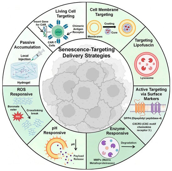
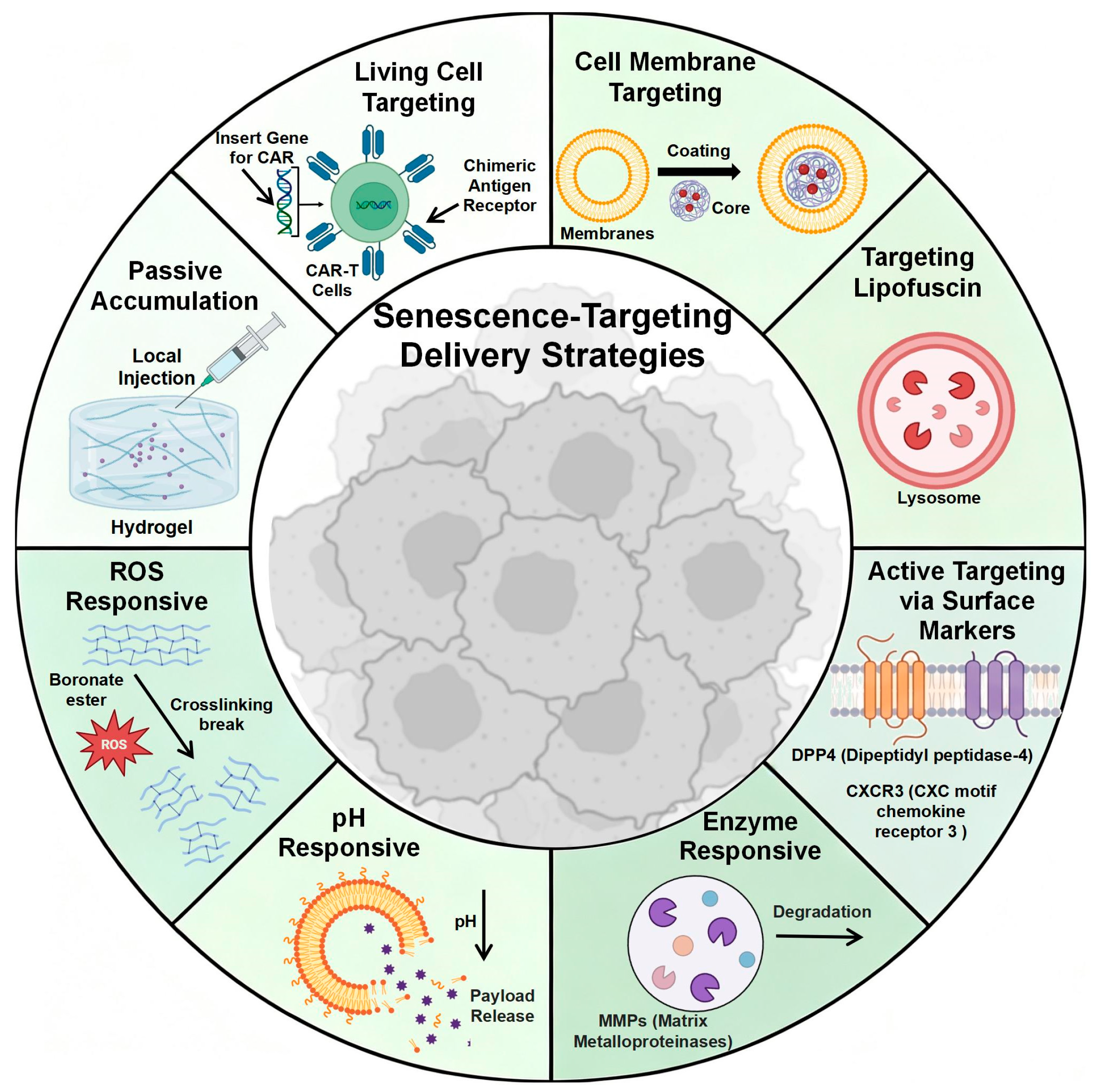
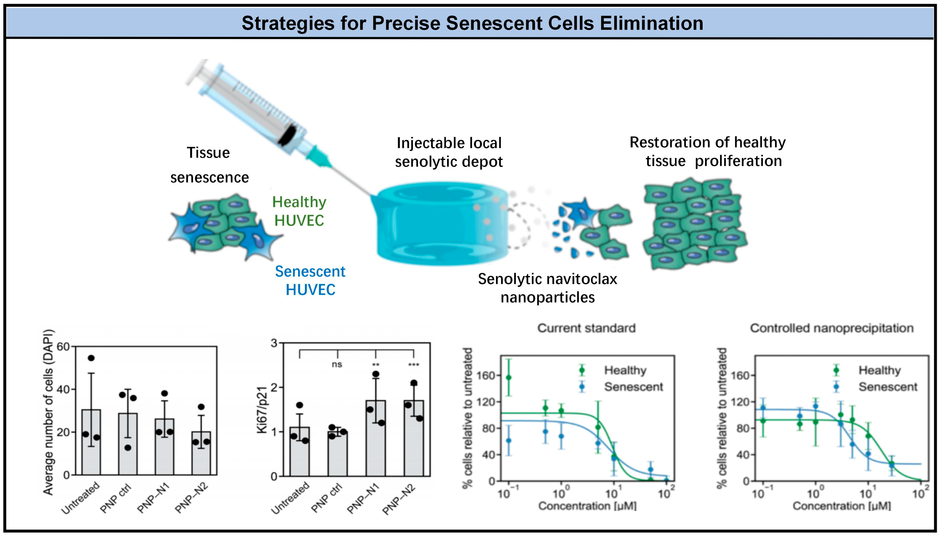
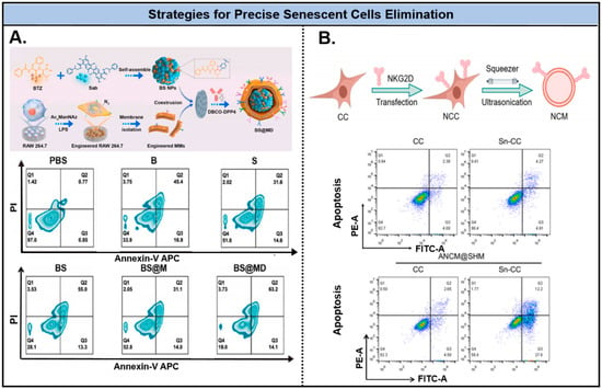
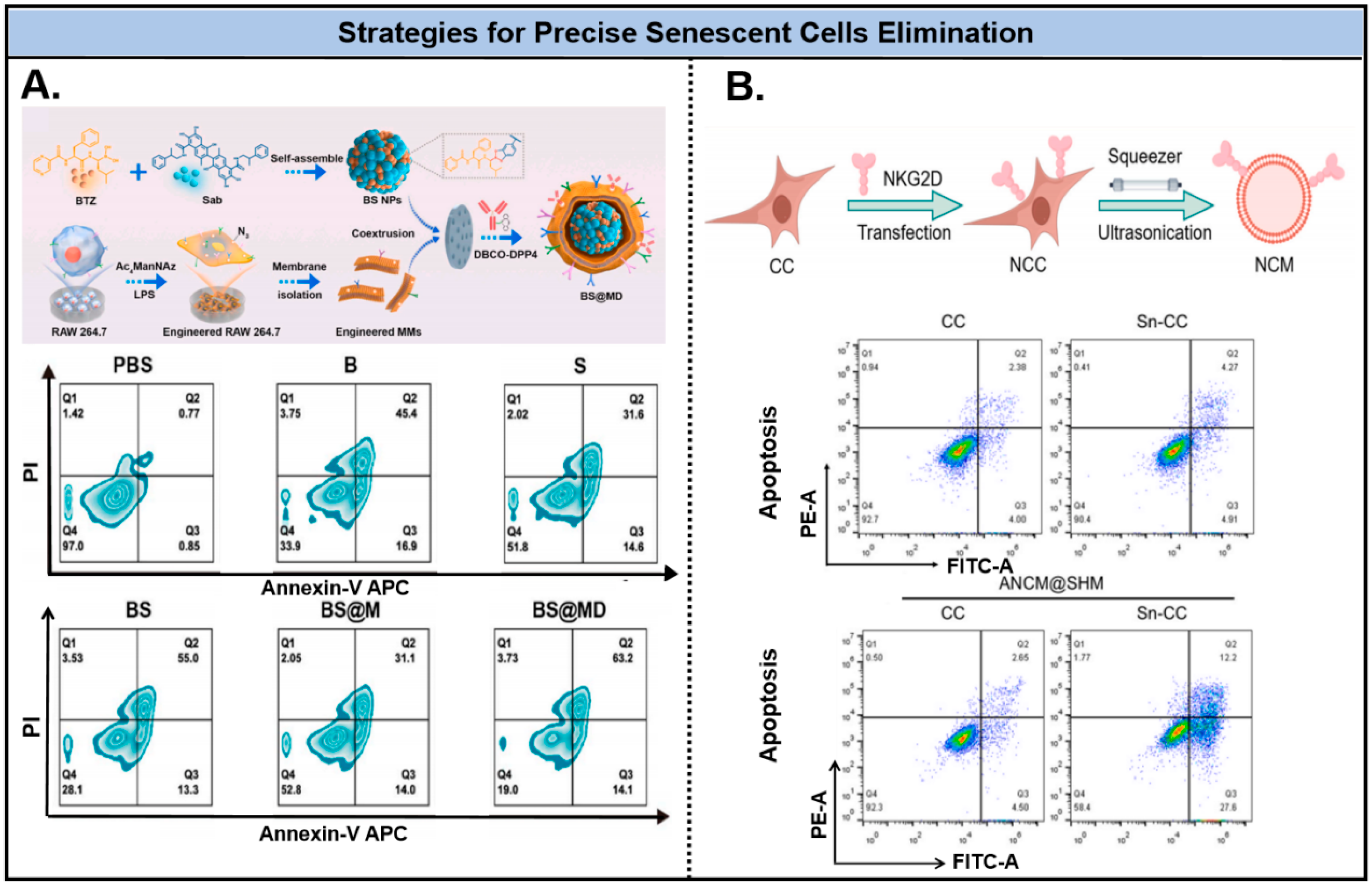
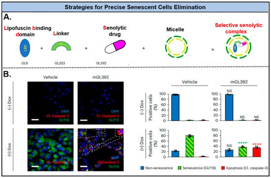
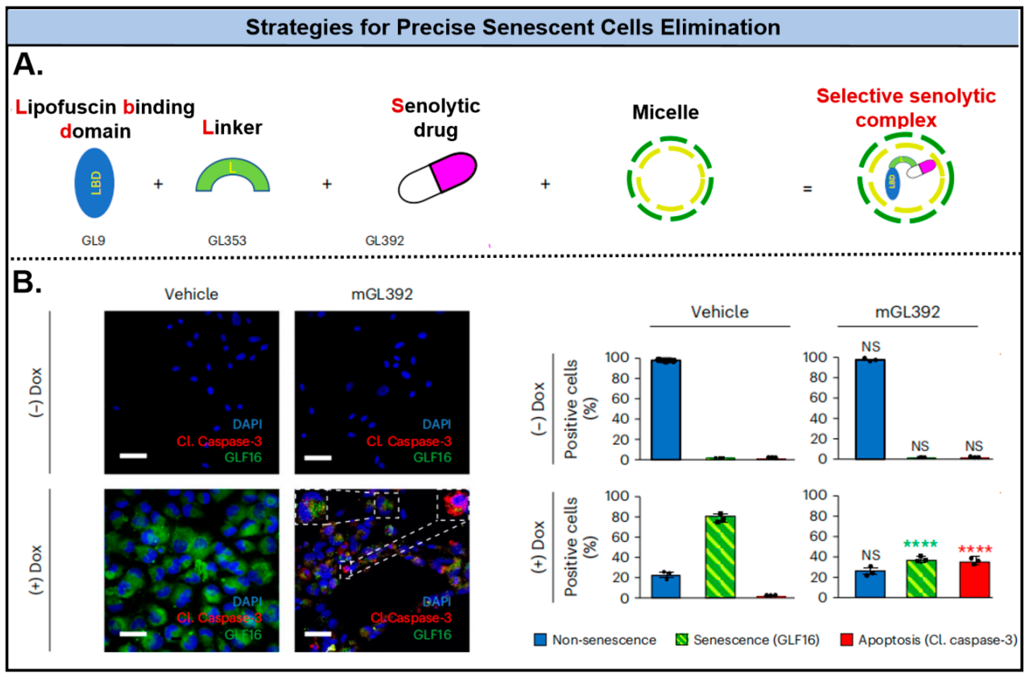
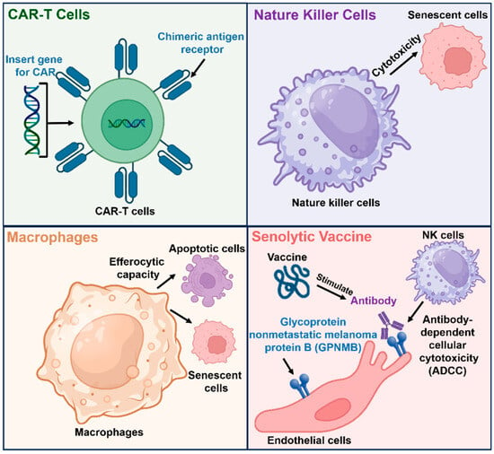
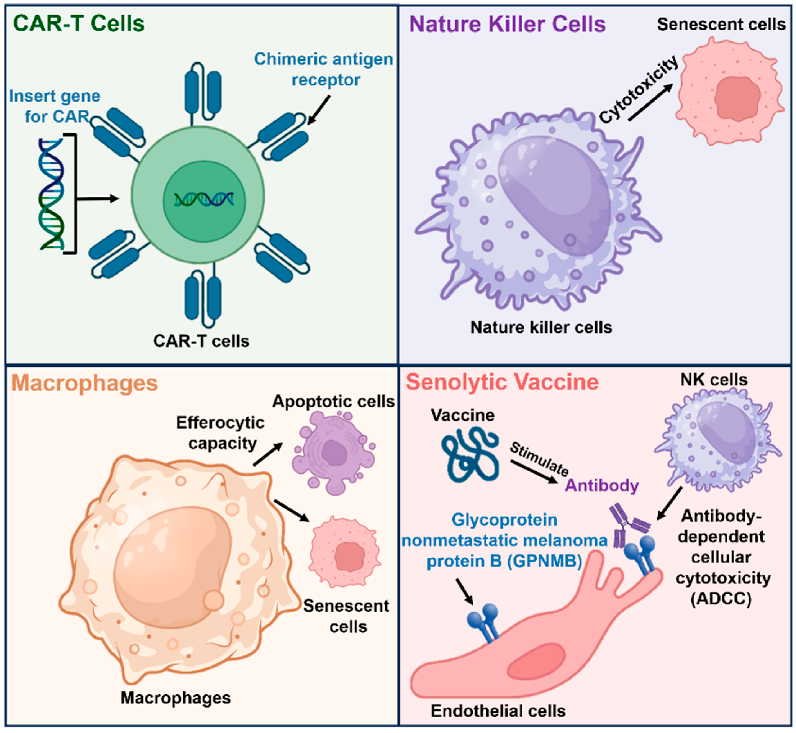
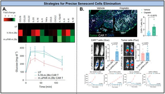
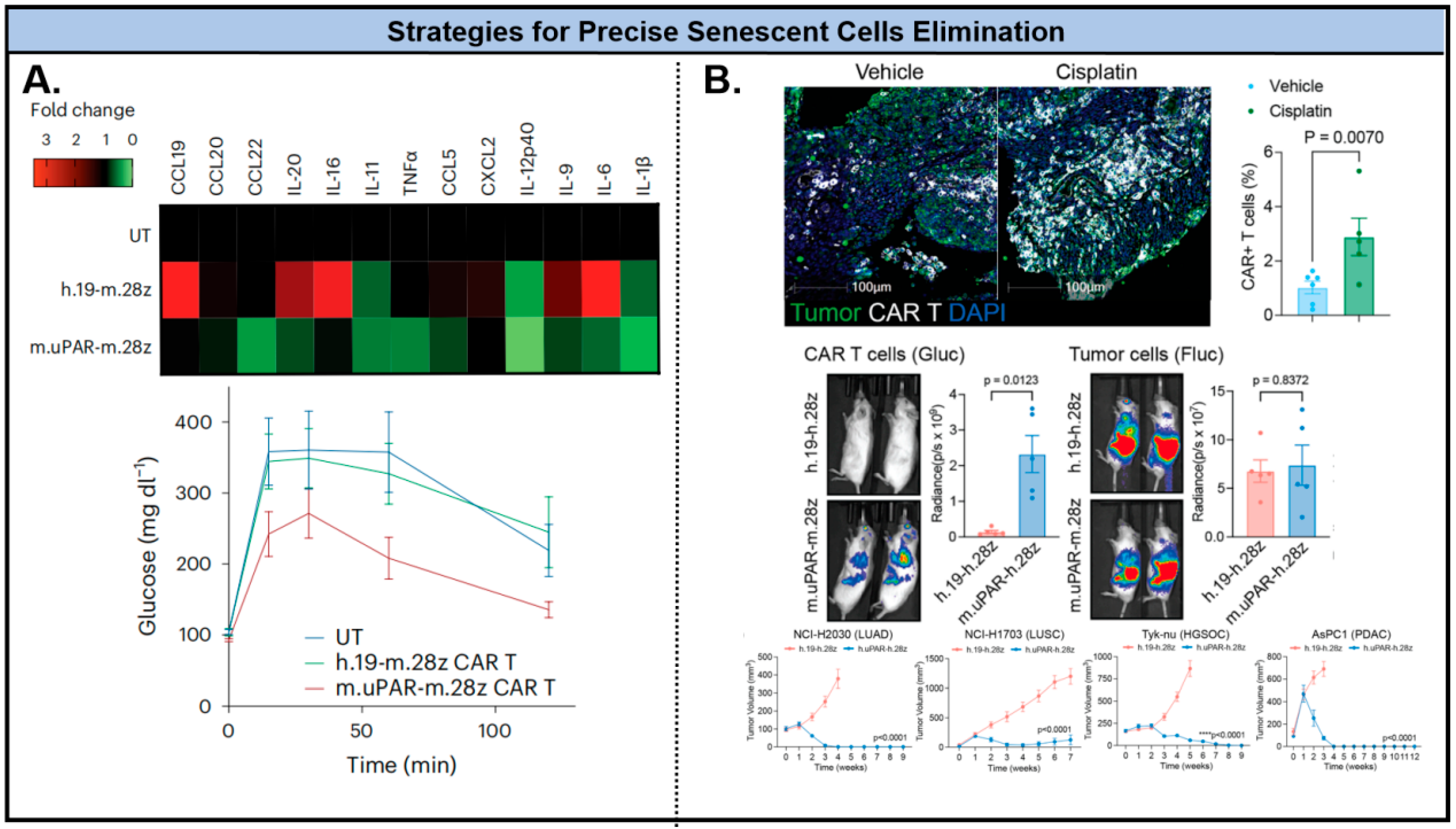
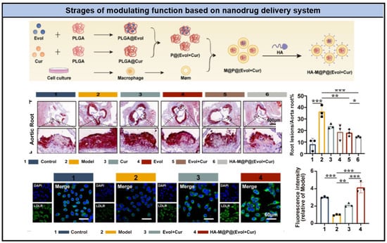
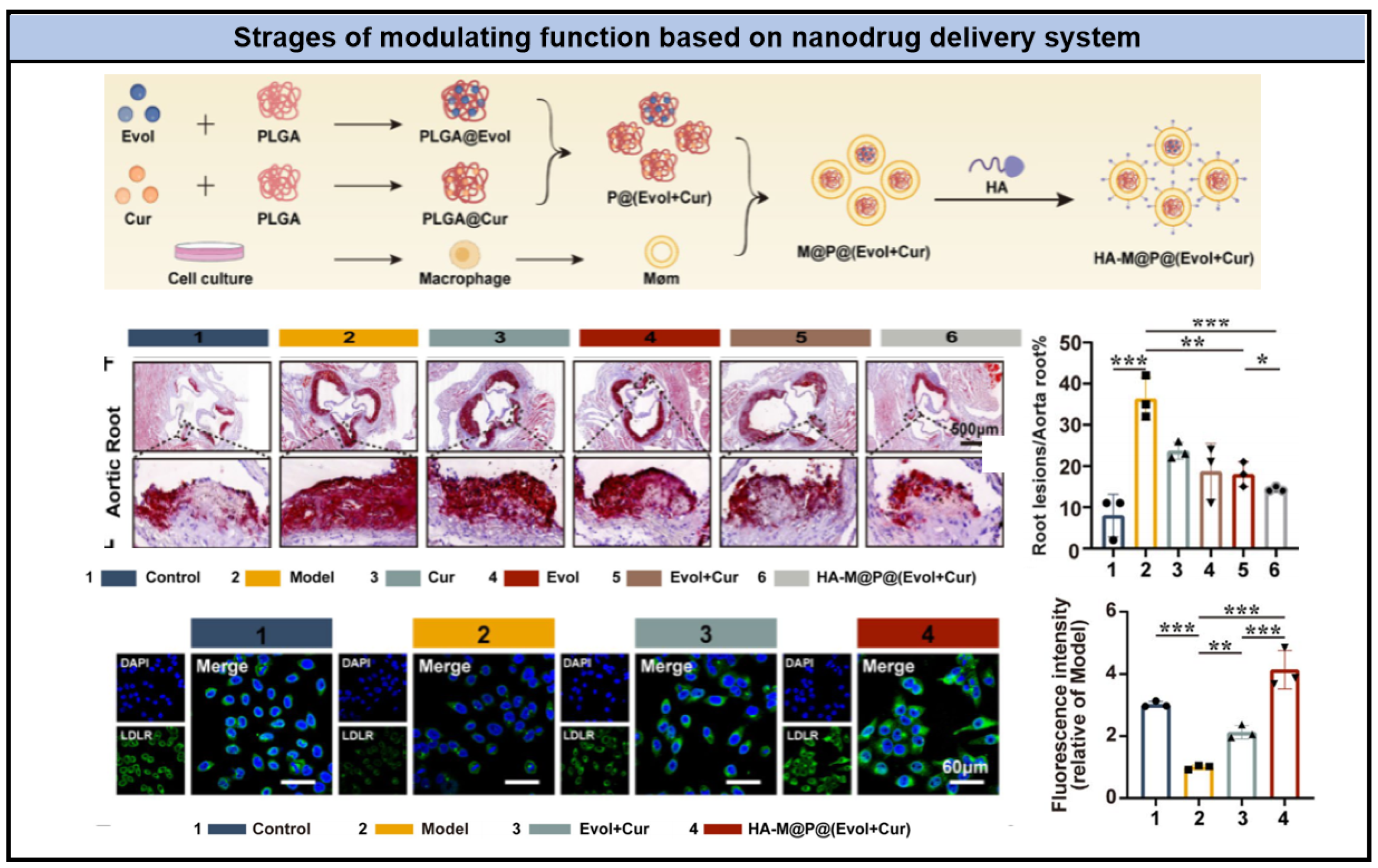
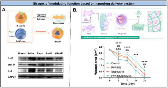
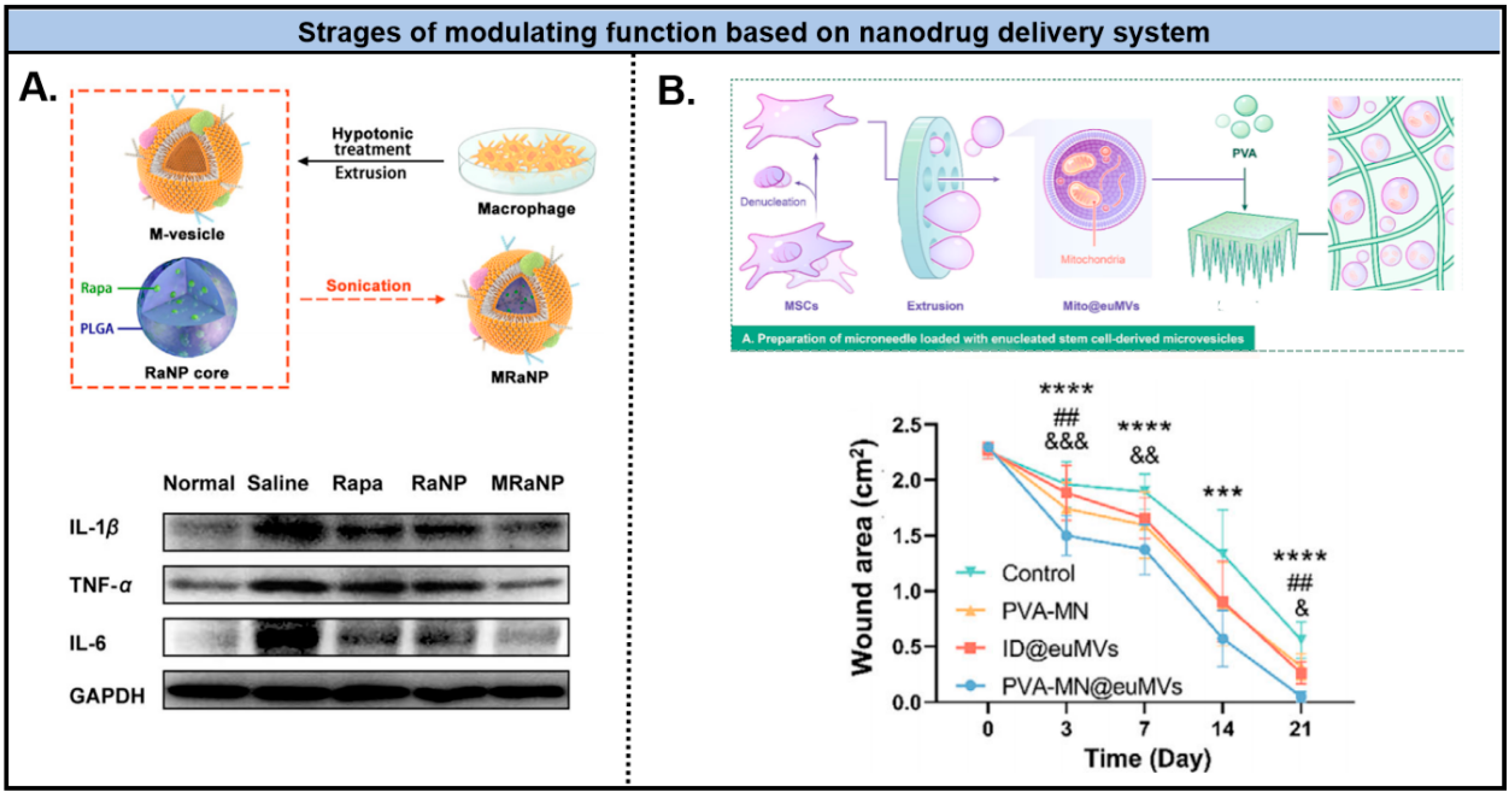
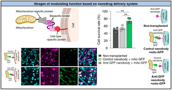
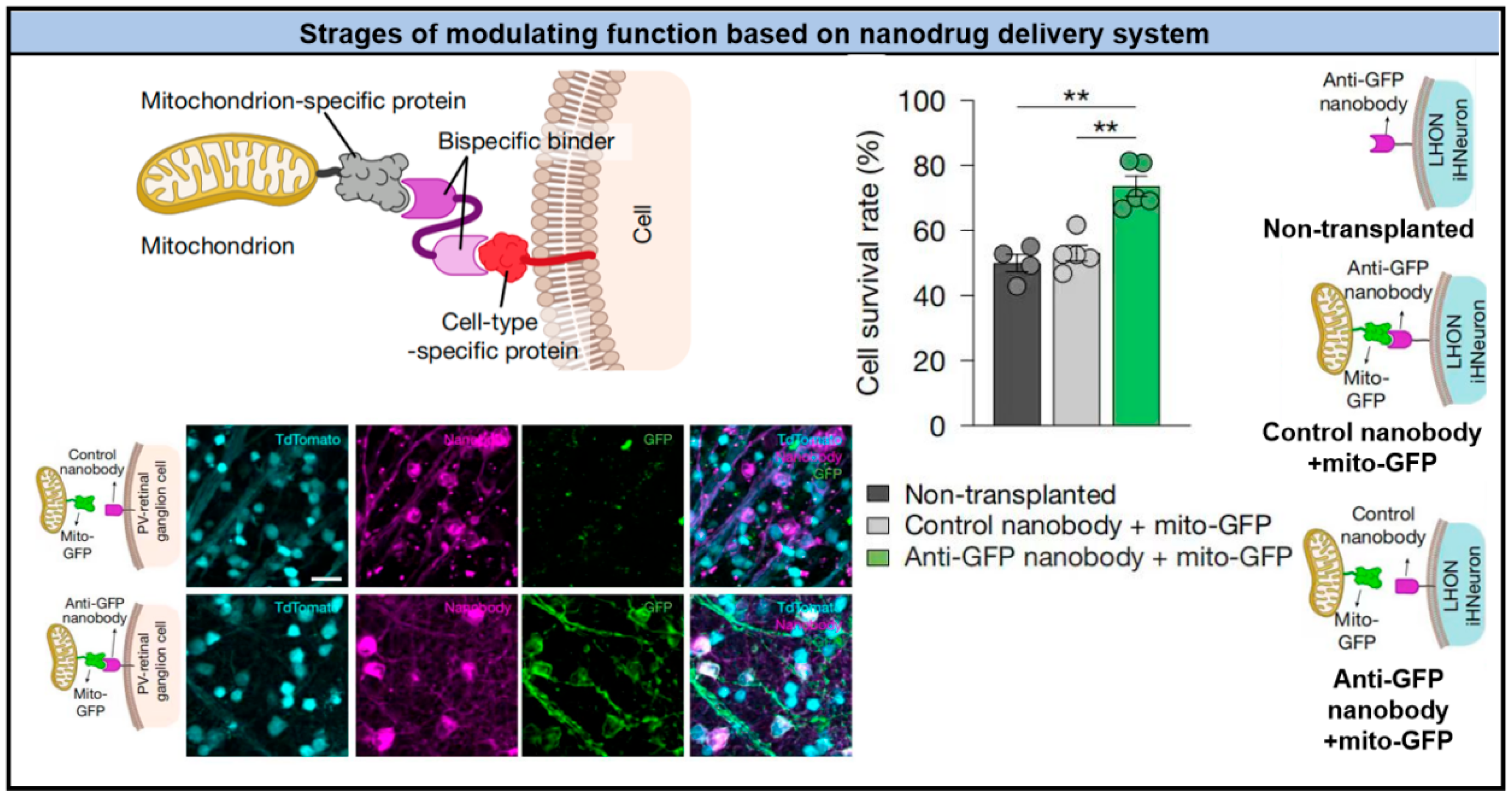
Disclaimer/Publisher’s Note: The statements, opinions and data contained in all publications are solely those of the individual author(s) and contributor(s) and not of MDPI and/or the editor(s). MDPI and/or the editor(s) disclaim responsibility for any injury to people or property resulting from any ideas, methods, instructions or products referred to in the content. |
© 2026 by the authors. Licensee MDPI, Basel, Switzerland. This article is an open access article distributed under the terms and conditions of the Creative Commons Attribution (CC BY) license.