


Abstract
Background: Oral candidiasis, predominantly caused by Candida albicans, presents significant challenges in treatment due to increasing antifungal resistance and biofilm formation. Antimicrobial photodynamic therapy (aPDT) using natural photosensitizers like riboflavin and hypericin offers a potential alternative to conventional antifungal therapies. Material and Methods: A systematic review was conducted to evaluate the efficacy of riboflavin- and hypericin-mediated aPDT in reducing Candida infections. The PRISMA framework guided the selection and analysis of 16 eligible studies published between 2014 and 2024. Data on light parameters, photosensitizer concentrations, and outcomes were extracted to assess antifungal effects. Results: Both riboflavin- and hypericin-mediated aPDT demonstrated significant antifungal activity, achieving substantial reductions in Candida biofilm and planktonic cell viability. Riboflavin activated by blue light and hypericin activated by yellow or orange light effectively targeted fluconazole-resistant Candida strains with minimal cytotoxicity to host tissues. However, complete biofilm eradication remained challenging, and variations in protocols highlighted the need for standardization. Conclusions: Riboflavin- and hypericin-mediated aPDT present promising, biocompatible alternatives for managing antifungal resistance in Candida infections. Further clinical trials and standardized protocols are essential to optimize outcomes and confirm efficacy in broader clinical settings.
1. Introduction
The oral cavity of humans is colonized by a diverse microbial community, predominantly composed of bacteria, with fungi constituting a smaller fraction [1]. Among these, Candida species are a natural part of the oral flora in healthy individuals, with Candida albicans being the most prevalent, accounting for 60–70% of cases, followed by Candida tropicalis and Candida glabrata [2]. While typically commensal, these yeasts can become pathogenic under specific conditions, leading to oral candidiasis [3]. C. albicans is the primary causative agent of oral candidiasis, responsible for up to 95% of cases [4]. Host factors such as xerostomia, smoking, oral prostheses, dental caries, diabetes, and cancer treatments can accelerate the disease process [5]. The immune response of the host mucosa plays a critical role in controlling C. albicans. Adaptive immune mechanisms, especially those mediated by Th1 and Th17 cellular responses, are essential for maintaining tissue homeostasis and combating fungal proliferation. Dendritic cells are pivotal in initiating these defenses by presenting antigens and producing cytokines [6,7]. However, systemic diseases or periods of immunosuppression may compromise these defenses, allowing Candida to exploit its virulence factors to establish and propagate infections [8]. The management of oral candidiasis typically involves topical or systemic antifungal agents, such as azoles and polyenes. However, the extensive use of these agents has led to the emergence of fluconazole-resistant Candida species [9]. Additionally, the formation of microbial biofilms provides a protective barrier, enhancing resistance to antifungal treatments and complicating disease management [10]. Given the rising prevalence of antifungal-resistant pathogens and the associated toxicity of conventional therapies, there is a pressing need for alternative strategies to control yeast infections [11]. Antimicrobial photodynamic therapy (aPDT) is emerging as a promising alternative. This technique involves the administration of a photosensitizing agent, which is activated by visible light at an appropriate wavelength to generate Reactive Oxygen Species (ROS) with antimicrobial effects [12]. The underlying mechanism of aPDT lies in its ability to induce oxidative stress, leading to microbial cell damage without significantly affecting host tissues. Importantly, aPDT has demonstrated efficacy against biofilm-associated infections and resistant strains, highlighting its clinical potential in managing recalcitrant fungal infections [13]. A critical aspect of aPDT is the selection of photosensitizers (PSs). In recent years, natural polyphenolic compounds have garnered interest due to their unique chemical structures, inherent antimicrobial properties, and favorable safety profiles. Curcumin, methylene blue, hypericin, and riboflavin have demonstrated strong antifungal activity in various studies [12,13,14]. Hypericin, derived from Hypericum perforatum (commonly known as St. John’s Wort), and riboflavin (vitamin B2) have shown promise against fluconazole-resistant strains of Candida [15,16,17,18,19]. Riboflavin, activated by blue light, produces ROS that induce oxidative damage to microbial cells, offering a natural and effective solution [20,21]. Given the increasing challenge of antifungal resistance and the need for innovative solutions, this review aims to systematically summarize the evidence supporting the use of polyphenolic natural products as photosensitizers in aPDT. By exploring their potential to counteract biofilm-related infections and resistant pathogens, this study seeks to establish a foundation for future clinical applications of aPDT in antifungal therapy.
2. Materials and Methods
2.1. Focused Question and Null Hypothesis
A systematic review was conducted following the PICO framework [22], as follows: In patients with Candida infections (Population), does treatment with riboflavin- or hypericin-mediated antimicrobial photodynamic therapy (Intervention), compared to blue light irradiation alone, the use of riboflavin or hypericin as photosensitizers alone, or other pharmacological treatments (Comparison), result in more effective eradication or reduction of Candida infections (Outcome)? The null hypothesis for our article stated that there is no significant difference in the eradication or elimination effectiveness of Candida strains when subjected to riboflavin- or hypericin-mediated antimicrobial photodynamic therapy compared to blue light irradiation alone, riboflavin or hypericin as photosensitizers alone, or other pharmacological treatments.
2.2. Search Strategy
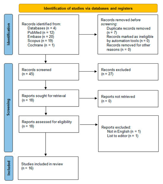
This review has been registered with PROSPERO under the ID CRD42024617727. The review was conducted following the Preferred Reporting Items for Systematic Reviews and Meta-Analyses (PRISMA) 2020 guidelines [23]. An electronic search was performed through PubMed/Medline, Embase, Scopus, and Cochrane Library databases (search phrases detailed in Figure 1). The databases were searched by three authors independently (M.Ł., J.F.-R. and R.W.) using the same search terms. Additional electronic filters were applied to include only articles published between 1 January 2014 and 3 December 2024 and restricted to publications in English. After initial screening, potential studies were selected based on titles and abstracts to determine if they met all inclusion criteria (Table 1). Two authors (J.F.-R. and R.W.) then conducted a full-text review of the selected studies to collate the desired data. Additionally, a snowball search was performed by examining the reference lists of articles deemed eligible for full-text review to identify further relevant studies. This research proposed that hypericin and riboflavin-mediated antimicrobial photodynamic therapy could serve as an effective approach for reducing Candida strains, potentially functioning as a complementary or alternative treatment for oral candidiasis compared to conventional pharmacological methods. Articles included in this review were selected based on specific inclusion and exclusion criteria.
Figure 1.
PRISMA 2020 flow diagram.
Table 1.
Search syntax used in the study.
2.3. Selection of Studies
During the study selection process for this systematic review, reviewers independently evaluated the titles and abstracts of the identified articles to minimize bias. Any disagreements regarding the eligibility of studies were resolved through collaborative discussions until a unanimous decision was achieved. This rigorous methodology, adhering to PRISMA guidelines, helped ensure that only the most relevant and high-quality studies were included in the analysis, thereby strengthening the review’s reliability and reproducibility [23].
2.4. Risk of Bias in Individual Studies
During the preliminary stage of study selection, reviewers independently examined titles and abstracts to minimize potential bias in the evaluation. Inter-reviewer agreement was measured using Cohen’s kappa statistic to ensure consistency in decision-making [24]. Disagreements concerning the inclusion or exclusion of studies were resolved through thorough discussions among the authors until a unanimous decision was achieved.
2.5. Quality Assessment
The quality of the included studies was independently assessed by two reviewers (J.F-R. and R.W.). The evaluation centered on critical elements of aPDT design, execution, and data analysis, with an emphasis on ensuring objectivity and result validation. The risk of bias was determined by assigning a score of 1 toeach “yes” response and 0 to each “no” response to a predefined set of evaluation criteria, as outlined below:
- Was a specific concentration of riboflavin or hypericin as the photosensitizer indicated?
- Was the origin or source of the photosensitizer (riboflavin or hypericin) provided?
- Was the incubation time for the photosensitizer clearly stated?
- Were detailed parameters of the light source (such as type, wavelength, output power, fluence, and power density) provided?
- Was a power meter used in the study?
- Was a negative control group included in the experimental design?
- Were numerical results reported, including relevant statistics?
- Was there no missing outcome data?
- Was the study independent from its source of funding?
The data extracted from each study were analysed and categorized based on the total count of affirmative (“yes”) responses to the specified criteria. The level of bias was determined using the following scoring thresholds: high risk: 0–3; moderate risk: 4–6; and low risk: 7–9. Each study’s scores were compiled, and a corresponding level of bias risk—classified as low, moderate, or high—was determined in accordance with the guidelines specified in the Cochrane Handbook for Systematic Reviews of Interventions [25].
2.6. Risk of Bias Across Studies
The results of the quality assessment and risk of bias across the studies are presented in Section 3.3.
2.7. Data Extraction
After reaching a consensus on the selection of included articles, the two reviewers (J.F.-R. and R.W.) extracted data on various aspects, including the citation details (first author and publication year), type of study, Candida strains used, test and control groups, follow-up period, outcomes, type and parameters of the light source, concentration of riboflavin or hypericin, and the use of nanocarriers, additional substances, as well as incubation and irradiation times.
3. Results
3.1. Study Selection
Figure 1 illustrates the detailed research methodology per PRISMA guidelines [23]. The initial search yielded 52 articles, which were narrowed down to 45 after duplicate removal. Following the screening of titles and abstracts, 18 studies qualified for full-text evaluation. Of these, two were excluded. One study was excluded for not including C. albicans. One study was omitted as it was primarily written in a non-English language, with only the title and abstract available in English, and another was excluded for being a letter to the editor. Ultimately, 16 studies were included in the final review, all published within the last 10 years.
3.2. Quality Assessment Presentation
The risk of bias assessment for the 16 studies included after a full-text review is detailed in Table 2. Studies were required to score at least six points to be included in the analysis. Among these, all studies were identified as having a low risk of bias, with three achieving the maximum score of 9. None of the studies were rated as high or moderate risk.
Table 2.
Selection criteria for papers included in the systematic review.
3.3. Data Presentation
The extracted data from the 16 studies that met the eligibility criteria and were included in the review are summarized in Table 3, Table 4 and Table 5. These include an overview of the general study characteristics, the specifications of the light sources used, and the attributes of hypericin or riboflavin as a photosensitizer in aPDT protocols. The results of the quality assessment and risk of bias across the studies are presented in Table 3.
Table 3.
The results of the quality assessment and risk of bias across the studies.
A comprehensive overview of the included studies is presented in Table 4.
Table 4.
A general overview of the studies.
Main outcomes and details from each study are presented in Table 5.
Table 5.
Main outcomes and details from each study.
3.4. General Characteristics of the Included Studies
The general characteristics of the 16 studies that were included are shown in Table 4.
3.5. Main Study Outcomes
The reviewed studies collectively highlight the potential of aPDT using natural photosensitizers like hypericin or riboflavin as effective alternatives for managing Candida infections, especially in the context of rising antifungal resistance. Agut et al. demonstrated the broad-spectrum antifungal efficacy of hypericin, achieving a 3-log reduction in C. albicans and other yeast strains at low hypericin concentrations (0.625–40 μM) with a 37 J/cm2 fluence, with no cytotoxic effects on human keratinocytes or fibroblasts [26]. Alam et al. further validated hypericin’s effectiveness, showing significant inhibition of C. albicans and Gram-positive bacteria when activated by orange light, with combination therapy also enhancing outcomes for resistant strains like P. aeruginosa [27]. Alshehri et al. explored riboflavin-mediated photodynamic therapy (RF-PDT) on acrylic denture materials and found it highly effective for reducing C. albicans biofilm viability, outperforming other treatments and maintaining the mechanical properties of the materials [28]. Arboleda et al. demonstrated that rose bengal aPDT was more effective than RF-PDT at inhibiting C. albicans, while Ardakani et al. found that aPDT using riboflavin and other natural photosensitizers significantly reduced biofilm mass and C. albicans CFU counts in a multispecies biofilm model, showing promise for biofilm-related infections [29,30]. Bernala et al. highlighted hypericin fungicidal efficacy even at low concentrations, achieving selective Candida inactivation without harming host cells [31]. Sakita et al. enhanced hypericin’s efficacy by encapsulating it in micelles, leading to complete biofilm inhibition for multiple Candida species, with synergy observed when combined with fluconazole against resistant strains [33]. Sato et al. advanced these findings in vivo, showing that hypericin-loaded nanostructured lipid carriers combined with aPDT significantly reduced C. albicans colonies in a mouse model with minimal host tissue damage [39]. Troichenko et al. demonstrated the potential of combined aPDT and collagen cross-linking for inhibiting C. albicans biofilms in ophthalmic applications, while Morelato et al. found that riboflavin–PDT with blue LED and methylene blue–PDT with red LED both significantly reduced biofilm formation in peri-implantitis models [37]. Rezusta et al. showed that hypericin-mediated aPDT was effective against azole-resistant C. albicans, achieving a 3-log reduction at low fungal concentrations. These studies collectively underscore PDT’s potential as a minimally invasive, biocompatible, and effective treatment for fungal infections, particularly those associated with biofilms or resistant strains [36]. While many studies demonstrate the efficacy of aPDT, limitations such as incomplete biofilm eradication, variability in the reduction of fungal cell viability, and the potential for regrowth under suboptimal conditions must be acknowledged. Differences in light source parameters, such as wavelength and energy density, were observed to significantly influence outcomes, with some protocols achieving higher efficacy due to optimized settings. These findings highlight the importance of standardized methodologies to ensure reproducibility and to maximize the therapeutic potential of aPDT [28,29,30,31,32,33,34,35,36,37,38,39,40]. However, differences in experimental protocols, including photosensitizer concentrations, light parameters, and fungal strains, highlight the need for standardization. The lack of large-scale clinical trials is a limitation, as most evidence comes from in vitro or small animal studies. Future research should prioritize clinical validation and explore standardized aPDT protocols, innovative delivery systems like micelles or hydrogels, and combinations with antifungal drugs to optimize treatment outcomes and expand aPDT’s applicability across clinical settings. These findings suggest that aPDT, particularly with natural photosensitizers like hypericin and riboflavin, could offer a safe, sustainable, and effective alternative to traditional antifungal therapies [26,27,28,29,30,31,32,33,34,35,36,37,38,39,40,41,42].
3.6. Characteristics of Light Sources Used in aPDT
Table 6 outlines the physical parameters of the light sources used in studies that satisfied the inclusion criteria.
Table 6.
Light sources’ physical parameters of studies that fulfilled the eligibility criteria.
Table 7 summarises the concentration and incubation time of the photosenistisors used i the studies.
Table 7.
Characteristics of PS used in studies meeting eligibility criteria.
4. Discussion
4.1. Results in the Context of Other Evidence
This systematic review provides compelling evidence to reject our null hypothesis, which posited no significant difference in the efficacy of riboflavin- and hypericin-mediated aPDT compared to conventional antifungal treatments for managing Candida infections, demonstrating that aPDT is a promising alternative for reducing fungal load, particularly in cases where conventional antifungals face challenges due to resistance or biofilm formation. Both riboflavin and hypericin exhibited substantial antifungal activity against Candida spp. in vitro, with multiple studies achieving complete eradication of planktonic cells under optimized conditions [27,28,31,36], and were effective in reducing biofilm mass and viability, although biofilms remained more resistant than planktonic cells due to their structural complexity and extracellular matrix [32,33,35,36]. The versatility of riboflavin, activated by blue light, and hypericin, which responds to yellow or orange light, underscores their clinical potential, supported by safety profiles showing minimal cytotoxicity to human keratinocytes and fibroblasts, indicating their suitability for clinical applications without damaging host tissues [31,36,39]. Riboflavin- and hypericin-mediated aPDT effectively targeted fluconazole-resistant Candida strains, offering a promising strategy to address antifungal drug resistance, while their combination with conventional antifungal agents like fluconazole enhanced effects, particularly against biofilm-associated infections, suggesting a dual-action therapeutic approach [32,33,36]. Despite significant reductions in Candida spp. cell counts, complete biofilm eradication was not consistently achieved due to the biofilm’s protective extracellular matrix emphasizing the difficulty of treating biofilm-associated infections, and the findings remain limited by the predominance of in vitro studies and the variability in study designs, including differences in light parameters, photosensitizer concentrations, and incubation times, complicating direct comparisons and underscoring the need for standardized protocols [35,36,37]. Nonetheless, in vivo studies confirmed the effectiveness of hypericin-loaded hydrogels in reducing Candida albicans vaginal colonies and riboflavin-mediated aPDT in decreasing fungal loads on dental surfaces and prosthetic materials, with both photosensitizers identified as promising natural alternatives due to their biocompatibility, low toxicity, and sustainable use in aPDT though the scarcity of in vivo or clinical trials limits the generalizability of results, requiring future research with large-scale studies to establish standardized protocols and optimize outcomes [26,36]. These findings align with prior evidence emphasizing the potential of photodynamic therapy in overcoming antimicrobial resistance and managing biofilm-associated infections, underscoring its promise for clinical application in drug-resistant Candida infections and biofilm-associated conditions. Numerous studies have highlighted the promising role of photodynamic therapy in managing oral candidiasis, strongly supporting its potential as an effective alternative or adjunct to traditional antifungal treatments. For instance, Hu et al. evaluated the efficacy of PDT in comparison to conventional antifungal drugs and found it to be superior to nystatin in reducing Candida colonies [42]. Their findings also suggested that PDT might offer greater effectiveness than other antifungals, such as fluconazole and miconazole. Rodríguez-Cerdeira et al. similarly reported that PDT is a promising treatment option due to its broad antimicrobial spectrum and the ability to target drug-resistant strains of Candida, making it a viable alternative where traditional antifungal treatments might fail [43]. Supporting this, D’Amico et al. demonstrated that PDT significantly reduces Candida albicans biofilm without exerting cytotoxic effects on gingival cells. Their study compared various photosensitizers and reinforced PDT’s potential as both a primary and adjunctive therapy for oral candidiasis [44]. The mechanisms underlying PDT’s effectiveness were further explored by Kashef and Hamblin, who noted its ability to alter Candida cell permeability, ultimately leading to fungal death [45]. They also highlighted PDT’s wide antibacterial spectrum, short therapeutic course, and strong targeting capabilities, which make it particularly advantageous in antifungal treatment strategies. However, the application of PDT shows variability depending on the site of treatment. For example, Agut et al. observed that PDT was more effective in reducing Candida colonies on the palate compared to the denture area [26]. This difference is attributed to the porous and irregular surfaces of dentures, which facilitate microbial adhesion and recolonization. The limited action of PDT on denture surfaces compared to broader oral areas treated with systemic drugs further underscores this finding. Studies by Pérez-Laguna et al. and others have emphasized that mechanical cleaning of dentures is essential to achieve optimal outcomes, as PDT alone is insufficient to address fungal growth on such surfaces [46]. Comparative analyses have further clarified PDT’s position relative to other antifungaltreatments. For example, while PDT showed similar efficacy to fluconazole, miconazole was found to be more effective [47]. However, PDT’s ability to target drug-resistant Candida strains gives it a distinct advantage over azoles in specific cases. Furthermore, systemic antifungals like amphotericin B demonstrated less effectiveness in removing fungal colonies from denture surfaces, further reinforcing the critical role of regular denture cleaning in managing oral candidiasis [48]. Despite these advancements, recurrence rates of candidiasis remain a significant concern. Studies by Mima et al., Macial et al., and Schwingel et al. have reported high recurrence rates, primarily due to inadequate denture cleaning and Candida recolonization [49,50,51]. Nevertheless, the combination of PDT with traditional antifungal agents has shown promise in mitigating these challenges [42]. Notably, the synergistic effect of PDT and nystatin has been demonstrated to significantly reduce Candida colonies and lower recurrence rates. This is attributed to their complementary mechanisms of action, which enhance overall treatment efficacy [42]. Studies by Paz-Cristobal et al. and Agut et al. highlighted that combination therapy with PDT and nystatin is both effective and safe, with adverse reactions reported as mild and self-limiting [26,38]. Common side effects, such as nausea and burning tongue, were mostly observed in immunodeficient patients and did not pose significant risks [42,52,53]. Overall, PDT presents itself as a valuable adjunct to traditional antifungal therapies due to its broad spectrum, strong targeting capability, and short therapeutic course. Combining PDT with antifungal agents like nystatin is recommended for achieving improved clinical outcomes and reducing the recurrence of oral candidiasis [42]. These findings collectively underline the potential of PDT as an innovative and effective approach tothe management of this challenging condition [36,37,38,39,40,41,42].
4.2. Limitations of the Evidence
The primary limitation lies in the significant variability of protocols across studies. Light source parameters, including wavelength, energy density, and fluence, as well as photosensitizer concentrations and incubation times, differed widely, making it difficult to compare outcomes or establish standardized guidelines. Furthermore, while many studies reported efficacy against Candida spp., most were conducted in vitro, with limited in vivo evidence or clinical trials available to confirm these findings in human populations. Additionally, while aPDT was effective in reducing biofilm mass, complete eradication of biofilms was rarely achieved, highlighting a persistent challenge in biofilm-associated infections. Finally, the studies included in this review often relied on subjective outcome measures, such as CFU reduction and visual biofilm assessment, without integrating advanced diagnostic tools like imaging or molecular analyses to substantiate their results.
4.3. Limitations of the Review Process
The lack of homogeneity among the included studies led to a narrative synthesis of results. The variability in study designs, intervention protocols, and outcome measures may have introduced bias in evaluating the overall efficacy of riboflavin- and hypericin-mediated aPDT. Moreover, the significant differences in the parameters used across studies prevented the authors from applying the GRADE tool, and as such, heterogeneity makes it challenging to formulate clear recommendations. To address this, future research should focus on conducting well-designed randomized controlled trials to directly compare specific parameters and establish standardized protocols. Furthermore, the exclusion of non-English studies and gray literature may have restricted the scope of the review, potentially overlooking relevant data. These limitations highlight the need for more rigorous research in this field to enable systematic comparisons and quantitative analyses.
4.4. Implications for Practice, Policy, and Future Research
Riboflavin- and hypericin-mediated aPDT shows promise as a safe and effective alternative or adjunct to conventional antifungal therapies, particularly for managing drug-resistant or recurrent Candida infections. Clinicians should consider incorporating aPDT into treatment protocols for conditions such as denture stomatitis and other mucosal infections. However, for this to become routine practice, standardized treatment protocols—including consistent light parameters, photosensitizer concentrations, and application techniques—must be developed and validated. Policymakers and research funding agencies should prioritize support for large-scale, multicenter clinical trials to confirm the efficacy and safety of aPDT in diverse patient populations. Furthermore, future research should explore the synergistic potential of combining aPDT with existing antifungal drugs, particularly for biofilm-associated infections where monotherapies often fall short. Innovations such as nanocarrier systems and hydrogels could enhance the delivery and stability of photosensitizers, improving therapeutic efficacy. While the current evidence highlights the potential of aPDT in combating oral candidiasis and related infections, addressing the identified gaps is crucial for its integration into routine clinical practice.
Future research should prioritize the standardization of parameters to enable more reliable and meaningful comparisons across studies. Moreover, further investigation is needed to examine the variations in efficacy between different regions of the mouth, such as tooth versus palate applications, to deepen our understanding of site-specific outcomes.
5. Conclusions
This systematic review demonstrates the potential of riboflavin- and hypericin-mediated antimicrobial photodynamic therapy as promising, non-invasive alternatives for managing Candida infections, particularly in cases resistant to conventional antifungal therapies. Significant reductions in fungal viability and biofilm mass, coupled with minimal cytotoxic effects, highlight the clinical promise of these photosensitizers. Riboflavin and hypericin, activated by blue and yellow/orange light, respectively, offer versatility and efficacy, particularly against fluconazole-resistant strains. Additionally, the reviewed evidence indicates that aPDT can achieve outcomes comparable to traditional antifungal agents while reducing systemic side effects and potentially enhancing efficacy through synergistic combinations. However, the lack of standardization in study protocols, such as variations in light parameters and photosensitizer concentrations, alongside a scarcity of large-scale clinical trials, limits the applicability of these findings. To address these limitations, future research should prioritize large-scale, multicenter studies with consistent methodologies and explore optimal treatment parameters to enhance the reproducibility and effectiveness of aPDT. Furthermore, focusing on areas where aPDT could have the most immediate clinical impact, such as the treatment of antifungal-resistant Candida strains and biofilm-associated infections, would maximize its relevance and utility. These targeted applications address critical gaps in current antifungal therapies and could accelerate the integration of aPDT into clinical practice.
Author Contributions
Conceptualization, M.Ł., W.N. and R.W.; methodology, W.N. and R.W.; software, M.Ł. and J.F.-R.; formal analysis, J.F.-R., W.N. and M.Ł.; investigation, J.F.-R., W.N. and M.Ł.; writing—original draft preparation, J.F.-R., W.N., R.W. and M.Ł.; writing—review and editing, W.N., J.F.-R., M.Ł., R.W. and D.S.; supervision, R.W. and D.S.; funding acquisition, R.W. and D.S. All authors have read and agreed to the published version of the manuscript.
Funding
This research received no external funding.
Data Availability Statement
Data are contained within the article.
Conflicts of Interest
The authors declare no conflicts of interest.
References
- Rapala-Kozik, M.; Surowiec, M.; Juszczak, M.; Wronowska, E.; Kulig, K.; Bednarek, A.; Gonzalez-Gonzalez, M.; Karkowska-Kuleta, J.; Zawrotniak, M.; Satała, D.; et al. Living Together: The Role of Candida albicans in the Formation of Polymicrobial Biofilms in the Oral Cavity. Yeast 2023, 40, 303–317. [Google Scholar] [CrossRef] [Scilit] [PubMed]
- Stenderup, A. Oral Mycology. Acta Odontol. Scand. 1990, 48, 3–10. [Google Scholar] [CrossRef] [Scilit] [PubMed]
- Fumes, A.C.; Da Silva Telles, P.D.; Corona, S.A.M.; Borsatto, M.C. Effect of APDT on Streptococcus Mutans and Candida albicans Present in the Dental Biofilm: Systematic Review. Photodiagnosis Photodyn. Ther. 2018, 21, 363–366. [Google Scholar] [CrossRef] [Scilit] [PubMed]
- Vila, T.; Sultan, A.S.; Montelongo-Jauregui, D.; Jabra-Rizk, M.A. Oral Candidiasis: A Disease of Opportunity. J. Fungi 2020, 6, 15. [Google Scholar] [CrossRef] [Scilit]
- Patel, M. Oral Cavity and Candida albicans: Colonisation to the Development of Infection. Pathogens 2022, 11, 335. [Google Scholar] [CrossRef] [Scilit]
- Zhou, Y.; Cheng, L.; Lei, Y.L.; Ren, B.; Zhou, X. The Interactions Between Candida albicans and Mucosal Immunity. Front. Microbiol. 2021, 12, 652725. [Google Scholar] [CrossRef] [Scilit]
- Talapko, J.; Juzbašić, M.; Matijević, T.; Pustijanac, E.; Bekić, S.; Kotris, I.; Škrlec, I. Candida albicans—The Virulence Factors and Clinical Manifestations of Infection. J. Fungi 2021, 7, 79. [Google Scholar] [CrossRef] [Scilit]
- Sanglard, D.; Coste, A.; Ferrari, S. Antifungal Drug Resistance Mechanisms in Fungal Pathogens from the Perspective of Transcriptional Gene Regulation. FEMS Yeast Res. 2009, 9, 1029–1050. [Google Scholar] [CrossRef] [Scilit]
- Seneviratne, C.J.; Silva, W.J.; Jin, L.J.; Samaranayake, Y.H.; Samaranayake, L.P. Architectural Analysis, Viability Assessment and Growth Kinetics of Candida albicans and Candida glabrata Biofilms. Arch. Oral Biol. 2009, 54, 1052–1060. [Google Scholar] [CrossRef] [Scilit]
- Carmello, J.C.; Alves, F.G.; Basso, F.; De Souza Costa, C.A.; Bagnato, V.S.; Mima, E.G.D.O.; Pavarina, A.C. Treatment of Oral Candidiasis Using Photodithazine®—Mediated Photodynamic Therapy In Vivo. PLoS ONE 2016, 11, e0156947. [Google Scholar] [CrossRef] [Scilit]
- Pereira Gonzales, F.; Maisch, T. Photodynamic Inactivation for Controlling Candida albicans Infections. Fungal Biol. 2012, 116, 1–10. [Google Scholar] [CrossRef] [Scilit] [PubMed]
- Kubizna, M.; Dawiec, G.; Wiench, R. Efficacy of Curcumin-Mediated Antimicrobial Photodynamic Therapy on Candida spp.—A Systematic Review. Int. J. Mol. Sci. 2024, 25, 8136. [Google Scholar] [CrossRef] [Scilit] [PubMed]
- Suwannasom, N.; Kao, I.; Pruß, A.; Georgieva, R.; Bäumler, H. Riboflavin: The Health Benefits of a Forgotten Natural Vitamin. Int. J. Mol. Sci. 2020, 21, 950. [Google Scholar] [CrossRef] [Scilit] [PubMed]
- Barroso, R.A.; Navarro, R.; Tim, C.R.; De Paula Ramos, L.; De Oliveira, L.D.; Araki, Â.T.; Fernandes, K.G.C.; Macedo, D.; Assis, L. Antimicrobial Photodynamic Therapy Against Propionibacterium acnes Biofilms Using Hypericin (Hypericum perforatum) Photosensitizer: In Vitro Study. Lasers Med. Sci. 2021, 36, 1235–1240. [Google Scholar] [CrossRef] [Scilit]
- Boen, M.; Brownell, J.; Patel, P.; Tsoukas, M.M. The Role of Photodynamic Therapy in Acne: An Evidence-Based Review. Am. J. Clin. Dermatol. 2017, 18, 311–321. [Google Scholar] [CrossRef] [Scilit]
- Li, S.-S.; Zhang, L.-L.; Nie, S.; Lv, T.; Wang, H.-W. Severe Acne in Monozygotic Twins Treated with Photodynamic Therapy. Photodiagnosis Photodyn. Ther. 2018, 23, 235–236. [Google Scholar] [CrossRef] [Scilit]
- Galeotti, N. Hypericum Perforatum (St John’s Wort) beyond Depression: A Therapeutic Perspective for Pain Conditions. J. Ethnopharmacol. 2017, 200, 136–146. [Google Scholar] [CrossRef] [Scilit]
- Napoli, E.; Siracusa, L.; Ruberto, G.; Carrubba, A.; Lazzara, S.; Speciale, A.; Cimino, F.; Saija, A.; Cristani, M. Phytochemical Profiles, Phototoxic and Antioxidant Properties of Eleven Hypericum Species—A Comparative Study. Phytochemistry 2018, 152, 162–173. [Google Scholar] [CrossRef] [Scilit]
- Polat, E.; Kang, K. Natural Photosensitizers in Antimicrobial Photodynamic Therapy. Biomedicines 2021, 9, 584. [Google Scholar] [CrossRef] [Scilit]
- Kubrak, T.P.; Kołodziej, P.; Sawicki, J.; Mazur, A.; Koziorowska, K.; Aebisher, D. Some natural photosensitizers and their Medicinal properties for Use in Photodynamic Therapy. Molecules 2022, 27, 1192. [Google Scholar] [CrossRef] [Scilit]
- Wang, X.; Wang, L.; Fekrazad, R.; Zhang, L.; Jiang, X.; He, G.; Wen, X. Polyphenolic Natural Products as Photosensitizers for Antimicrobial Photodynamic Therapy: Recent Advances and Future Prospects. Front. Immunol. 2023, 14, 1275859. [Google Scholar] [CrossRef] [Scilit] [PubMed]
- Schardt, C.; Adams, M.B.; Owens, T.; Keitz, S.; Fontelo, P. Utilization of the PICO Framework to Improve Searching PubMed for Clinical Questions. BMC Med. Inform. Decis. Making. 2007, 7, 16. [Google Scholar] [CrossRef] [Scilit]
- Page, M.J.; McKenzie, J.E.; Bossuyt, P.M.; Boutron, I.; Hoffmann, T.C.; Mulrow, C.D.; Shamseer, L.; Tetzlaff, J.M.; Akl, E.A.; Brennan, S.E.; et al. The PRISMA 2020 Statement: An Updated Guideline for Reporting Systematic Reviews. BMJ 2021, 372, n71. [Google Scholar] [CrossRef] [Scilit] [PubMed]
- Watson, P.F.; Petrie, A. Method Agreement Analysis: A Review of Correct Methodology. Theriogenology 2010, 73, 1167–1179. [Google Scholar] [CrossRef] [Scilit] [PubMed]
- Higgins, J.; Thomas, J.; Chandler, J.; Cumpston, M.; Li, T.; Page, M. Welch Cochrane Handbook for Systematic Reviews of Interventions Version 6.4; Cochrane: London, UK, 2023; Available online: http://www.training.cochrane.org/handbook (accessed on 20 October 2024).
- Agut, M.; Rezusta, A.; Paz-Cristobal, M.P.; Vicente, P.M.; Díez, D.R.; López-Chicón, P.; Alemany, M.; Semino, C.; Gilaberte, Y.; Nonell, S. Photodynamic effect in vitro of hypericin on yeasts and evaluation of its photocytotoxicity in keratinocytes and fibroblasts. Photodiagnosis Photodyn. Ther. 2011, 8, 224. [Google Scholar] [CrossRef] [Scilit]
- Alam, S.T.; Le, T.A.N.; Park, J.-S.; Kwon, H.C.; Kang, K. Antimicrobial Biophotonic Treatment of Ampicillin-Resistant Pseudomonas aeruginosa with Hypericin and Ampicillin Cotreatment Followed by Orange Light. Pharmaceutics 2019, 11, 641. [Google Scholar] [CrossRef] [Scilit]
- Alshehri, A.H. Mechanical and antimicrobial effects of riboflavin-mediated photosensitization of in vitro C. albicans formed on polymethyl methacrylate resin. Photodiagnosis Photodyn. Ther. 2021, 36, 102488. [Google Scholar] [CrossRef] [Scilit]
- Arboleda, A.; Miller, D.; Cabot, F.; Taneja, M.; Aguilar, M.C.; Alawa, K.; Amescua, G.; Yoo, S.H.; Parel, J.M. Assessment of rose bengal versus riboflavin photodynamic therapy for inhibition of fungal keratitis isolates. Am. J. Ophthalmol. 2014, 158, 64–70.e2. [Google Scholar] [CrossRef] [Scilit]
- Shahi Ardakani, A.; Benedicenti, S.; Solimei, L.; Shahabi, S.; Afrasiabi, S. Reduction of Multispecies Biofilms on an Acrylic Denture Base Model by Antimicrobial Photodynamic Therapy Mediated by Natural Photosensitizers. Pharmaceuticals 2024, 17, 1232. [Google Scholar] [CrossRef] [Scilit]
- Bernal, C.; Rodrigues, J.A.O.; Guimarães, A.P.P.; Ribeiro, A.O.; de Oliveira, K.T.; Imasato, H.; Perussi, J.R. Selective photoinactivation of C. albicans and C. dubliniensis with hypericin. Laser Phys. 2011, 21, 245–249. [Google Scholar] [CrossRef] [Scilit]
- Troichenko, L.F.; Drozhzhyna, G.I.; Moloda, A.L.; Dolenko, L.V. In vitro investigation of the effect of photosensitizer-mediated 365-nm UV light and 630–670-nm low-energy laser irradiation on the fungal flora, Candida albicans and Fusarium spp. Oftalmol. Zhurnal 2021, 92, 34–40. [Google Scholar] [CrossRef] [Scilit]
- Sakita, K.M.; Conrado, P.C.; Faria, D.R.; Arita, G.S.; Capoci, I.R.; Rodrigues-Vendramini, F.A.; Pieralisi, N.; Cesar, G.B.; Gonçalves, R.S.; Caetano, W.; et al. Copolymeric micelles as efficient inert nanocarrier for hypericin in the photodynamic inactivation of Candida species. Future Microbiol. 2019, 14, 519–531. [Google Scholar] [CrossRef] [Scilit] [PubMed]
- Sato, M.R.; Oshiro-Junior, J.A.; Rodero, C.F.; Boni, F.I.; Araújo, V.H.S.; Bauab, T.M.; Nicholas, D.; Callan, J.F.; Chorilli, M. Photodynamic therapy-mediated hypericin-loaded nanostructured lipid carriers against vulvovaginal candidiasis. J. Med. Mycol. 2022, 32, 101296. [Google Scholar] [CrossRef] [Scilit] [PubMed]
- Güzel Tunçcan, Ö.; Kalkancı, A.; Unal, E.A.; Abdulmajed, O.; Erdoğan, M.; Dizbay, M.; Çağlar, K. The in vitro effect of antimicrobial photodynamic therapy on Candida and Staphylococcus biofilms. Turk. J. Med. Sci. 2018, 48, 873–879. [Google Scholar] [CrossRef] [Scilit]
- Rezusta, A.; López-Chicón, P.; Paz-Cristobal, M.P.; Alemany-Ribes, M.; Royo-Díez, D.; Agut, M.; Semino, C.; Nonell, S.; Revillo, M.J.; Aspiroz, C.; et al. In vitro fungicidal photodynamic effect of hypericin on Candida species. Photochem. Photobiol. 2012, 88, 613–619. [Google Scholar] [CrossRef] [Scilit]
- Morelato, L.; Budimir, A.; Smojver, I.; Katalinić, I.; Vuletić, M.; Ajanović, M.; Gabrić, D. A Novel Technique for Disinfection Treatment of Contaminated Dental Implant Surface Using 0.1% Riboflavin and 445 nm Diode Laser—An In Vitro Study. Bioengineering 2022, 9, 308. [Google Scholar] [CrossRef] [Scilit]
- Paz-Cristobal, M.P.; Royo, D.; Rezusta, A.; Andrés-Ciriano, E.; Alejandre, M.C.; Meis, J.F.; Revillo, M.J.; Aspiroz, C.; Nonell, S.; Gilaberte, Y. Photodynamic fungicidal efficacy of hypericin and dimethyl methylene blue against azole-resistant Candida albicans strains. Mycoses 2014, 57, 35–42. [Google Scholar] [CrossRef] [Scilit]
- Sato, M.R.; Oshiro-Junior, J.A.; Rodero, C.F.; Boni, F.I.; Araújo, V.H.S.; Bauab, T.M.; Nicholas, D.; Callan, J.F.; Chorilli, M. Enhancing Antifungal Treatment of Candida albicans with Hypericin-Loaded Nanostructured Lipid Carriers in Hydrogels: Characterization, In Vitro, and In Vivo Photodynamic Evaluation. Pharmaceuticals 2023, 16, 1094. [Google Scholar] [CrossRef] [Scilit] [PubMed] [PubMed Central]
- Kashiwabuchi, R.T.; Carvalho, F.R.; Khan, Y.A.; Hirai, F.; Campos, M.S.; McDonnell, P.J. Assessment of fungal viability after long-wave ultraviolet light irradiation combined with riboflavin administration. Graefes Arch. Clin. Exp. Ophthalmol. 2013, 251, 521–527. [Google Scholar] [CrossRef] [Scilit]
- Sousa, V.; Gomes, A.T.P.C.; Freitas, A.; Faustino, M.A.F.; Neves, M.G.P.M.S.; Almeida, A. Photodynamic Inactivation of Candida albicans in Blood Plasma and Whole Blood. Antibiotics 2019, 8, 221. [Google Scholar] [CrossRef] [Scilit]
- Hu, Q.; Li, T.; Yang, J.; Peng, Y.; Liu, Q.; Liu, N. Efficacy of photodynamic therapy in the treatment of oral candidiasis: A systematic review and meta-analysis. BMC Oral Health 2023, 23, 802. [Google Scholar] [CrossRef] [Scilit] [PubMed]
- Rodríguez-Cerdeira, C.; Martínez-Herrera, E.; Fabbrocini, G.; Sanchez-Blanco, B.; López-Barcenas, A.; El-Samahy, M.; Juárez-Durán, E.R.; González-Cespón, J.L. New Applications of Photodynamic Therapy in the Management of Candidiasis. J. Fungi 2021, 7, 1025. [Google Scholar] [CrossRef] [Scilit] [PubMed]
- D’Amico, E.; Di Lodovico, S.; Pierfelice, T.V.; Tripodi, D.; Piattelli, A.; Iezzi, G.; Petrini, M.; D’Ercole, S. What Is the Impact of Antimicrobial Photodynamic Therapy on Oral Candidiasis? An In Vitro Study. Gels 2024, 10, 110. [Google Scholar] [CrossRef] [Scilit]
- Kashef, N.; Hamblin, M.R. Can microbial cells develop resistance to oxidative stress in antimicrobial photodynamic inactivation? Drug Resist. Updates 2017, 31, 31–42. [Google Scholar] [CrossRef] [Scilit]
- Pérez-Laguna, V.; Gilaberte, Y.; Millán-Lou, M.I.; Agut, M.; Nonell, S.; Rezusta, A.; Hamblin, M.R. A combination of photodynamic therapy and antimicrobial compounds to treat skin and mucosal infections: A systematic review. Photochem. Photobiol. Sci. 2019, 18, 1020–1029. [Google Scholar] [CrossRef] [Scilit]
- Goldman, G.H.; da Silva Ferreira, M.E.; dos Reis Marques, E.; Savoldi, M.; Perlin, D.; Park, S.; Martinez, P.C.G.; Goldman, M.H.S.; Colombo, A.L. Evaluation of fluconazole resistance mechanisms in Candida albicans clinical isolates from HIV- infected patients in Brazil. Diagn. Microbiol. Infect. Dis. 2004, 50, 25–32. [Google Scholar] [CrossRef] [Scilit]
- Yanxuan, X.; Yutong, S.; Yiming, X.; Xiaoting, D.; Xianwen, W.; Qianming, C.; Lu, J. Meta-analysis of the efficacy and safety of topical antifungal drugs in the treatment of oral candidiasis. In Proceedings of the 13th National Oral Mucosal Pathology and the 11th National Conference on Combination of Chinese and Western Medicine in Stomatology of the Chinese Society of Stomatology, Wuhan, China, 15 October 2021. [Google Scholar] [CrossRef]
- Mima, E.G.; Vergani, C.E.; Machado, A.L.; Massucato, E.M.S.; Colombo, A.L.; Bagnato, V.S.; Pavarina, A.C. Comparison of Photodynamic Therapy versus conventional antifungal therapy for the treatment of denture stomatitis: A randomized clinical trial. Clin. Microbiol. Infect. 2012, 18, E380–E388. [Google Scholar] [CrossRef] [Scilit]
- Maciel, C.M.; Piva, M.R.; Ribeiro, M.A.; de Santana Santos, T.; Ribeiro, C.F.; Martins-Filho, P.R. Methylene Blue-Mediated Photodynamic Inactivation Followed by Low-Laser Therapy versus Miconazole Gel in the Treatment of Denture Stomatitis. J. Prosthodont. 2016, 25, 28–32. [Google Scholar] [CrossRef] [Scilit]
- Scwingel, A.R.; Barcessat, A.R.; Núñez, S.C.; Ribeiro, M.S. Antimicrobial photodynamic therapy in the treatment of oral candidiasis in HIV-infected patients. Photomed. Laser Surg. 2012, 30, 429–432. [Google Scholar] [CrossRef] [Scilit]
- Zhao, Y. Efficacy of Photodynamic Antimicrobial Therapy on Oral Candidiasis in AIDS Patients with Combined Oral Cavity. Master’s Thesis, Guangxi Medical University, Guangxi, China, 2018. [Google Scholar]
- Qin, J.M. Study on the Antibacterial Effect of Photodynamic Therapy on Oral Candidiasis in Patients with AIDS. Master’s Thesis, Guangxi Medical University, Guangxi, China, 2018. [Google Scholar]

Disclaimer/Publisher’s Note: The statements, opinions and data contained in all publications are solely those of the individual author(s) and contributor(s) and not of MDPI and/or the editor(s). MDPI and/or the editor(s) disclaim responsibility for any injury to people or property resulting from any ideas, methods, instructions or products referred to in the content. |
© 2024 by the authors. Licensee MDPI, Basel, Switzerland. This article is an open access article distributed under the terms and conditions of the Creative Commons Attribution (CC BY) license (https://creativecommons.org/licenses/by/4.0/).