


Abstract
Rift Valley fever virus (RVFV) and Crimean-Congo hemorrhagic fever virus (CCHFV) are designated by the World Health Organization as priority pathogens due to their epidemic potential, zoonotic transmission, and the absence of licensed therapeutics or vaccines. The development of effective antivirals critically relies on robust in vitro and in vivo models; however, progress is limited by the requirement for high-containment facilities. In this review, we provide a comprehensive overview of the experimental models currently available for RVFV and CCHFV, ranging from cell-based assays to animal models, and discuss their respective advantages, limitations, and translational relevance. We further highlight strategies allowing for BSL-2 experimentations, thereby expanding research accessibility, and accelerating the development of countermeasures against these high-priority pathogens.
1. Introduction
The emergence of viral diseases represents a major threat to public health, as illustrated by the recent SARS-CoV-2 pandemic, which resulted in substantial morbidity and mortality as well as profound socio-economic disruption worldwide. To improve preparedness for future epidemics, the World Health Organization (WHO) has published a global list of priority pathogens to guide the development and implementation of medical countermeasures [1]. Among these priority pathogens are members of the large viral families Nairoviridae and Phenuiviridae, within the order Hareavirales and the class Bunyaviricetes [2]. These families include highly pathogenic viruses affecting both humans and animals, notably Rift Valley fever virus (RVFV) and Crimean-Congo hemorrhagic fever virus (CCHFV).
Currently, no licensed therapeutics or vaccines exist for human use against RVFV or CCHFV [3,4], emphasizing the urgent need to pursue research in this area. Antiviral drug development is a lengthy, costly, and strictly regulated process, consisting of several key stages: discovery, preclinical testing, clinical trials, and regulatory approval. During the preclinical phase, candidate compounds are assessed both in vitro and in vivo for their efficacy, toxicity, and pharmacokinetic and pharmacodynamic properties. This phase critically depends on the use of appropriate study models, integrating both cell-based and animal models to efficiently screen candidates and characterize their properties before advancing them to clinical trials [5]. The availability of appropriate models remains a major challenge because work with these viruses requires high-containment biosafety level (BSL) laboratories (BSL-3 for RVFV and BSL-4 for CCHFV), due to their ability to cause severe outbreaks and their potential misuse as biological weapons. These laboratories require specialized infrastructure, multiple regulatory authorizations, and highly qualified personnel to operate, resulting in significant financial costs and posing major challenges for the research and development of such models.
This review introduces RVFV and CCHFV and provides an overview of available in vitro and in vivo models for antiviral development. We discuss their respective advantages, limitations, and translational relevance, and highlight solutions proposed by researchers to overcome challenges such as limited access to high-biosafety laboratories and the need for representative study models.
2. Rift Valley Fever Virus
2.1. General Aspects
RVFV is a member of the Bunyaviricetes class, Phenuiviridae family, and Phlebovirus genus. It is a mosquito-borne zoonotic arbovirus endemic to Africa, particularly East Africa, and has more recently been reported in the Arabian Peninsula. Since its identification in 1931 [6], RVFV has caused numerous outbreaks, the most recent in humans being reported in Senegal and Mauritania in 2025 [7].
Among animals, RVFV primarily infects domestic animals such as cattle, sheep, goats, and camels with sheep and goats being the most susceptible to severe disease. Infection in animals often results in high rates of abortion and mortality, leading to significant socioeconomic impact, particularly in sub-Saharan Africa, where livestock farming is a major source of income and nutrition. RVFV is primarily maintained in the environment through Aedes mosquitoes. These mosquitoes become infected either by feeding on infected animals or via vertical transmission to their eggs, which allows the virus to persist across mosquito generations during inter-epizootic periods. During outbreaks, other mosquitoes, such as Culex, also serve as horizontal vectors, amplifying transmission among animals and to humans. Environmental factors, particularly heavy rainfall, contribute to the transition from endemic maintenance to epizootic or epidemic outbreaks [8].
In humans, RVFV is transmitted through mosquito bites or by direct or indirect contact with blood or tissues of infected animals, placing individuals in close contact with livestock, such as farmers, veterinarians, and slaughterhouse workers, at higher risk [8]. Consumption of raw or unpasteurized animal products is also a risk of being infected with RVFV. Finally, vertical transmission in humans has been reported [9,10], but no human-to-human horizontal transmission. Most human cases are asymptomatic or present with mild flu-like symptoms; however, severe disease occurs in approximately 5% of symptomatic forms [4].
2.2. Virological Aspects
By electron microscopy, the RVFV virion is enveloped and exhibits a spherical or pleomorphic morphology with an average diameter of approximately 100 nm [11]. Its genome consists of three single-stranded RNA segments (Figure 1). The S-segment has an ambisense organization, encoding the nucleoprotein N in the antisense orientation and the non-structural protein NSs in the sense orientation. Through alternative start codons and co-translational cleavage, the M-segment, a negative-sense RNA segment, encodes the envelope glycoproteins Gn and Gc, as well as the non-structural proteins NSm (also known as P14) and NSm-Gn (or P78) [12]. Finally, the L-segment is a negative-sense RNA segment that encodes the RNA-dependent RNA polymerase L. Untranslated regions, at each segment termini, serve as promoters for transcription and replication by the viral polymerase, and facilitate segment circularization [13,14]. Circularized segments, together with N and L proteins, form ribonucleoprotein (RNP) complexes that are packaged into progeny virions via interactions with Gc [14,15].
Figure 1.
Structure and genomic organization of RVFV. The S segment has an ambisense organization, encoding the nucleoprotein N and the non-structural protein NSs. The M segment contains five alternative start codons (indicated by black symbols) that can be used for protein translation. Translation from the first start codon produces a polyprotein comprising the 78 kDa NSm-Gn (P78) and the glycoprotein Gc, while translation from the second start codon yields a polyprotein containing NSm, Gc, and Gn. These polyproteins are subsequently processed by cellular proteases at cleavage sites indicated by red symbols to generate the functional proteins. The L segment encodes the viral RNA-dependent RNA polymerase (L). Created in BioRender. Render4, B. (2026). BioRender.com/uqc5egw.
The envelope glycoproteins Gn and Gc can adopt different conformations and allow the attachment of the virion to the host cell through interactions with diverse receptors [15]. These include C-type lectins, such as DC-SIGN and L-SIGN [16,17], heparan sulfate-based proteoglycans [18,19], and receptors from the low-density lipoprotein receptor family, such as LRP1 [20,21]. RVFV exhibits a broad cellular tropism, suggesting the involvement of other cellular factors remaining to be characterized [22]. Following attachment, entry occurs via a pH-dependent membrane fusion mediated by Gc, which functions as a class II fusion protein [22,23]. The nucleoprotein N encapsidates the viral genome, protecting it from degradation and immune recognition [24,25].
The non-structural proteins contribute to RVFV pathogenesis. NSs is a major virulence factor that inhibits type I interferon transcription, notably by forming a repressive complex on the interferon-beta (IFN-β) promoter [26,27,28,29]. NSs also seems to suppress host RNA transcription and prevents translational shutdown through multiple mechanisms, thereby facilitating viral protein synthesis [29]. NSm contributes by inhibiting apoptosis in infected cells [30]. Despite these insights, several aspects of the functions and mechanisms of RVFV non-structural proteins remain to be fully elucidated.
2.3. Human Clinical Features and Physiopathology
RVFV infection is typically mild or asymptomatic in most human cases, with an incubation period of 2 to 6 days. The disease typically presents as a non-specific viral syndrome, including fever, fatigue, myalgias, headache and chills, often accompanied by gastrointestinal symptoms such as nausea, vomiting, and diarrhea. Laboratory findings of infection typically include a usually moderate elevation of liver transaminases and lactate dehydrogenase (LDH) and leukoneutropenia. These symptoms generally resolve spontaneously within 4 to 7 days, although they may persist longer in some individuals [4,31,32,33,34,35]. RVFV exhibits broad cell tropism and can be detected in various organs including, but not limited to, the liver, brain, gastro-intestinal tract, kidneys, lungs, and spleen. Dendritic cells and macrophages are primary target cells, facilitating systemic viral dissemination once infected [36].
Approximately 5% of symptomatic cases develop complications, which are categorized into three severe syndromes: hemorrhagic fever (<1%), meningoencephalitis (<1%), and severe ocular manifestations (0.5–2%) [4,32]. The prevalence of these complications varies across outbreaks, and some patients may present with combination of these syndromes [4]. The severity of the symptoms is correlated with blood viral loads [37].
2.3.1. Hepatitis and Hemorrhagic Fever
Hemorrhagic fever typically arises within the first few weeks, with lethality reaching up to 65% [4,34]. It is characterized by hemorrhagic signs including petechiae, gingival bleeding, hematemesis, and ecchymoses, and is invariably associated with thrombocytopenia and reduced hemoglobin levels. This syndrome is often accompanied by hepato-renal failure, disseminated intravascular coagulation (DIC) and/or encephalitis [4,31]. RVFV replicates early and extensively in the liver, causing significant damage including necrotic lesions [36,38]. Infection of hepatocytes and Kupffer cells has been demonstrated, suggesting that liver necrosis is directly induced by the virus [36]. Lesions are mainly localized in the mid-to-central zones of hepatic lobules but can also be more diffuse. Mild inflammatory infiltrates composed of lymphocytes and macrophages have also been reported [33,36]. Major hepatic involvement is reflected by highly elevated liver transaminases and LDH levels, with levels of elevation strongly correlated with severe disease. The resulting hepatocellular failure leads to a decreased synthesis of clotting factors [31,32,33,34,35].
2.3.2. Meningo-Encephalitis
The meningoencephalitis form typically appears 1 to 4 weeks after the initial symptoms [4,32]. It can present as an acute, often fatal but short-lived form, or a subacute form, which is less severe but prolonged and frequently leaves sequelae [4]. The most common acute-phase symptoms include headache, neck stiffness, delirium, and retro-orbital pain, whereas the subacute form may involve dizziness, disorientation, and hallucinations [4,31,39,40,41,42,43]. Patients with this condition exhibit cerebrospinal fluid (CSF) enriched in proteins and white blood cells [35,39,41,43], as well as necrotic lesions and infiltration of lymphocytes and macrophages in the central nervous system (CNS) [39,43]. Neural cell infection has been demonstrated in animal models [44,45], but the mechanisms by which RVFV invades the nervous system remain incompletely understood [43]. Recently, Quellec et al. demonstrated in an in vitro human blood–brain barrier model that RVFV crosses the barrier via direct infection of the cells, without disrupting tight junction integrity, suggesting a mechanism of direct viral invasion [46].
2.3.3. Ocular Form
The ocular form may lead to impairment or loss of central visual acuity, occurring from the onset of the disease up to several months later [31,47,48,49]. It typically develops 1 to 3 weeks after the initial symptoms, most commonly presenting with macular or paramacular edema [31,35]. Clinical manifestations include photophobia, reduced vision, scotomas, uveitis, retinitis, and retinal hemorrhages [31,35,47,48,49]. Many patients do not fully regain their vision, and scarring may persist [31,35,47,50]; however, partial improvement has been observed in some patients several months after infection [31,48,49]. The pathogenesis at the ocular level remains poorly understood [51].
2.3.4. Immune Response
The human immune response to RVFV remains to be thoroughly characterized. With regard to adaptive immunity, seroepidemiological studies in endemic areas and follow-up of infected patients have demonstrated the presence of RVFV-specific antibodies [52,53,54]. High titers of neutralizing antibodies targeting the Gn glycoprotein indicate a post-infection humoral response directed against the virion surface glycoproteins [52,55].
Concerning innate immunity, RVFV inhibits the IFN-I antiviral response through its non-structural protein NSs, facilitating high viral loads that correlate with more severe disease [37,56]. Cytokine profiling in infected patients has revealed elevated levels of IL-10, an immunosuppressive cytokine [54,56,57]. However, findings regarding pro-inflammatory cytokines are inconsistent: one study associates them with survival [56], while another associates them with a fatal outcome [57]. The discrepancy between these findings is likely attributable to differences in the markers utilized or the number of samples examined. Regardless of the outcome of the disease, elevated levels of IL-6 and IL-8 have been observed in infected patients compared to uninfected individuals, indicating an inflammatory response [57]. Additionally, the chemokine CCL5, involved in leukocyte recruitment, is found at lower levels in fatal cases than in less severe infections [57].
Although the underlying mechanisms through which these immune factors influence disease progression remain unclear, current data suggest that, in addition to the inhibition of immune factors by the RVFV, dysregulation of the immune system may contribute to severe RVFV infection, while a well-regulated response may limit viral replication and reduce disease severity.
2.4. Virus Models: Strain, Engineered Virus, and Surrogate
2.4.1. Wild-Type RVFV Strains
Most in vitro and in vivo efficacy studies use original clinical isolates and laboratory or recombinant strains. The ZH501 laboratory strain, also known as Zagazig Hospital 501, isolated during the 1977 outbreak in Egypt from a severe and fatal human case, is the most employed. Its widespread use facilitates comparison across different models, as viral virulence and pathology can vary by strain. However, these characteristics may also differ between viral batches, since ZH501 strain has been produced using various methods over time, likely selecting for distinct subpopulations [58]. Others used strains can be found in Table 1 and Table 2.
Table 1.
In vitro antiviral studies against RVFV or surrogate PTV.
Table 2.
Animal models used for antiviral development or pathogenesis studies on RVFV.
2.4.2. Attenuated Viral Strains and Surrogates
Due to its high pathogenicity and the absence of licensed vaccines or treatments, RVFV must be handled under BSL-3 containment. In some countries, it is further classified as a select agent, requiring enhanced BSL-3 measures. Attenuated strains or surrogate viruses provide safer alternatives that can be used under BSL-2 conditions. Punta Toro virus (PTV), a related phlebovirus used as a RVFV surrogate, and the MP-12 strain have both been employed in in vitro and in vivo studies for the initial screening and characterization of antiviral compounds (Table 1 and Table 2) [74,75,76,77,100,101,103,115]. The MP-12 strain was generated through serial passage of the RVFV ZH548 strain in the presence of 5′-fluorouracil [116]. MP-12 carries 23 mutations throughout its genome [117], resulting in a significantly attenuated phenotype that permits its use under BSL-2 conditions, depending on national regulations.
2.4.3. Engineered Viruses
The development of reverse genetics has led to the creation of tools such as fluorescent reporter strains, virus-like particles (VLPs) and pseudoviruses. Several of these systems have been used in vitro for the development of antiviral compounds against RVFV (Table 1).
Islam et al. replaced the NSs protein in ZH548 strain with the fluorescent protein Katushka to screen compound libraries and optimize the efficacy of antiviral candidates [59,60,61]. Using the same approach, other teams also employed the red fluorescent protein (RFP) [67] or luciferase [71] reporters. Keck et al. used the MP-12 strain, tagging the NSs gene with a Flag, to better understand the mechanism of action of Bortezomib while retaining all viral proteins [88]. In an effort to conserve viral proteins while using a wild-type strain (56/74), Nogales et al. recently developed and characterized a reporter RVFV by inserting the luciferase gene into the S-segment. They demonstrated that the insertion did not affect the viral kinetics in vitro and then conducted a proof-of-concept for an antiviral assay in vitro using ribavirin [91].
VLPs are non-infectious systems that can also be used depending on the experimental objective. Piper and Gerrard developed a minigenome system based on the ZH501 strain, enabling the production of T7 polymerase dependent VLPs upon transfection with plasmids expressing glycoproteins, N and L proteins in trans. This system incorporates a luciferase reporter for read-outs, allowing the evaluation of compounds such as ribavirin under safer conditions, including BSL-2 laboratories when permitted by national regulations [79]. Other non-replicative systems, such as those using S and L segments of the 35/74 strain with the GFP gene replacing NSs, similarly facilitate the study of antiviral dose–response [64,118].
Pseudoviruses can also be generated using the envelop proteins of other viruses, such as the vesicular stomatitis virus (VSV). Koehler et al. replaced the VSV glycoprotein gene with a luciferase gene and provided the RVFV glycoprotein in trans, enabling the testing of antiviral peptides targeting the glycoproteins [18,82]. Similarly, fluorescent VSV pseudoviruses incorporating RVFV glycoproteins have been used to study antiviral mechanisms, including fusion and cell attachment [90].
These engineered and surrogate systems are modular, facilitating compound screening and mechanistic studies, and in some cases allow BSL-3 bypass when non-replicative or non-infectious systems are used in accordance with national regulations.
2.5. In Vitro Models
In vitro cell models provide a first step for compound selection before their evaluation in in vivo models. They enable high-throughput screening, assessment of efficacy and toxicity, and elucidation of mechanisms of action. Table 1 summarizes the cell models used for antiviral development and evaluation against RVFV.
To date, the most used cell lines for RVFV are immortalized epithelial kidney cells derived from monkeys, including Vero and its derivatives, Vero 76 and Vero E6. Human cell lines are also used, including human embryonic kidney cells (HEK293) [70], pulmonary epithelial cells (A549) [59,60,61,62], skin-derived epithelial cells (Mel-JuSo) [64], retinal pigment epithelial cells (RPE) [67], osteosarcoma epithelial cells (U2OS) [68,90], hepatocellular carcinoma epithelial cells (Huh7.5) [92], and cervical carcinoma epithelial cells (HeLa) [80]. They are relatively easy to culture and cost-effective, with well-established techniques. However, they undergo genetic alterations, lack interactions with the extracellular matrix, and do not establish contact with other cell types, reducing their representativeness of in vivo conditions. Additionally, these cells may not accurately reflect the target tissue of replication in patients. Therefore, complementing assays in immortalized cells with primary cells and 3D models is recommended to improve the reliability of findings.
Primary cells can be used in antiviral development, and various cell types have already been tested for RVFV, including human small airway epithelial cells (HSAEC) [80,83,88,89], human brain microvascular endothelial cells (HBMEC) [68], and human umbilical vein endothelial cells (HUVEC) [84] (Table 1). Although primary cells are more expensive and have a limited lifespan, they are directly derived from donors and remain unmodified, making them more representative of in vivo responses. However, a key limitation is their availability, which varies depending on the tissue of origin [119]. These cells are typically cultured in two dimensions but can also be grown in three dimensions to generate more complex models, such as spheroids or organ-on-chip systems. Co-culture approaches are also feasible. For RVFV, one such model has been applied for antiviral testing: human primary hepatic spheroids composed of hepatocytes, endothelial cells, and Kupffer cells, available in 96-well plates [92] (Table 1). Additionally, the blood–brain barrier model employed to study RVFV pathogenesis in the brain could be adapted for drug development. This model employs a transwell system with immortalized human pericytes on the lower side, human endothelial brain-like endothelial cells derived from primary CD34+ human umbilical cord blood endothelial cells on the upper side, and commercially available human primary astrocytes within the well [46]. Beyond standard readouts, these complex systems also allow assessment of the innate immune response [46,120].
2.6. In Vivo Models
2.6.1. Mice
Mice are widely used for studying RVFV pathogenesis and evaluating antiviral compounds (Table 2), offering advantages such as relatively low cost compared to other animal models. BALB/c mice are the most documented for RVFV infection, particularly for compound efficacy testing. In this model, RVFV causes severe disease with high lethality from 2 days post-infection (dpi) to 11 dpi following intraperitoneal (IP), subcutaneous (SC), or aerosol inoculation (Table 2). The disease initially presents with signs of fulminant hepatitis, including elevated liver enzymes and reduced albumin levels. Viral loads are high, particularly in the liver, but also in the kidneys, brain, and lungs. Mice succumbing later show encephalitic signs with neurological symptoms [44,62,66,93,94,95]. Histologically, lesions have been reported in the liver, intestines, and brain [44,62,66,93]. Overall, this model recapitulates severe human RVF, reflecting two severe manifestations: hepatitis, which may be associated with hemorrhagic fever, and later encephalitis. The ocular form is less characterized, but the virus has been detected in mouse eyes [44]. While this model is important due to its similarity to the severe human pathology, the disease is not uniform among individuals therefore requires the use of a larger number of animals.
Some mice strains exhibit only the hepatitis when infected with RVFV, including C57BL/6, 129Sv, and A/J [97,98]. Liver susceptibility appears to be partly determined by genetic factors, as specific genomic loci have been associated with the development of acute liver disease in MBT mice [121,122]. Upon SC or footpad (FP) exposure, C57BL6 mice are more susceptible than BALB/c, with total mortality before 4 dpi [97,98]. High viral loads are observed in the liver, spleen, blood and brain. Hematological and biochemical alterations, as well as liver and spleen damage, are comparable to those seen in BALB/c mice. With SC inoculation, no brain lesions or viral antigen are detected, likely due to early mortality [97].
To study neurological involvement, an encephalitis model was developed using the NSs-deleted ZH501 strain with intranasal (IN) inoculation in C57BL/6 mice. This strain does not cause overt disease when administered via SC or FP but induces encephalitic manifestation after IN exposure. However, its attenuated virulence may limit its utility for antiviral testing in certain contexts [123]. Using a wild-type strain, Cartwright et al. infected collaborative cross (CC) mice [99]. The CC panel is a genetically recombinant mouse population obtained through systematic crossbreeding of several strains, including five inbred and three outbred lines. The resulting mice were then inbred to generate mostly homozygous and genetically defined lines [124]. In their study, aimed at identifying a mouse model of RVFV-induced encephalitis, Cartwright et al. selected twenty CC strains carrying a functional wild-derived Mx1 locus, since the human homolog MxA has been shown to inhibit RVFV replication in vitro [99,125]. As a result, they demonstrated that CC057/Unc mice develop exclusively encephalitic manifestations when infected via FP injection [99].
To work under safer conditions and circumvent BSL-3 constraints, depending on national regulations, murine models using the attenuated MP-12 strain have been developed. This strain is non-virulent in immunocompetent mice, so immunodeficient IFNAR −/− [74] or STAT-1 KO [101] mice have been used (Table 2). These models lack efficient immune responses but allow initial compound screening under BSL-2 conditions before testing with virulent strains, reducing BSL-3-associated costs. Additionally, the surrogate Punta Toro virus (PTV) can be used in immunocompetent mice to evaluate antivirals targeting conserved proteins or functions shared by the two viruses [75,76,115]. Finally, the use of live-attenuated strains may also result in different phenotypes. Notably, in a study involving intranasal administration to immunocompetent BALB/c mice, the Clone 13 and Smithburn strains, used for livestock vaccination and obtained respectively through serial passages in mouse brains and through a natural deletion of the NSs protein, were associated with the development of encephalitis [96] (Table 2).
2.6.2. Rats
Rats can also serve as models for developing RVFV countermeasures. Two comparative studies evaluated different strains of rats after SC inoculation or aerosols exposure [106], showing that clinical manifestations varied according to both strain and inoculation route (Table 2).
Several strains were highly susceptible to RVFV. Wistar and Brown Norway rats developed fulminant hepatitis with 100% lethality by 4 dpi following SC or aerosol infection [106,108]. In these rats, clinical symptoms, viral titers, and histological lesions closely resembled those observed in susceptible mice [44,97]. The Wistar model was notably employed to evaluate the antiviral efficacy of favipiravir [107]. In contrast, ACI and MAXX rats survived the first week after SC infection but subsequently developed clinical and histological signs of encephalitis, with high viral loads in brain and approximately 50% lethality between 11 and 16 dpi [108]. Aerosol infection in ACI rats accelerated disease progression, leading to more severe encephalitis, shortened survival to around 6 dpi and 100% lethality [106]. Therefore, this model of infection provides a more stringent encephalitis model, potentially useful for evaluating therapeutics.
Finally, other strains appeared more resistant to SC infection. This is the case for Lewis rats, which initially did not develop any notable clinical signs [108]. However, upon aerosol exposure, they exhibited 100% lethality with encephalitic manifestations similar to ACI rats [106], supporting their relevance as an encephalitis model (Table 2).
2.6.3. Other Small Animals
Other small animal models have also been employed (Table 2). Subcutaneous RVFV infection in hamsters results in severe, fulminant hepatic disease with 100% mortality at 3–4 dpi, similar to that observed in C57BL6 mice. Post-mortem analyses revealed systemic viral dissemination, with severe hepatic and splenic involvement, and histopathological lesions resembling those observed in human and rodent hepatitis [36,38,97,106]. This model enabled antiviral testing of favipiravir, galidesivir, and ribavirin [73,81]. Additionally, it was also applied for BSL-2 studies, using the surrogate PTV [76,77], and the MP-12 attenuated strain in an immunodeficient STAT-2 KO hamster model [103] (Table 2).
The gerbil was also evaluated as a RVFV infection model via SC inoculation with encephalitis causing consistent mortality across inbred and outbred strains. However, age influenced susceptibility with three-week-old gerbils showing 100% lethality, while those over seven weeks had 61–90% survival [105]. Histopathological analyses revealed lesions similar to those reported in other animal models and in humans [39,43,44,105,106]. Therefore, young gerbils offer a relevant encephalitis model for antiviral evaluation, though reagent availability remains limited.
The ferret was recently investigated as a RVFV infection model, with IN inoculation inducing acute encephalitis. Although, they did not develop severe hepatitis, ferrets showed elevated liver enzymes and viral detection in multiple organs suggested systemic dissemination [104] (Table 2). Therefore, as a cost-effective, space-efficient, and non-rodent alternative to non-human primates (NHP), this model could complement rodent studies for assessing countermeasures targeting RVFV-induced encephalitis.
2.6.4. Non-Human Primates
NHP models are critical for late-stage antiviral development due to their physiological similarity to humans. Various NHP models have been used for RVFV, as reviewed recently by Ebisine et al. [126] and are presented in Table 2.
The rhesus macaque has been the most widely used, exhibiting human-like RVFV pathology, with the most severe manifestations occurring particularly after intravenous (IV) inoculation [111,112,113,126]. Some macaques remain asymptomatic or develop a mild form of the disease, while others develop severe forms, with a fatality rate of 17%. Among the severe cases, two macaques died from hepatitis within the first week post-infection, while another succumbed later to acute encephalitis. The clinical symptoms and histopathological lesions observed in the liver and brain closely resemble those reported in humans and other animal models [36,39,111]. Furthermore, hemorrhagic manifestations, such as petechiae and DIC, have been reported [111].
Other NHP species have also been evaluated as models for RVFV infection. Smith et al. evaluated the common marmoset by testing several routes of exposure (IV, SC and IN). Marmosets proved more sensitive than rhesus macaques, exhibiting severe clinical signs, viremia, and more pronounced biochemical and hematological abnormalities. Pathology was route-dependent with IV exposure inducing a hemorrhagic form, characterized by 75% lethality, DIC, thrombocytopenia, and fibrin thrombi; SC exposure resulting in either hepatitis or late-stage encephalitis with 50% mortality; and IN infection leading to uniformly lethal late-stage encephalitis and interstitial pneumonia. Notably, all animals infected via the IN route showed viral presence in retina, suggesting this model could also be valuable for studying ocular manifestations. A subsequent study confirmed marmoset sensitivity, with infection via aerosols. The hematological changes were comparable to those following IN inoculation, with the addition of reduction in platelet count and changes in platelet size, suggesting platelet turnover and a thrombotic process. Encephalitic forms were only observed at the highest doses and in animals that succumbed to the infection [114]. Therefore, marmosets offer a model of severe disease with tunable clinical presentations based on inoculation route.
The African green monkeys (AGM) were also found to be susceptible to aerosol infection, although at higher doses than marmosets, with 5 of 6 animals succumbing to encephalitis [114]. Hematological parameters in AGMs were comparable to those in marmosets, although no elevation in liver enzymes (ALT and ALP) was observed, suggesting limited hepatic involvement [114]. Aerosol-infected AGMs therefore constitute another relevant NHP model for studying the encephalitic forms of RVFV and evaluating antiviral countermeasures.
3. Crimean Congo Hemorrhagic Fever Virus
3.1. General Aspects
CCHFV belongs to the class Bunyaviricetes, family Nairoviridae and genus Orthonairovirus. It was identified in 1969 as the etiological agent of both Crimean fever (first described in 1944) and Congo fever (1956) [127,128]. It is a globally distributed tick-borne virus, present in Africa, the Middle East, Southern Asia, and Southern and Eastern Europe. Its distribution closely follows that of its primary vector, Hyalomma spp. ticks.
CCHFV is the arbovirus with the highest known genetic diversity. Multiple genotypes can be distinguished, each with a characteristic geographical distribution [129]. Traditionally, seven genotypes have been recognized: Africa 1 to 3, Asia 1 and 2, and Europe 1 and 2 [130]. However, the International Committee on taxonomy of viruses (ICTV) has recommended reclassifying clade Europe 2 as a distinct species within the genus Orthonairovirus, now designated Orthonairovirus parahaemorrhagiae (Aigai virus) [2,131].
Humans are most commonly infected with CCHFV through tick bites, but infection can also occur via direct contact with infected livestock or direct exposure to infected blood, organs, or body fluids [3]. A wide range of wild and domestic animals serve as amplifying hosts, supporting viral transmission between ticks; however, humans are the only species that may develop symptoms [3]. In humans, CCHFV is responsible for severe hemorrhagic fever, with a case-fatality rate ranging from 5 to 30% depending on studies and region [3,132].
3.2. Virological Aspects
When observed by electron microscopy, CCHFV particles display a spherical morphology with a diameter of 90 to 105 nm and surface projections of 8 to 10 nm [133]. The viral genome consists of three single-stranded RNA segments (Figure 2). The S segment encodes the nucleoprotein N in the antisense orientation and the non-structural protein NSs in the sense orientation [134,135]. The M segment encodes in the negative sense the nonstructural protein NSm, the envelope glycoproteins Gn and Gc, the nonstructural glycoprotein GP38, and a mucin-like domain (MLD) [3,136,137,138]. The L segment encodes in the negative sense the RNA-dependent RNA polymerase (L protein) [3,139].
Figure 2.
Structure and genomic organization of CCHFV. Schematic representation of the CCHFV virion and its genome organization. The S segment has an ambisense organization and encodes the nucleoprotein N and the non-structural protein NSs. The M segment encodes the precursor GPC that will be proteolytically processed to produce the envelope glycoproteins Gn and Gc, the non-structural protein NSm, the glycoprotein GP38, and a mucin-like domain (MLD). The L segment encodes the viral RNA-dependent RNA polymerase (L). Created in BioRender. Render4, B. (2026). BioRender.com/z55r13g.
As in RVFV, the untranslated regions at both termini of each segment contain signals required for transcription, replication, and packaging into nascent virions [14,140,141]. The nucleoprotein N protects viral RNA and drives formation of ribonucleoprotein (RNP) complexes that interact with glycoproteins to ensure encapsidation [142,143,144]. By binding the 5′ untranslated region and host ribosomes, N enhances translation of viral mRNAs over cellular mRNAs [145]. It also exhibits in vitro endonuclease activity of unclear function [146] and contains a conserved caspase-3 cleavage site, which may regulate RNA synthesis and apoptosis [142,147,148].
Gc and Gn mediate receptor binding, membrane fusion, and viral entry, a process dependent on low pH and clathrin-mediated endocytosis [149]. Identified host receptors include the low-density lipoprotein receptor (LDL-R) [150,151,152], the C-type lectin DC-SIGN [153], and potentially nucleolin [154]. The functions of MLD and GP38 remain incompletely understood. When still bound to GP38, MLD seems to regulate Golgi accumulation of glycoproteins, reducing particle numbers while enriching envelope glycoproteins, whereas GP38 is essential for infectious particle formation by enabling Gn/Gc maturation and incorporation [136].
The role of NSm is not fully elucidated. It appears to facilitate Gc maturation and the release of infectious particles [136], although it is dispensable for replication and virulence in interferon receptor-deficient (IFNAR −/−) mice [155]. Nevertheless, NSm-deficient strains show slower replication kinetics in interferon-competent cells compared with interferon-deficient cells [155]. Moreover, in a CCHFV strain adapted to an immunocompetent mouse model, a nonsynonymous mutation emerged within the NSm coding region, supporting a role in viral adaptation to the murine host and possibly in the evasion of the murine innate immune response [156]. In contrast, NSs is critical for modulating apoptosis to favor replication, either by triggering caspase activation or disrupting mitochondrial membrane potential, though the precise mechanisms remain unclear [135].
Finally, the viral polymerase provides RNA-dependent RNA polymerase activity, mediates “cap-snatching” and possesses an ovarian tumor-like (OTU) protease domain which blocks the type I interferon antiviral response mediated by RIG-I [3,157].
3.3. Human Clinical Features and Physiopathology
3.3.1. Crimean-Congo Fever
Clinical manifestations of CCHFV infection range from asymptomatic or mild illness to a severe disease, named Crimean-Congo hemorrhagic fever (CCHF). Reported case-fatality rates average around 30%, but vary widely depending on study size, geographic region, and time period, as reviewed by Nasirian [132]. Importantly, underdiagnosis of mild or asymptomatic cases leads to overestimation of the true case-fatality rates.
CCHF typically progresses through four phases: incubation, pre-hemorrhagic, hemorrhagic, and convalescent. The incubation period usually ranges from 1 to 9 days, being shorter following tick bites than after contact with infected blood or tissues [3], although prolonged incubation up to 53 days has been reported [158]. The pre-hemorrhagic phase typically lasts 2 to 5 days and is characterized by nonspecific symptoms including fever, headache, myalgia, nausea, and vomiting. The subsequent hemorrhagic phase generally lasts 2 to 3 days but can extend up to 2 weeks. This stage is marked by bleeding manifestations, such as petechiae, ecchymoses, and mucosal hemorrhages (epistaxis, hematemesis, melena, and hematuria). Laboratory findings during CCHF typically include elevated liver transaminases, increased LDH levels, and leukoneutropenia, sometimes associated with renal failure and hepatocellular insufficiency. Hemorrhagic symptoms are particularly pronounced in CCHF compared with some other viral hemorrhagic fevers, such as RVF. Severe hepatic and renal dysfunction are common, and in critical cases, disseminated intravascular coagulation, shock, and multi-organ failure may occur [3,159,160,161].
The convalescent phase usually begins between days 9 and 20 post-infection, with gradual normalization of laboratory parameters. However, full recovery may require several months, during which patients frequently experience fatigue, memory impairment, alopecia, cardiovascular abnormalities (hypotension, tachycardia, or bradycardia), and sensory disturbances [3,159,160,161].
Several clinical and laboratory markers have been associated with disease severity, including high viral load [162,163,164], thrombocytopenia, elevated levels of liver transaminases, LDH, and creatine phosphokinase levels, as well as prolonged clotting time [161,165,166,167].
3.3.2. Pathogenesis and Immune Response
Immune cells appear to be early targets of CCHFV infection, particularly monocytes, activated macrophages, and dendritic cells (reviewed in [168]). The innate immune response represents the first line of defense, primarily mediated by type I interferon response, which restricts viral dissemination [3,168]. Its protective role has been demonstrated both in vitro [169] and in vivo, as inactivation of the interferon pathway in mice results in rapid fatality, whereas immunocompetent mice remain asymptomatic [170,171,172]. In humans, certain polymorphisms in toll-like receptor genes, involved in viral RNA sensing and interferon induction, are associated with increased disease severity [173,174]. CCHFV has evolved several immune evasion strategies, notably through the OTU protease domain of the L protein and via the apoptosis regulation mediated by NSs and N proteins [135,142,147,148,157]. Dysregulation of the immune response, characterized by excessive production of pro-inflammatory cytokines such as MCP-1, IL-6, and IL-8, is associated with severe disease [175].
The adaptive immune response contributes to viral control. IgM antibodies arise rapidly after infection and then disappear within a few months, whereas IgG antibodies appear later and may persist for years [176]. Early responses predominantly target the N protein, followed by glycoproteins. The presence of anti-N IgM correlates with improved viral clearance [177], and overall antibody production is associated with survival, as antibodies are often undetectable in fatal cases [168,176]. A temporal association between IgM/IgG emergence and viremia decline further supports their role in infection control [168,176]. Nonetheless, antibody titers and neutralizing capacity have not been clearly linked to disease severity in murine or primate models [178,179]. In humans, neutralizing antibodies are rare and usually detected at low levels in convalescent individuals, suggesting they are not essential for recovery [176]. Thus, the precise contribution of humoral immunity to protection against CCHFV remains uncertain.
T-cell responses also play a critical role. While early activation and strong inflammatory responses correlate with severe disease [162,167], both CD4+ and CD8+ T cells are required for viral control in IFN-I-deficient mouse models [179]. In humans, antigen-specific CD8+ memory responses, primarily directed against the nucleoprotein, can persist for over a decade post-infection [180]. As with antibody, the balance between protective T-cell activity and immunopathology appears critical in determining clinical outcome [168].
3.4. Virus Models: Strain, Engineered Virus, and Surrogate
3.4.1. Wild-Type CCHFV Strains and Surrogates
The IbAr 10200 laboratory strain, isolated from a tick in 1966 in Nigeria, is the most widely used reference for in vitro and in vivo CCHFV research (Table 3 and Table 4). This strain is valuable because of its widespread use, and its extensive characterization facilitates reproducibility and comparison of experimental findings. However, its human pathogenicity remains unclear [181,182] and studies have highlighted its divergence from pathogenic strains currently circulating in humans [183]. In addition, multiple laboratory passages, including in newborn mice brain, may have introduced adaptations to culture models further increasing its differences from circulating strains. Therefore, inclusion of strains circulating in humans is essential as strain-specific differences in antiviral susceptibility can affect the outcomes. Several clinical isolates from human cases that are more recent, with minimal passaging, and no adaptation in mice, have also been employed both in vitro and in vivo, including Kosova Hoti [184], Afg-09 2990 [185], and YL16070 (GenBank KY354082) (Table 3 and Table 4).
Table 3.
In vitro antiviral studies against CCHFV or surrogate HAZV.
Table 4.
Animal models used for antiviral development or pathogenesis studies on CCHFV.
3.4.2. BSL-2 Surrogate and Engineered Viruses
A major challenge in studying CCHFV is the requirement for BSL-4 facilities. As with RVFV, several surrogate systems have been developed to enable research under BSL-2 conditions, which are more accessible.
The Hazara virus (HAZV), an Orthonairovirus closely related to CCHFV, serves as one such surrogate model. HAZV can be used in BSL-2 laboratories as a fully infectious virus to evaluate therapeutic candidates in vitro and in vivo [188,189,197,213] (Table 3). This model has been applied to characterize different antiviral compounds [189,197], as well as to validate screening results and dose–response data obtained with CCHFV and HAZV minigenome systems [188]. Furthermore, an in vivo mouse model for HAZV infection has also been established and will be discussed later in this review [213].
Minigenome systems provide a powerful, non-infectious tool for assessing antiviral activity against CCHFV under BSL-2 conditions (Table 3). Hirano et al. developed a CCHFV minigenome by co-transfecting plasmids encoding the viral L and N proteins together with a construction containing the 3′ and 5′ ends from the L segment with a reporter (luciferase). This system enabled high-throughput compound screening and dose–response evaluation [188]. Similarly, Liu et al. employed a GFP-based minigenome to elucidate the mechanism of action of baloxavir against CCHFV [198].
Finally, pseudotyped viruses have been used in vitro to assess potential CCHFV inhibitors targeting viral entry and fusion (Table 3) [183,186,192]. These pseudoviruses are typically based on VSV envelop carrying CCHFV glycoproteins, and engineered to express a reporter gene, such as GFP [186] or luciferase [183,192]. Notably, Zivcec et al. (2017) [192] demonstrated that pseudoviruses enable the evaluation of cross-neutralization activity of antibodies against multiple CCHFV strains by expressing strain-specific glycoproteins, thereby circumventing the need to manipulate all these infectious viral strains in BSL-4 facilities.
3.5. In Vitro Models
Immortalized cell lines are widely used to assess antiviral compounds against CCHFV. In vitro, SW-13 cells, derived from human adrenal cortical carcinoma, are frequently employed [186,187,188,192,193]. Other human cell lines used include HEK293 kidney cells [199], A549 lung epithelial cells [194,197], and Huh7.5 hepatoma cells [186,187,195] (Table 3). Immortalized cell lines from other animal species, commonly used in virology, have also been applied in CCHFV research, including Vero E6 cells, derived from African green monkey kidney epithelium [189,191,195,196], and BHK-21 cells, originating from newborn hamster kidney fibroblasts [186,188] (Table 3). While these cell lines are useful, complementary models that better recapitulate human physiology are valuable for validating and refining results.
Few studies have employed primary cells or more complex in vitro models to evaluate antiviral compounds against CCHFV. Müller et al. used primary human hepatocytes to determine dose–response relationships and cytotoxicity for CR-31-B (-) and silvestrol against CCHFV and Lassa virus (Arenaviridae), another hemorrhagic fever virus [190]. Liu et al. assessed baloxavir in vitro using primary human umbilical vein endothelial cells (HUVEC), establishing dose–response curves, cytotoxicity and investigating its mechanism of action via time-of-addition experiments [198]. Additionally, primary human macrophages were used to compare the infectivity of VLPs bearing glycoproteins from different CCHFV strains [183].
To date, organotypic models have not been applied for CCHFV antiviral development. However, in recent a study identifying the low-density lipoprotein receptor (LDLR) as a receptor for CCHFV attachment and entry, findings were confirmed using blood vessel organoids [152]. Such organoid model could be explored to validate the activity of antiviral compounds initially identified in immortalized or primary cells, providing a more physiologically relevant context prior to in vivo experimentation.
3.6. In Vivo Models
Progress in understanding CCHFV infection has been limited by the lack of suitable animal models. For many years, only newborn mice were available for evaluating antiviral strategies. However, their immature immune system restricted their utility for such studies. It was not until 2010 that more relevant alternative models were developed. These models are summarized in Table 4.
3.6.1. Mice
Several immunodeficient mouse models have been employed to study CCHFV infection, including C57BL6 IFNAR −/−, 129Sv IFNAR −/− et 129S6/Sv IFNAR −/−, and C57BL6 mice transiently immunocompromised with anti-IFNAR antibodies (Table 4). All these models develop severe disease resembling human CCHF, with marked liver and spleen damage. The most affected cell types include hepatocytes, endothelial cells, and macrophages, including Kupffer cells [170,172,191,196,214]. Hematological abnormalities, such as thrombocytopenia, prolonged coagulation times, and elevated liver enzymes are reported, consistently with disease markers of severity in humans. Leukopenia is also observed in mice and frequently reported in human CCHF cases [161,165,166,167,170,172]. Cytokine analyses reveal a pronounced pro-inflammatory response, similar to that in patients [167,170,172,196,215]. Various inoculation routes have been used, including intraperitoneal, subcutaneous, and intradermal injections (Table 4). The uniform lethality within 2 to 7 days, combined with the severity and similarity to human pathology, makes these models valuable for antiviral testing, despite limitations due to immunosuppression.
A humanized female mouse model has also been described [208] (Table 4). These mice were infected with two CCHFV strains, of which only the Turkey-200406546 strain caused uniformly lethal infection, with death occurring between 12 and 23 days post-infection. Liver and spleen pathology resembled that in immunodeficient mice, although only rare hepatocytes were positive for viral antigen. Notably, brain involvement was also observed, a feature not reported in other models, suggesting different viral tropism. High viral titers and viral antigen were detected in astrocytes, glial cells, and some neurons [208]. Rare neurological symptoms have been reported in humans [216,217], suggesting this model could be useful for studying CCHFV human neurotropism and neuropathogenicity. However, the availability of other mouse models that recapitulate human disease limits the utility of humanized mice for antiviral development, given the practical constraints of using humanized mice.
In general, immunocompetent mice do not develop diseases following CCHFV infection. However, Hawman et al. (2021) reported an immunocompetent mouse model by adapting the Hoti strain through eleven passages in the mouse liver [156]. The resulting strain, MA-CCHFV, caused lethal infection in 83% of C57BL6 mice at 105 TCID50 [207] (Table 4). A marked sex-dependent difference in susceptibility was observed with female exhibited only mild disease, whereas males developed a severe form resembling that seen in immunodeficient mice, although survival varied depending on the inoculated dose [156,207]. The basis for this sex difference remains unexplained, and the adapted strain does not correspond to circulating strains. Nevertheless, this model is unique in reproducing severe disease in immunocompetent rodents and provides a valuable alternative to immunodeficient models. Male mice from this model have notably been used to evaluate the efficacy of several antiviral compounds against CCHFV [207] (Table 4).
A BSL-2 surrogate model has also been described, using 129Sv IFNAR −/− mice infected with HAZV. This model produces disease with characteristics similar to human CCHF, including liver and spleen pathology. The disease is uniformly lethal within 5 days depending on the inoculated dose of virus (ID inoculation) and presents a pathology very similar to CCHFV in immunodeficient mice. It includes significant weight loss, viral titers detected in the liver, spleen, and lymph nodes, and histological changes noted in the same organs [213]. These similarities make this model a useful surrogate for BSL-4 studies and evaluation of antiviral compounds.
3.6.2. Other Small Animals
To date, only one small animal model other than mice has been described for CCHFV therapeutics development: the STAT2 −/− Syrian golden hamster [209] (Table 4). SC infection in this model is uniformly lethal, and animals develop pathology similar to that seen in immunodeficient mice and humans, including liver and spleen lesions, elevated liver enzymes, and prolonged coagulation times [209]. However, some differences are observed, notably an increase in white blood cell counts and the absence of IL-6 or IL-8 upregulation, cytokines associated with disease severity in humans [175]. Furthermore, some infected hamsters exhibit hemorrhagic signs, including petechiae, which are absent in immunodeficient mouse models but reflect features of human CCHF pathology [3,209]. Therefore, this model could serve as a useful alternative to mouse models for therapeutic development.
3.6.3. Non-Human Primates
Rhesus macaques and African green monkeys have been evaluated as potential models for CCHFV infection, but infection in these species did not result in overt disease [178]. In contrast, cynomolgus macaques develop moderate to severe illness, with some animals succumbing to infection [178,210,211]. In the initial characterization of this model, Haddock et al. tested several exposure routes (SC, IV, and combined SC + IV). SC inoculation produced only mild disease, whereas IV and SC + IV routes resulted in more severe disease, with the highest mortality following IV administration, although pathology was similar between the two routes [178]. Mortality, when observed, generally occurred between 5 and 7 days after infection [178,210].
Subsequent studies reported variable outcomes reflecting inter-individual variability. Cross et al. observed only mild disease and 100% survival [211]. In the most severe cases, clinical signs included anorexia, dehydration, edema, gastrointestinal hemorrhage, petechiae, and internal bleeding. Hematological and biochemical analyses revealed leukopenia, thrombocytopenia, elevated liver enzymes, and increased inflammatory cytokines, consistent with human severe CCHF [178,210]. Histopathological lesions were primarily observed in the liver and spleen, with viral antigen detected in high amounts in blood, lymph nodes, liver, spleen, adrenal glands, and kidneys [178]. Although this model is not uniformly lethal and presents a spectrum of outcomes, necessitating larger animal cohorts, it remains the most relevant NHP model currently available and has been successfully used to evaluate antiviral efficacy, including favipiravir [210].
To enhance lethality, Hawman et al. (2024) adapted the Hoti strain of CCHFV through four serial passages in the liver of cynomolgus macaques [212]. Due to restricted access to cynomolgus macaques during the SARS-CoV-2 pandemic, the adapted strain was subsequently tested in rhesus macaques [212], a species typically resistant to wild-type strains [178]. Combined IV and SC inoculation resulted in viremia in all animals, with some exhibiting thrombocytopenia and elevated liver enzymes. Viral RNA was primarily detected in lymph nodes, liver, and spleen of animals exhibiting the most pronounced biochemical alterations [212]. Although the adapted strain induced only mild to moderate disease, this model offers a useful alternative for therapeutic evaluation, particularly when cynomolgus macaques are not available.
4. Conclusions
RVFV and CCHFV are two WHO-priority pathogens of particular concern due to their epidemic potential, their impact on humans and animals, and the lack of licensed treatments or vaccines. The development of antivirals relies critically on suitable in vitro and in vivo models. In this review, we present the currently available models for these two viruses, including approaches that enable initial evaluation under BSL-2 conditions, followed by confirmation with the pathogens of interest.
For both viruses, primary cell or organotypic models remain limited. Nonetheless, their physiological relevance and potential to reduce animal experimentation suggest an increasing role in the development of new therapies. Regarding animal models, numerous models are available for RVFV, but the diversity of clinical presentations in humans requires the use of complementary models. For CCHFV, the absence of uniformly severe disease in immunocompetent animals, particularly non-human primates, complicates antiviral evaluation. Despite this, the immunodeficient mouse models and the cynomolgus macaque model remain highly valuable and closely recapitulate human disease.
Finally, the choice of viral strains is also crucial. Laboratory strains facilitate standardization, but testing candidates against recent, low-passage isolates is essential to ensure translatability of results.
In conclusion, improving and diversifying preclinical models, alongside judicious use of the available approaches, is essential to accelerate the development of effective therapeutic strategies against RVFV and CCHFV. For these viruses, as well as for other emerging pathogens, sustained research efforts during inter-epidemic periods are crucial to maintain robust and validated tools that will enable rapid and effective responses to future outbreaks.
Author Contributions
S.C.: writing—original draft—review and editing, conceptualization. A.N.: writing—review and editing, conceptualization, funding acquisition. F.T.: writing—review and editing, conceptualization, supervision. All authors have read and agreed to the published version of the manuscript.
Funding
S.C. received a thesis scholarship funded by the French Ministry of Defense—Defense Innovation Agency and Aix-Marseille University. F.T. is supported by the French Research Institute for Developement (IRD) Chair “Antiviral strategy for emergence in the South” (Aix-Marseille University, Inserm and ANRS MIE).
Data Availability Statement
No new data were created or analyzed in this study. Data sharing is not applicable to this article.
Conflicts of Interest
The authors declare no conflicts of interest.
Abbreviations
The following abbreviations are used in this manuscript:
| AGM | African green monkey |
| ALP | Alkaline phosphatase |
| ALT | Alanine aminotransferase |
| AST | Aspartate aminotransferase |
| BSL | Biosafety laboratory level |
| CCHF | Crimean Congo hemorrhagic fever |
| CCHFV | Crimean Congo hemorrhagic fever virus |
| DIC | Disseminated intravascular coagulation |
| FP | Footpad |
| GFP | Green fluorescent protein |
| HAZV | Hazara virus |
| ID | Intradermal |
| IFN | Interferon |
| IN | Intranasal |
| IP | Intraperitoneal |
| IV | Intravenous |
| LDH | Lactate dehydrogenase |
| MLD | Mucin-like domain |
| NHP | Non-human primate |
| PTV | Punta Toro virus |
| RVFV | Rift Valley fever virus |
| SC | Subcutaneous |
| VLP | Virus-like particle |
| VSV | Vesicular stomatitis virus |
| WHO | World Health Organization |
References
- World Health Organization. Pathogen Prioritization: A Scientific Framework for Epidemic and Pandemic Research Preparedness; World Health Organization: Geneva, Switzerland, 2024. [Google Scholar]
- International Committee on Taxonomy of Viruses. ICTV Master Species List; International Committee on Taxonomy of Viruses: Birmingham, AL, USA, 2024. [Google Scholar]
- Hawman, D.W.; Feldmann, H. Crimean–Congo Haemorrhagic Fever Virus. Nat. Rev. Microbiol. 2023, 21, 463–477. [Google Scholar] [CrossRef] [Scilit]
- Javelle, E.; Lesueur, A.; Pommier de Santi, V.; de Laval, F.; Lefebvre, T.; Holweck, G.; Durand, G.A.; Leparc-Goffart, I.; Texier, G.; Simon, F. The Challenging Management of Rift Valley Fever in Humans: Literature Review of the Clinical Disease and Algorithm Proposal. Ann. Clin. Microbiol. Antimicrob. 2020, 19, 4. [Google Scholar] [CrossRef] [Scilit] [PubMed]
- Food and Drug Administration. Antiviral Product Development—Conducting and Submitting Virology Studies to the Agency; Food and Drug Administration: Silver Spring, MD, USA, 2006.
- Daubney, R.; Hudson, J.R.; Garnham, P.C. Enzootic Hepatitis or Rift Valley Fever. An Undescribed Virus Disease of Sheep Cattle and Man from East Africa. J. Pathol. Bacteriol. 1931, 34, 545–579. [Google Scholar] [CrossRef] [Scilit]
- Diagne, M.M.; Fall, G.; Sall, A.; Sow, B.; Ndiaye, N.A.; Gaye, A.; Ndao, M.S.; Gaye, A.; Ndiaye, E.H.; Ndiaye, M.; et al. Molecular Characterization of Rift Valley Fever Virus From the 2025 Outbreak in Northern Senegal Reveals Lineage H Persistence and Key Polymerase Mutations. J. Med. Virol. 2025, 97, e70734. [Google Scholar] [CrossRef] [Scilit]
- Linthicum, K.J.; Britch, S.C.; Anyamba, A. Rift Valley Fever: An Emerging Mosquito-Borne Disease. Annu. Rev. Entomol. 2016, 61, 395–415. [Google Scholar] [CrossRef] [Scilit] [PubMed]
- Arishi, H.M.; Aqeel, A.Y.; Al Hazmi, M.M. Vertical Transmission of Fatal Rift Valley Fever in a Newborn. Ann. Trop. Paediatr. 2006, 26, 251–253. [Google Scholar] [CrossRef] [Scilit]
- Adam, I.; Karsany, M.S. Case Report: Rift Valley Fever with Vertical Transmission in a Pregnant Sudanese Woman. J. Med. Virol. 2008, 80, 929. [Google Scholar] [CrossRef] [Scilit]
- Freiberg, A.N.; Sherman, M.B.; Morais, M.C.; Holbrook, M.R.; Watowich, S.J. Three-Dimensional Organization of Rift Valley Fever Virus Revealed by Cryoelectron Tomography. J. Virol. 2008, 82, 10341–10348. [Google Scholar] [CrossRef] [Scilit]
- Kreher, F.; Tamietti, C.; Gommet, C.; Guillemot, L.; Ermonval, M.; Failloux, A.-B.; Panthier, J.-J.; Bouloy, M.; Flamand, M. The Rift Valley Fever Accessory Proteins NSm and P78/NSm-GN Are Distinct Determinants of Virus Propagation in Vertebrate and Invertebrate Hosts. Emerg. Microbes Infect. 2014, 3, e71. [Google Scholar] [CrossRef] [Scilit]
- Gauliard, N.; Billecocq, A.; Flick, R.; Bouloy, M. Rift Valley Fever Virus Noncoding Regions of L, M and S Segments Regulate RNA Synthesis. Virology 2006, 351, 170–179. [Google Scholar] [CrossRef] [Scilit]
- Wichgers Schreur, P.J.; Kormelink, R.; Kortekaas, J. Genome Packaging of the Bunyavirales. Curr. Opin. Virol. 2018, 33, 151–155. [Google Scholar] [CrossRef] [Scilit]
- Huiskonen, J.T.; Overby, A.K.; Weber, F.; Grünewald, K. Electron Cryo-Microscopy and Single-Particle Averaging of Rift Valley Fever Virus: Evidence for GN-GC Glycoprotein Heterodimers. J. Virol. 2009, 83, 3762–3769. [Google Scholar] [CrossRef] [Scilit] [PubMed]
- Phoenix, I.; Nishiyama, S.; Lokugamage, N.; Hill, T.; Huante, M.; Slack, O.; Carpio, V.; Freiberg, A.; Ikegami, T. N-Glycans on the Rift Valley Fever Virus Envelope Glycoproteins Gn and Gc Redundantly Support Viral Infection via DC-SIGN. Viruses 2016, 8, 149. [Google Scholar] [CrossRef] [Scilit]
- Léger, P.; Tetard, M.; Youness, B.; Cordes, N.; Rouxel, R.N.; Flamand, M.; Lozach, P. Differential Use of the C-Type Lectins L- SIGN and DC-SIGN for Phlebovirus Endocytosis. Traffic 2016, 17, 639–656. [Google Scholar] [CrossRef] [Scilit]
- Riblett, A.M.; Blomen, V.A.; Jae, L.T.; Altamura, L.A.; Doms, R.W.; Brummelkamp, T.R.; Wojcechowskyj, J.A. A Haploid Genetic Screen Identifies Heparan Sulfate Proteoglycans Supporting Rift Valley Fever Virus Infection. J. Virol. 2016, 90, 1414–1423. [Google Scholar] [CrossRef] [Scilit] [PubMed]
- De Boer, S.M.; Kortekaas, J.; De Haan, C.A.M.; Rottier, P.J.M.; Moormann, R.J.M.; Bosch, B.J. Heparan Sulfate Facilitates Rift Valley Fever Virus Entry into the Cell. J. Virol. 2012, 86, 13767–13771. [Google Scholar] [CrossRef] [Scilit]
- Schwarz, M.M.; Ganaie, S.S.; Feng, A.; Brown, G.; Yangdon, T.; White, J.M.; Hoehl, R.M.; McMillen, C.M.; Rush, R.E.; Connors, K.A.; et al. Lrp1 Is Essential for Lethal Rift Valley Fever Hepatic Disease in Mice. Sci. Adv. 2023, 9, eadh2264. [Google Scholar] [CrossRef] [Scilit]
- Ganaie, S.S.; Schwarz, M.M.; McMillen, C.M.; Price, D.A.; Feng, A.X.; Albe, J.R.; Wang, W.; Miersch, S.; Orvedahl, A.; Cole, A.R.; et al. Lrp1 Is a Host Entry Factor for Rift Valley Fever Virus. Cell 2021, 184, 5163–5178.e24. [Google Scholar] [CrossRef] [Scilit]
- Ganaie, S.S.; Leung, D.W.; Hartman, A.L.; Amarasinghe, G.K. Host Entry Factors of Rift Valley Fever Virus Infection. Adv. Virus Res. 2023, 117, 121–136. [Google Scholar] [CrossRef] [Scilit]
- de Boer, S.M.; Kortekaas, J.; Spel, L.; Rottier, P.J.M.; Moormann, R.J.M.; Bosch, B.J. Acid-Activated Structural Reorganization of the Rift Valley Fever Virus Gc Fusion Protein. J. Virol. 2012, 86, 13642–13652. [Google Scholar] [CrossRef] [Scilit] [PubMed]
- Ferron, F.; Li, Z.; Danek, E.I.; Luo, D.; Wong, Y.; Coutard, B.; Lantez, V.; Charrel, R.; Canard, B.; Walz, T.; et al. The Hexamer Structure of Rift Valley Fever Virus Nucleoprotein Suggests a Mechanism for Its Assembly into Ribonucleoprotein Complexes. PLoS Pathog. 2011, 7, e1002030. [Google Scholar] [CrossRef] [Scilit]
- Raymond, D.D.; Piper, M.E.; Gerrard, S.R.; Smith, J.L. Structure of the Rift Valley Fever Virus Nucleocapsid Protein Reveals Another Architecture for RNA Encapsidation. Proc. Natl. Acad. Sci. USA 2010, 107, 11769–11774. [Google Scholar] [CrossRef] [Scilit]
- Bouloy, M.; Janzen, C.; Vialat, P.; Khun, H.; Pavlovic, J.; Huerre, M.; Haller, O. Genetic Evidence for an Interferon-Antagonistic Function of Rift Valley Fever Virus Nonstructural Protein NSs. J. Virol. 2001, 75, 1371–1377. [Google Scholar] [CrossRef] [Scilit] [PubMed]
- Billecocq, A.; Spiegel, M.; Vialat, P.; Kohl, A.; Weber, F.; Bouloy, M.; Haller, O. NSs Protein of Rift Valley Fever Virus Blocks Interferon Production by Inhibiting Host Gene Transcription. J. Virol. 2004, 78, 9798–9806. [Google Scholar] [CrossRef] [Scilit] [PubMed]
- May, N.L.; Mansuroglu, Z.; Léger, P.; Josse, T.; Blot, G.; Billecocq, A.; Flick, R.; Jacob, Y.; Bonnefoy, E.; Bouloy, M. A SAP30 Complex Inhibits IFN-β Expression in Rift Valley Fever Virus Infected Cells. PLoS Pathog. 2008, 4, e13. [Google Scholar] [CrossRef] [Scilit]
- Ly, H.J.; Ikegami, T. Rift Valley Fever Virus NSs Protein Functions and the Similarity to Other Bunyavirus NSs Proteins. Virol. J. 2016, 13, 118. [Google Scholar] [CrossRef] [Scilit]
- Won, S.; Ikegami, T.; Peters, C.J.; Makino, S. NSm Protein of Rift Valley Fever Virus Suppresses Virus-Induced Apoptosis. J. Virol. 2007, 81, 13335–13345. [Google Scholar] [CrossRef] [Scilit]
- Ikegami, T.; Makino, S. The Pathogenesis of Rift Valley Fever. Viruses 2011, 3, 493–519. [Google Scholar] [CrossRef] [Scilit] [PubMed]
- Laughlin, L.W.; Meegan, J.M.; Strausbaugh, L.J.; Morens, D.M.; Watten, R.H. Epidemic Rift Valley Fever in Egypt: Observations of the Spectrum of Human Illness. Trans. R. Soc. Trop. Med. Hyg. 1979, 73, 630–633. [Google Scholar] [CrossRef] [Scilit]
- El-Din Abdel-Wahab, K.S.; El Baz, L.M.; El Tayeb, E.M.; Omar, H.; Moneim Ossman, M.A.; Yasin, W. Rift Valley Fever Virus Infections in Egypt: Pathological and Virological Findings in Man. Trans. R. Soc. Trop. Med. Hyg. 1978, 72, 392–396. [Google Scholar] [CrossRef] [Scilit]
- Madani, T.A.; Al-Mazrou, Y.Y.; Al-Jeffri, M.H.; Mishkhas, A.A.; Al-Rabeah, A.M.; Turkistani, A.M.; Al-Sayed, M.O.; Abodahish, A.A.; Khan, A.S.; Ksiazek, T.G.; et al. Rift Valley Fever Epidemic in Saudi Arabia: Epidemiological, Clinical, and Laboratory Characteristics. Clin. Infect. Dis. 2003, 37, 1084–1092. [Google Scholar] [CrossRef] [Scilit]
- Al-Hazmi, M.; Ayoola, E.A.; Abdurahman, M.; Banzal, S.; Ashraf, J.; El-Bushra, A.; Hazmi, A.; Abdullah, M.; Abbo, H.; Elamin, A.; et al. Epidemic Rift Valley Fever in Saudi Arabia: A Clinical Study of Severe Illness in Humans. Clin. Infect. Dis. 2003, 36, 245–252. [Google Scholar] [CrossRef] [Scilit]
- Odendaal, L.; Davis, A.S.; Venter, E.H. Insights into the Pathogenesis of Viral Haemorrhagic Fever Based on Virus Tropism and Tissue Lesions of Natural Rift Valley Fever. Viruses 2021, 13, 709. [Google Scholar] [CrossRef] [Scilit]
- Njenga, M.K.; Paweska, J.; Wanjala, R.; Rao, C.Y.; Weiner, M.; Omballa, V.; Luman, E.T.; Mutonga, D.; Sharif, S.; Panning, M.; et al. Using a Field Quantitative Real-Time PCR Test to Rapidly Identify Highly Viremic Rift Valley Fever Cases. J. Clin. Microbiol. 2009, 47, 1166–1171. [Google Scholar] [CrossRef] [Scilit] [PubMed]
- Shieh, W.-J.; Paddock, C.D.; Lederman, E.; Rao, C.Y.; Gould, L.H.; Mohamed, M.; Mosha, F.; Mghamba, J.; Bloland, P.; Njenga, M.K.; et al. Pathologic Studies on Suspect Animal and Human Cases of Rift Valley Fever from an Outbreak in Eastern Africa, 2006–2007. Am. J. Trop. Med. Hyg. 2010, 83, 38. [Google Scholar] [CrossRef] [Scilit]
- van Velden, D.J.; Meyer, J.D.; Olivier, J.; Gear, J.H.; McIntosh, B. Rift Valley Fever Affecting Humans in South Africa: A Clinicopathological Study. S. Afr. Med. J. 1977, 51, 867–871. [Google Scholar]
- Alrajhi, A.A.; Al-Semari, A.; Al-Watban, J. Rift Valley Fever Encephalitis. Emerg. Infect. Dis. 2004, 10, 554–555. [Google Scholar] [CrossRef] [Scilit]
- Laughlin, L.W.; Girgis, N.I.; Meegan, J.M.; Strausbaugh, L.J.; Yassin, M.W.; Watten, R.H. Clinical Studies on Rift Valley Fever. Part 2: Ophthalmologic and Central Nervous System Complications. J. Egypt. Public Health Assoc. 1978, 53, 183–184. [Google Scholar] [PubMed]
- Riou, O.; Philippe, B.; Jouan, A.; Coulibaly, I.; Mondo, M.; Digoutte, J.P. Neurologic and neurosensory forms of Rift Valley fever in Mauritania. Bull. Soc. Pathol. Exot. Filiales 1989, 82, 605–610. [Google Scholar] [PubMed]
- Connors, K.A.; Hartman, A.L. Advances in Understanding Neuropathogenesis of Rift Valley Fever Virus. Annu. Rev. Virol. 2022, 9, 437–450. [Google Scholar] [CrossRef] [Scilit]
- Smith, D.R.; Steele, K.E.; Shamblin, J.; Honko, A.; Johnson, J.; Reed, C.; Kennedy, M.; Chapman, J.L.; Hensley, L.E. The Pathogenesis of Rift Valley Fever Virus in the Mouse Model. Virology 2010, 407, 256–267. [Google Scholar] [CrossRef] [Scilit] [PubMed]
- Caroline, A.L.; Kujawa, M.R.; Oury, T.D.; Reed, D.S.; Hartman, A.L. Inflammatory Biomarkers Associated with Lethal Rift Valley Fever Encephalitis in the Lewis Rat Model. Front. Microbiol. 2016, 6, 1509. [Google Scholar] [CrossRef] [Scilit] [PubMed]
- Quellec, J.; Piro-Megy, C.; Cannac, M.; Nisole, S.; Marty, F.H.; Gosselet, F.; Shimizu, F.; Kanda, T.; Cêtre-Sossah, C.; Salinas, S. Rift Valley Fever Virus Is Able to Cross the Human Blood–Brain Barrier in Vitro by Direct Infection with No Deleterious Effects. J. Virol. 2024, 98, e01267-24. [Google Scholar] [CrossRef] [Scilit]
- Deutman, A.F.; Klomp, H.J. Rift Valley Fever Retinitis. Am. J. Ophthalmol. 1981, 92, 38–42. [Google Scholar] [CrossRef] [Scilit]
- Schrire, L. Macular Changes in Rift Valley Fever. S. Afr. Med. J. 1951, 25, 926–930. [Google Scholar]
- Fouad, Y.A.; Mekkawy, M.O.; Sallam, A.B. Bilateral Macular Retinitis in Patients with Presumed Rift Valley Fever from Sudan: A Case Series. Eur. J. Ophthalmol. 2022, 33, 377381. [Google Scholar] [CrossRef] [Scilit]
- Siam, A.L.; Meegan, J.M.; Gharbawi, K.F. Rift Valley Fever Ocular Manifestations: Observations during the 1977 Epidemic in Egypt. Br. J. Ophthalmol. 1980, 64, 366–374. [Google Scholar] [CrossRef] [Scilit]
- Bird, B.H.; McElroy, A.K. Rift Valley Fever Virus: Unanswered Questions. Antivir. Res. 2016, 132, 274–280. [Google Scholar] [CrossRef] [Scilit] [PubMed]
- Nair, N.; Osterhaus, A.D.M.E.; Rimmelzwaan, G.F.; Prajeeth, C.K. Rift Valley Fever Virus-Infection, Pathogenesis and Host Immune Responses. Pathogens 2023, 12, 1174. [Google Scholar] [CrossRef] [Scilit] [PubMed]
- de Glanville, W.A.; Nyarobi, J.M.; Kibona, T.; Halliday, J.E.B.; Thomas, K.M.; Allan, K.J.; Johnson, P.C.D.; Davis, A.; Lankester, F.; Claxton, J.R.; et al. Inter-Epidemic Rift Valley Fever Virus Infection Incidence and Risks for Zoonotic Spillover in Northern Tanzania. PLoS Negl. Trop. Dis. 2022, 16, e0010871. [Google Scholar] [CrossRef] [Scilit]
- de St Maurice, A.; Harmon, J.; Nyakarahuka, L.; Balinandi, S.; Tumusiime, A.; Kyondo, J.; Mulei, S.; Namutebi, A.; Knust, B.; Shoemaker, T.; et al. Rift Valley Fever Viral Load Correlates with the Human Inflammatory Response and Coagulation Pathway Abnormalities in Humans with Hemorrhagic Manifestations. PLoS Negl. Trop. Dis. 2018, 12, e0006460. [Google Scholar] [CrossRef] [Scilit]
- Wright, D.; Allen, E.R.; Clark, M.H.A.; Gitonga, J.N.; Karanja, H.K.; Hulswit, R.J.G.; Taylor, I.; Biswas, S.; Marshall, J.; Mwololo, D.; et al. Naturally Acquired Rift Valley Fever Virus Neutralizing Antibodies Predominantly Target the Gn Glycoprotein. iScience 2020, 23, 101669. [Google Scholar] [CrossRef] [Scilit]
- McElroy, A.K.; Nichol, S.T. Rift Valley Fever Virus Inhibits a Pro-Inflammatory Response in Experimentally Infected Human Monocyte Derived Macrophages and a pro-Inflammatory Cytokine Response May Be Associated with Patient Survival during Natural Infection. Virology 2012, 422, 6–12. [Google Scholar] [CrossRef] [Scilit]
- Jansen van Vuren, P.; Shalekoff, S.; Grobbelaar, A.A.; Archer, B.N.; Thomas, J.; Tiemessen, C.T.; Paweska, J.T. Serum Levels of Inflammatory Cytokines in Rift Valley Fever Patients Are Indicative of Severe Disease. Virol. J. 2015, 12, 159. [Google Scholar] [CrossRef] [Scilit]
- Ikegami, T.; Balogh, A.; Nishiyama, S.; Lokugamage, N.; Saito, T.B.; Morrill, J.C.; Shivanna, V.; Indran, S.V.; Zhang, L.; Smith, J.K.; et al. Distinct Virulence of Rift Valley Fever Phlebovirus Strains from Different Genetic Lineages in a Mouse Model. PLoS ONE 2017, 12, e0189250. [Google Scholar] [CrossRef] [Scilit]
- Islam, M.K.; Baudin, M.; Eriksson, J.; Öberg, C.; Habjan, M.; Weber, F.; Överby, A.K.; Ahlm, C.; Evander, M. High-Throughput Screening Using a Whole-Cell Virus Replication Reporter Gene Assay to Identify Inhibitory Compounds against Rift Valley Fever Virus Infection. J. Biomol. Screen. 2016, 21, 354–362. [Google Scholar] [CrossRef] [Scilit]
- Islam, M.K.; Strand, M.; Saleeb, M.; Svensson, R.; Baranczewski, P.; Artursson, P.; Wadell, G.; Ahlm, C.; Elofsson, M.; Evander, M. Anti-Rift Valley Fever Virus Activity in Vitro, Pre-Clinical Pharmacokinetics and Oral Bioavailability of Benzavir-2, a Broad-Acting Antiviral Compound. Sci. Rep. 2018, 8, 1925. [Google Scholar] [CrossRef] [Scilit]
- Islam, K.; Carlsson, M.; Enquist, P.-A.; Qian, W.; Marttila, M.; Strand, M.; Ahlm, C.; Evander, M. Structural Modifications and Biological Evaluations of Rift Valley Fever Virus Inhibitors Identified from Chemical Library Screening. ACS Omega 2022, 7, 6854–6868. [Google Scholar] [CrossRef] [Scilit] [PubMed]
- Johnson, K.N.; Kalveram, B.; Smith, J.K.; Zhang, L.; Juelich, T.; Atkins, C.; Ikegami, T.; Freiberg, A.N. Tilorone-Dihydrochloride Protects against Rift Valley Fever Virus Infection and Disease in the Mouse Model. Microorganisms 2022, 10, 92. [Google Scholar] [CrossRef] [Scilit]
- More, G.K.; Makola, R.T.; Prinsloo, G. In Vitro Evaluation of Anti-Rift Valley Fever Virus, Antioxidant and Anti-Inflammatory Activity of South African Medicinal Plant Extracts. Viruses 2021, 13, 221. [Google Scholar] [CrossRef] [Scilit] [PubMed]
- Luteijn, R.D.; Praest, P.; Thiele, F.; Sadasivam, S.M.; Singethan, K.; Drijfhout, J.W.; Bach, C.; de Boer, S.M.; Lebbink, R.J.; Tao, S.; et al. A Broad-Spectrum Antiviral Peptide Blocks Infection of Viruses by Binding to Phosphatidylserine in the Viral Envelope. Cells 2020, 9, 1989. [Google Scholar] [CrossRef] [Scilit] [PubMed]
- Saikh, K.U.; Morazzani, E.M.; Piper, A.E.; Bakken, R.R.; Glass, P.J. A Small Molecule Inhibitor of MyD88 Exhibits Broad Spectrum Antiviral Activity by up Regulation of Type I Interferon. Antivir. Res. 2020, 181, 104854. [Google Scholar] [CrossRef] [Scilit]
- Gutjahr, B.; Keller, M.; Rissmann, M.; von Arnim, F.; Jäckel, S.; Reiche, S.; Ulrich, R.; Groschup, M.H.; Eiden, M. Two Monoclonal Antibodies against Glycoprotein Gn Protect Mice from Rift Valley Fever Challenge by Cooperative Effects. PLoS Negl. Trop. Dis. 2020, 14, e0008143. [Google Scholar] [CrossRef] [Scilit]
- Andersen, P.I.; Krpina, K.; Ianevski, A.; Shtaida, N.; Jo, E.; Yang, J.; Koit, S.; Tenson, T.; Hukkanen, V.; Anthonsen, M.W.; et al. Novel Antiviral Activities of Obatoclax, Emetine, Niclosamide, Brequinar, and Homoharringtonine. Viruses 2019, 11, 964. [Google Scholar] [CrossRef] [Scilit]
- Hackett, B.A.; Dittmar, M.; Segrist, E.; Pittenger, N.; To, J.; Griesman, T.; Gordesky-Gold, B.; Schultz, D.C.; Cherry, S. Sirtuin Inhibitors Are Broadly Antiviral against Arboviruses. mBio 2019, 10, e01446-19. [Google Scholar] [CrossRef] [Scilit]
- Borrego, B.; de Ávila, A.I.; Domingo, E.; Brun, A. Lethal Mutagenesis of Rift Valley Fever Virus Induced by Favipiravir. Antimicrob. Agents Chemother. 2019, 63, e00669-19. [Google Scholar] [CrossRef] [Scilit]
- Ellenbecker, M.; Lanchy, J.-M.; Lodmell, J.S. Inhibition of Rift Valley Fever Virus Replication and Perturbation of Nucleocapsid-RNA Interactions by Suramin. Antimicrob. Agents Chemother. 2014, 58, 7405–7415. [Google Scholar] [CrossRef] [Scilit]
- Bell, T.M.; Espina, V.; Lundberg, L.; Pinkham, C.; Brahms, A.; Carey, B.D.; Lin, S.-C.; Dahal, B.; Woodson, C.; de la Fuente, C.; et al. Combination Kinase Inhibitor Treatment Suppresses Rift Valley Fever Virus Replication. Viruses 2018, 10, 191. [Google Scholar] [CrossRef] [Scilit]
- Ahmed, E.M.; Boseila, A.A.; Hanora, A.S.; Solyman, S.M. Antiviral and Protective Effect of Small Interfering RNAs against Rift Valley Fever Virus in Vitro. Mol. Biol. Rep. 2023, 50, 5837–5848. [Google Scholar] [CrossRef] [Scilit]
- Westover, J.B.; Mathis, A.; Taylor, R.; Wandersee, L.; Bailey, K.W.; Sefing, E.J.; Hickerson, B.T.; Jung, K.-H.; Sheridan, W.P.; Gowen, B.B. Galidesivir Limits Rift Valley Fever Virus Infection and Disease in Syrian Golden Hamsters. Antivir. Res. 2018, 156, 38–45. [Google Scholar] [CrossRef] [Scilit]
- Smee, D.F.; Jung, K.-H.; Westover, J.; Gowen, B.B. 2′-Fluoro-2′-Deoxycytidine Is a Broad-Spectrum Inhibitor of Bunyaviruses in Vitro and in Phleboviral Disease Mouse Models. Antivir. Res. 2018, 160, 48–54. [Google Scholar] [CrossRef] [Scilit] [PubMed]
- Sidwell, R.W.; Huffman, J.H.; Barnett, B.B.; Pifat, D.Y. In Vitro and in Vivo Phlebovirus Inhibition by Ribavirin. Antimicrob. Agents Chemother. 1988, 32, 331–336. [Google Scholar] [CrossRef] [Scilit]
- Gowen, B.B.; Wong, M.-H.; Jung, K.-H.; Sanders, A.B.; Mendenhall, M.; Bailey, K.W.; Furuta, Y.; Sidwell, R.W. In Vitro and In Vivo Activities of T-705 against Arenavirus and Bunyavirus Infections. Antimicrob. Agents Chemother. 2007, 51, 3168–3176. [Google Scholar] [CrossRef] [Scilit]
- Gowen, B.B.; Wong, M.-H.; Jung, K.-H.; Blatt, L.M.; Sidwell, R.W. Prophylactic and Therapeutic Intervention of Punta Toro Virus (Phlebovirus, Bunyaviridae) Infection in Hamsters with Interferon Alfacon-1. Antivir. Res. 2008, 77, 215–224. [Google Scholar] [CrossRef] [Scilit] [PubMed]
- Selvam, P.; Vijayalakshimi, P.; Smee, D.F.; Gowen, B.B.; Julander, J.G.; Day, C.W.; Barnard, D.L. Novel 3-Sulphonamido-Quinazolin-4(3H)-One Derivatives: Microwave-Assisted Synthesis and Evaluation of Antiviral Activities against Respiratory and Biodefense Viruses. Antivir. Chem. Chemother. 2007, 18, 301–305. [Google Scholar] [CrossRef] [Scilit]
- Piper, M.E.; Gerrard, S.R. A Novel System for Identification of Inhibitors of Rift Valley Fever Virus Replication. Viruses 2010, 2, 731–747. [Google Scholar] [CrossRef] [Scilit]
- Mudhasani, R.; Kota, K.P.; Retterer, C.; Tran, J.P.; Whitehouse, C.A.; Bavari, S. High Content Image-Based Screening of a Protease Inhibitor Library Reveals Compounds Broadly Active against Rift Valley Fever Virus and Other Highly Pathogenic RNA Viruses. PLoS Negl. Trop. Dis. 2014, 8, e3095. [Google Scholar] [CrossRef] [Scilit]
- Scharton, D.; Bailey, K.W.; Vest, Z.; Westover, J.B.; Kumaki, Y.; Van Wettere, A.; Furuta, Y.; Gowen, B.B. Favipiravir (T-705) Protects against Peracute Rift Valley Fever Virus Infection and Reduces Delayed-Onset Neurologic Disease Observed with Ribavirin Treatment. Antivir. Res. 2014, 104, 84–92. [Google Scholar] [CrossRef] [Scilit]
- Koehler, J.W.; Smith, J.M.; Ripoll, D.R.; Spik, K.W.; Taylor, S.L.; Badger, C.V.; Grant, R.J.; Ogg, M.M.; Wallqvist, A.; Guttieri, M.C.; et al. A Fusion-Inhibiting Peptide against Rift Valley Fever Virus Inhibits Multiple, Diverse Viruses. PLoS Negl. Trop. Dis. 2013, 7, e2430. [Google Scholar] [CrossRef] [Scilit]
- Narayanan, A.; Kehn-Hall, K.; Senina, S.; Lundberg, L.; Van Duyne, R.; Guendel, I.; Das, R.; Baer, A.; Bethel, L.; Turell, M.; et al. Curcumin Inhibits Rift Valley Fever Virus Replication in Human Cells. J. Biol. Chem. 2012, 287, 33198–33214, Correction in J. Biol. Chem. 2014, 289, 22671. [Google Scholar] [CrossRef] [Scilit]
- Li, S.; Ye, M.; Chen, Y.; Zhang, Y.; Li, J.; Liu, W.; Li, H.; Peng, K. Screening of a Small Molecule Compound Library Identifies Toosendanin as an Inhibitor Against Bunyavirus and SARS-CoV-2. Front. Pharmacol. 2021, 12, 735223. [Google Scholar] [CrossRef] [Scilit]
- Kryshchyshyn-Dylevych, A.; Kaminskyy, D.; Lesyk, R. In-Vitro Antiviral Screening of Some Thiopyranothiazoles. Chem. Biol. Interact. 2023, 386, 110738. [Google Scholar] [CrossRef] [Scilit] [PubMed]
- Shimojima, M.; Fukushi, S.; Tani, H.; Yoshikawa, T.; Fukuma, A.; Taniguchi, S.; Suda, Y.; Maeda, K.; Takahashi, T.; Morikawa, S.; et al. Effects of Ribavirin on Severe Fever with Thrombocytopenia Syndrome Virus In Vitro. Jpn. J. Infect. Dis. 2014, 67, 423–427. [Google Scholar] [CrossRef] [Scilit]
- Alkan, C.; O’Brien, T.; Kenyon, V.; Ikegami, T. Computer-Selected Antiviral Compounds: Assessing In Vitro Efficacies against Rift Valley Fever Virus. Viruses 2024, 16, 88. [Google Scholar] [CrossRef] [Scilit]
- Keck, F.; Amaya, M.; Kehn-Hall, K.; Roberts, B.; Bailey, C.; Narayanan, A. Characterizing the Effect of Bortezomib on Rift Valley Fever Virus Multiplication. Antivir. Res. 2015, 120, 48–56. [Google Scholar] [CrossRef] [Scilit]
- Anderson, C.A.; Barrera, M.D.; Boghdeh, N.A.; Smith, M.; Alem, F.; Narayanan, A. Brilacidin as a Broad-Spectrum Inhibitor of Enveloped, Acutely Infectious Viruses. Microorganisms 2023, 12, 54. [Google Scholar] [CrossRef] [Scilit] [PubMed]
- Griesman, T.; McMillen, C.M.; Negatu, S.G.; Hulahan, J.J.; Whig, K.; Dohnalová, L.; Dittmar, M.; Thaiss, C.A.; Jurado, K.A.; Schultz, D.C.; et al. The Lipopeptide Pam3CSK4 Inhibits Rift Valley Fever Virus Infection and Protects from Encephalitis. PLoS Pathog. 2024, 20, e1012343. [Google Scholar] [CrossRef] [Scilit] [PubMed]
- Nogales, A.; Alonso, C.; Moreno, S.; Lorenzo, G.; Borrego, B.; Martinez-Sobrido, L.; Brun, A. Novel Replication-Competent Reporter-Expressing Rift Valley Fever Viruses for Molecular Studies. J. Virol. 2024, 99, e0178224. [Google Scholar] [CrossRef] [Scilit] [PubMed]
- Chaput, S.; Driouich, J.-S.; de Lamballerie, X.; Nougairède, A.; Touret, F. Assessing Human Liver Spheroids as a Model for Antiviral Drug Evaluation Against BSL-3 Haemorrhagic Fever Viruses. Antivir. Res. 2025, 239, 106188. [Google Scholar] [CrossRef] [Scilit] [PubMed]
- Reed, C.; Lin, K.; Wilhelmsen, C.; Friedrich, B.; Nalca, A.; Keeney, A.; Donnelly, G.; Shamblin, J.; Hensley, L.E.; Olinger, G.; et al. Aerosol Exposure to Rift Valley Fever Virus Causes Earlier and More Severe Neuropathology in the Murine Model, Which Has Important Implications for Therapeutic Development. PLoS Negl. Trop. Dis. 2013, 7, e2156. [Google Scholar] [CrossRef] [Scilit] [PubMed]
- Allen, E.R.; Krumm, S.A.; Raghwani, J.; Halldorsson, S.; Elliott, A.; Graham, V.A.; Koudriakova, E.; Harlos, K.; Wright, D.; Warimwe, G.M.; et al. A Protective Monoclonal Antibody Targets a Site of Vulnerability on the Surface of Rift Valley Fever Virus. Cell Rep. 2018, 25, 3750–3758.e4. [Google Scholar] [CrossRef] [Scilit] [PubMed]
- Wichgers Schreur, P.J.; van de Water, S.; Harmsen, M.; Bermúdez-Méndez, E.; Drabek, D.; Grosveld, F.; Wernike, K.; Beer, M.; Aebischer, A.; Daramola, O.; et al. Multimeric Single-Domain Antibody Complexes Protect against Bunyavirus Infections. eLife 2020, 9, e52716. [Google Scholar] [CrossRef] [Scilit]
- Lacote, S.; Tamietti, C.; Chabert, M.; Confort, M.-P.; Conquet, L.; Pulido, C.; Aurine, N.; Baquerre, C.; Thiesson, A.; Pain, B.; et al. Intranasal Exposure to Rift Valley Fever Virus Live-Attenuated Strains Leads to High Mortality Rate in Immunocompetent Mice. Viruses 2022, 14, 2470. [Google Scholar] [CrossRef] [Scilit] [PubMed]
- Gray, K.K.; Worthy, M.N.; Juelich, T.L.; Agar, S.L.; Poussard, A.; Ragland, D.; Freiberg, A.N.; Holbrook, M.R. Chemotactic and Inflammatory Responses in the Liver and Brain Are Associated with Pathogenesis of Rift Valley Fever Virus Infection in the Mouse. PLoS Negl. Trop. Dis. 2012, 6, e1529. [Google Scholar] [CrossRef] [Scilit]
- Cartwright, H.N.; Barbeau, D.J.; McElroy, A.K. Rift Valley Fever Virus Is Lethal in Different Inbred Mouse Strains Independent of Sex. Front. Microbiol. 2020, 11, 1962. [Google Scholar] [CrossRef] [Scilit]
- Cartwright, H.N.; Barbeau, D.J.; Doyle, J.D.; Klein, E.; Heise, M.T.; Ferris, M.T.; McElroy, A.K. Genetic Diversity of Collaborative Cross Mice Enables Identification of Novel Rift Valley Fever Virus Encephalitis Model. PLoS Pathog. 2022, 18, e1010649. [Google Scholar] [CrossRef] [Scilit]
- Lang, Y.; Henningson, J.; Jasperson, D.; Li, Y.; Lee, J.; Ma, J.; Li, Y.; Cao, N.; Liu, H.; Wilson, W.; et al. Mouse Model for the Rift Valley Fever Virus MP12 Strain Infection. Vet. Microbiol. 2016, 195, 70–77. [Google Scholar] [CrossRef] [Scilit]
- Lang, Y.; Li, Y.; Jasperson, D.; Henningson, J.; Lee, J.; Ma, J.; Li, Y.; Duff, M.; Liu, H.; Bai, D.; et al. Identification and Evaluation of Antivirals for Rift Valley Fever Virus. Vet. Microbiol. 2019, 230, 110–116. [Google Scholar] [CrossRef] [Scilit] [PubMed]
- Scharton, D.; Van Wettere, A.J.; Bailey, K.W.; Vest, Z.; Westover, J.B.; Siddharthan, V.; Gowen, B.B. Rift Valley Fever Virus Infection in Golden Syrian Hamsters. PLoS ONE 2015, 10, e0116722. [Google Scholar] [CrossRef] [Scilit]
- Hickerson, B.T.; Westover, J.B.; Van Wettere, A.J.; Rigas, J.D.; Miao, J.; Conrad, B.L.; Motter, N.E.; Wang, Z.; Gowen, B.B. Pathogenesis of Rift Valley Fever Virus Aerosol Infection in STAT2 Knockout Hamsters. Viruses 2018, 10, 651. [Google Scholar] [CrossRef] [Scilit] [PubMed]
- Barbeau, D.J.; Albe, J.R.; Nambulli, S.; Tilston-Lunel, N.L.; Hartman, A.L.; Lakdawala, S.S.; Klein, E.; Duprex, W.P.; McElroy, A.K. Rift Valley Fever Virus Infection Causes Acute Encephalitis in the Ferret. mSphere 2020, 5, e00798-20. [Google Scholar] [CrossRef] [Scilit] [PubMed]
- Anderson, G.W.; Slone, T.W.; Peters, C.J. The Gerbil, Meriones Unguiculatus, a Model for Rift Valley Fever Viral Encephalitis. Arch. Virol. 1988, 102, 187–196. [Google Scholar] [CrossRef] [Scilit]
- Bales, J.; Powell, D.; Bethel, L.; Reed, D.; Hartman, A. Choice of Inbred Rat Strain Impacts Lethality and Disease Course after Respiratory Infection with Rift Valley Fever Virus. Front. Cell. Infect. Microbiol. 2012, 2, 105. [Google Scholar] [CrossRef] [Scilit]
- Caroline, A.L.; Powell, D.S.; Bethel, L.M.; Oury, T.D.; Reed, D.S.; Hartman, A.L. Broad Spectrum Antiviral Activity of Favipiravir (T-705): Protection from Highly Lethal Inhalational Rift Valley Fever. PLoS Negl. Trop. Dis. 2014, 8, e2790. [Google Scholar] [CrossRef] [Scilit]
- Peters, C.J.; Slone, T.W. Inbred Rat Strains Mimic the Disparate Human Response to Rift Valley Fever Virus Infection. J. Med. Virol. 1982, 10, 45–54. [Google Scholar] [CrossRef] [Scilit]
- Walters, A.W.; Kujawa, M.R.; Albe, J.R.; Reed, D.S.; Klimstra, W.B.; Hartman, A.L. Vascular Permeability in the Brain Is a Late Pathogenic Event during Rift Valley Fever Virus Encephalitis in Rats. Virology 2019, 526, 173–179. [Google Scholar] [CrossRef] [Scilit]
- Schwarz, M.M.; Connors, K.A.; Davoli, K.A.; McMillen, C.M.; Albe, J.R.; Hoehl, R.M.; Demers, M.J.; Ganaie, S.S.; Price, D.A.; Leung, D.W.; et al. Rift Valley Fever Virus Infects the Posterior Segment of the Eye and Induces Inflammation in a Rat Model of Ocular Disease. J. Virol. 2022, 96, e01112-22. [Google Scholar] [CrossRef] [Scilit] [PubMed]
- Morrill, J.C.; Jennings, G.B.; Johnson, A.J.; Cosgriff, T.M.; Gibbs, P.H.; Peters, C.J. Pathogenesis of Rift Valley Fever in Rhesus Monkeys: Role of Interferon Response. Arch. Virol. 1990, 110, 195–212. [Google Scholar] [CrossRef] [Scilit]
- Morrill, J.C.; Jennings, G.B.; Cosgriff, T.M.; Gibbs, P.H.; Peters, C.J. Prevention of Rift Valley Fever in Rhesus Monkeys with Interferon-Alpha. Rev. Infect. Dis. 1989, 11, S815–S825. [Google Scholar] [CrossRef] [Scilit] [PubMed]
- Smith, D.R.; Bird, B.H.; Lewis, B.; Johnston, S.C.; McCarthy, S.; Keeney, A.; Botto, M.; Donnelly, G.; Shamblin, J.; Albariño, C.G.; et al. Development of a Novel Nonhuman Primate Model for Rift Valley Fever. J. Virol. 2012, 86, 2109–2120. [Google Scholar] [CrossRef] [Scilit]
- Hartman, A.L.; Powell, D.S.; Bethel, L.M.; Caroline, A.L.; Schmid, R.J.; Oury, T.; Reed, D.S. Aerosolized Rift Valley Fever Virus Causes Fatal Encephalitis in African Green Monkeys and Common Marmosets. J. Virol. 2014, 88, 2235–2245. [Google Scholar] [CrossRef] [Scilit]
- Smee, D.F.; Sidwell, R.W.; Huffman, J.H.; Huggins, J.W.; Kende, M.; Verbiscar, A.J. Antiviral Activities of Tragacanthin Polysaccharides on Punta Toro Virus Infections in Mice. Chemotherapy 1996, 42, 286–293. [Google Scholar] [CrossRef] [Scilit] [PubMed]
- Caplen, H.; Peters, C.J.; Bishop, D.H. Mutagen-Directed Attenuation of Rift Valley Fever Virus as a Method for Vaccine Development. J. Gen. Virol. 1985, 66, 2271–2277. [Google Scholar] [CrossRef] [Scilit]
- Lokugamage, N.; Freiberg, A.N.; Morrill, J.C.; Ikegami, T. Genetic Subpopulations of Rift Valley Fever Virus Strains ZH548 and MP-12 and Recombinant MP-12 Strains. J. Virol. 2012, 86, 13566–13575. [Google Scholar] [CrossRef] [Scilit] [PubMed]
- Kortekaas, J.; Oreshkova, N.; Cobos-Jiménez, V.; Vloet, R.P.M.; Potgieter, C.A.; Moormann, R.J.M. Creation of a Nonspreading Rift Valley Fever Virus. J. Virol. 2011, 85, 12622–12630. [Google Scholar] [CrossRef] [Scilit] [PubMed]
- Ekert, J.E.; Deakyne, J.; Pribul-Allen, P.; Terry, R.; Schofield, C.; Jeong, C.G.; Storey, J.; Mohamet, L.; Francis, J.; Naidoo, A.; et al. Recommended Guidelines for Developing, Qualifying, and Implementing Complex In Vitro Models (CIVMs) for Drug Discovery. SLAS Discov. 2020, 25, 1174–1190. [Google Scholar] [CrossRef] [Scilit]
- Kermanizadeh, A.; Brown, D.M.; Moritz, W.; Stone, V. The Importance of Inter-Individual Kupffer Cell Variability in the Governance of Hepatic Toxicity in a 3D Primary Human Liver Microtissue Model. Sci. Rep. 2019, 9, 7295. [Google Scholar] [CrossRef] [Scilit]
- Tokuda, S.; Do Valle, T.Z.; Batista, L.; Simon-Chazottes, D.; Guillemot, L.; Bouloy, M.; Flamand, M.; Montagutelli, X.; Panthier, J.-J. The Genetic Basis for Susceptibility to Rift Valley Fever Disease in MBT/Pas Mice. Genes Immun. 2015, 16, 206–212. [Google Scholar] [CrossRef] [Scilit]
- Batista, L.; Jouvion, G.; Simon-Chazottes, D.; Houzelstein, D.; Burlen-Defranoux, O.; Boissière, M.; Tokuda, S.; do Valle, T.Z.; Cumano, A.; Flamand, M.; et al. Genetic Dissection of Rift Valley Fever Pathogenesis: Rvfs2 Locus on Mouse Chromosome 11 Enables Survival to Early-Onset Hepatitis. Sci. Rep. 2020, 10, 8734. [Google Scholar] [CrossRef] [Scilit] [PubMed]
- Dodd, K.A.; McElroy, A.K.; Jones, T.L.; Zaki, S.R.; Nichol, S.T.; Spiropoulou, C.F. Rift Valley Fever Virus Encephalitis Is Associated with an Ineffective Systemic Immune Response and Activated T Cell Infiltration into the CNS in an Immunocompetent Mouse Model. PLoS Negl. Trop. Dis. 2014, 8, e2874. [Google Scholar] [CrossRef] [Scilit] [PubMed]
- Collaborative Cross Consortium. The Genome Architecture of the Collaborative Cross Mouse Genetic Reference Population. Genetics 2012, 190, 389–401. [Google Scholar] [CrossRef] [Scilit]
- Frese, M.; Kochs, G.; Feldmann, H.; Hertkorn, C.; Haller, O. Inhibition of Bunyaviruses, Phleboviruses, and Hantaviruses by Human MxA Protein. J. Virol. 1996, 70, 915–923. [Google Scholar] [CrossRef] [Scilit]
- Ebisine, K.; Quist, D.; Findlay-Wilson, S.; Kennedy, E.; Dowall, S. A Review of Nonhuman Primate Models of Rift Valley Fever Virus Infection: Progress, Challenge Strains, and Future Directions. Pathogens 2024, 13, 856. [Google Scholar] [CrossRef] [Scilit] [PubMed]
- Casals, J. Antigenic Similarity between the Virus Causing Crimean Hemorrhagic Fever and Congo Virus. Exp. Biol. Med. 1969, 131, 233–236. [Google Scholar] [CrossRef] [Scilit] [PubMed]
- Whitehouse, C.A. Crimean–Congo Hemorrhagic Fever. Antivir. Res. 2004, 64, 145–160. [Google Scholar] [CrossRef] [Scilit]
- Bente, D.A.; Forrester, N.L.; Watts, D.M.; McAuley, A.J.; Whitehouse, C.A.; Bray, M. Crimean-Congo Hemorrhagic Fever: History, Epidemiology, Pathogenesis, Clinical Syndrome and Genetic Diversity. Antivir. Res. 2013, 100, 159–189. [Google Scholar] [CrossRef] [Scilit]
- Atkinson, B.; Latham, J.; Chamberlain, J.; Logue, C.; O’Donoghue, L.; Osborne, J.; Carson, G.; Brooks, T.; Carroll, M.; Jacobs, M.; et al. Sequencing and Phylogenetic Characterisation of a Fatal Crimean—Congo Haemorrhagic Fever Case Imported into the United Kingdom, October 2012. Eurosurveillance 2012, 17, 20327. [Google Scholar] [CrossRef] [Scilit]
- Papa, A.; Marklewitz, M.; Paraskevopoulou, S.; Garrison, A.R.; Alkhovsky, S.V.; Avšič-Županc, T.; Bente, D.A.; Bergeron, É.; Burt, F.; Di Paola, N.; et al. History and Classification of Aigai Virus (Formerly Crimean–Congo Haemorrhagic Fever Virus Genotype VI). J. Gen. Virol. 2022, 103, 001734. [Google Scholar] [CrossRef] [Scilit] [PubMed]
- Nasirian, H. New Aspects about Crimean-Congo Hemorrhagic Fever (CCHF) Cases and Associated Fatality Trends: A Global Systematic Review and Meta-Analysis. Comp. Immunol. Microbiol. Infect. Dis. 2020, 69, 101429. [Google Scholar] [CrossRef] [Scilit]
- Korolev, M.B.; Donets, M.A.; Rubin, S.G.; Chumakov, M.P. Morphology and Morphogenesis of Crimean Hemorrhagic Fever Virus. Arch. Virol. 1976, 50, 169–172. [Google Scholar] [CrossRef] [Scilit]
- Hewson, R.; Chamberlain, J.; Mioulet, V.; Lloyd, G.; Jamil, B.; Hasan, R.; Gmyl, A.; Gmyl, L.; Smirnova, S.E.; Lukashev, A.; et al. Crimean-Congo Haemorrhagic Fever Virus: Sequence Analysis of the Small RNA Segments from a Collection of Viruses World Wide. Virus Res. 2004, 102, 185–189. [Google Scholar] [CrossRef] [Scilit]
- Barnwal, B.; Karlberg, H.; Mirazimi, A.; Tan, Y.-J. The Non-Structural Protein of Crimean-Congo Hemorrhagic Fever Virus Disrupts the Mitochondrial Membrane Potential and Induces Apoptosis. J. Biol. Chem. 2016, 291, 582–592. [Google Scholar] [CrossRef] [Scilit] [PubMed]
- Freitas, N.; Enguehard, M.; Denolly, S.; Levy, C.; Neveu, G.; Lerolle, S.; Devignot, S.; Weber, F.; Bergeron, E.; Legros, V.; et al. The Interplays between Crimean-Congo Hemorrhagic Fever Virus (CCHFV) M Segment-Encoded Accessory Proteins and Structural Proteins Promote Virus Assembly and Infectivity. PLoS Pathog. 2020, 16, e1008850. [Google Scholar] [CrossRef] [Scilit]
- Sanchez, A.J.; Vincent, M.J.; Nichol, S.T. Characterization of the Glycoproteins of Crimean-Congo Hemorrhagic Fever Virus. J. Virol. 2002, 76, 7263–7275. [Google Scholar] [CrossRef] [Scilit] [PubMed]
- Sanchez, A.J.; Vincent, M.J.; Erickson, B.R.; Nichol, S.T. Crimean-Congo Hemorrhagic Fever Virus Glycoprotein Precursor Is Cleaved by Furin-Like and SKI-1 Proteases to Generate a Novel 38-Kilodalton Glycoprotein. J. Virol. 2006, 80, 514–525. [Google Scholar] [CrossRef] [Scilit]
- Honig, J.E.; Osborne, J.C.; Nichol, S.T. Crimean–Congo Hemorrhagic Fever Virus Genome L RNA Segment and Encoded Protein. Virology 2004, 321, 29–35. [Google Scholar] [CrossRef] [Scilit] [PubMed]
- Flick, K.; Katz, A.; Överby, A.; Feldmann, H.; Pettersson, R.F.; Flick, R. Functional Analysis of the Noncoding Regions of the Uukuniemi Virus (Bunyaviridae) RNA Segments. J. Virol. 2004, 78, 11726–11738. [Google Scholar] [CrossRef] [Scilit]
- Barr, J.N.; Wertz, G.W. Bunyamwera Bunyavirus RNA Synthesis Requires Cooperation of 3′- and 5′-Terminal Sequences. J. Virol. 2004, 78, 1129–1138. [Google Scholar] [CrossRef] [Scilit]
- Carter, S.D.; Surtees, R.; Walter, C.T.; Ariza, A.; Bergeron, É.; Nichol, S.T.; Hiscox, J.A.; Edwards, T.A.; Barr, J.N. Structure, Function, and Evolution of the Crimean-Congo Hemorrhagic Fever Virus Nucleocapsid Protein. J. Virol. 2012, 86, 10914–10923. [Google Scholar] [CrossRef] [Scilit]
- Överby, A.K.; Pettersson, R.F.; Neve, E.P.A. The Glycoprotein Cytoplasmic Tail of Uukuniemi Virus (Bunyaviridae) Interacts with Ribonucleoproteins and Is Critical for Genome Packaging. J. Virol. 2007, 81, 3198–3205. [Google Scholar] [CrossRef] [Scilit]
- Estrada, D.F.; De Guzman, R.N. Structural Characterization of the Crimean-Congo Hemorrhagic Fever Virus Gn Tail Provides Insight into Virus Assembly. J. Biol. Chem. 2011, 286, 21678–21686. [Google Scholar] [CrossRef] [Scilit]
- Jeeva, S.; Cheng, E.; Ganaie, S.S.; Mir, M.A. Crimean-Congo Hemorrhagic Fever Virus Nucleocapsid Protein Augments mRNA Translation. J. Virol. 2017, 91, e00636-17. [Google Scholar] [CrossRef] [Scilit]
- Guo, Y.; Wang, W.; Ji, W.; Deng, M.; Sun, Y.; Zhou, H.; Yang, C.; Deng, F.; Wang, H.; Hu, Z.; et al. Crimean–Congo Hemorrhagic Fever Virus Nucleoprotein Reveals Endonuclease Activity in Bunyaviruses. Proc. Natl. Acad. Sci. USA 2012, 109, 5046–5051. [Google Scholar] [CrossRef] [Scilit]
- Karlberg, H.; Tan, Y.-J.; Mirazimi, A. Induction of Caspase Activation and Cleavage of the Viral Nucleocapsid Protein in Different Cell Types during Crimean-Congo Hemorrhagic Fever Virus Infection. J. Biol. Chem. 2011, 286, 3227–3234. [Google Scholar] [CrossRef] [Scilit]
- Wang, Y.; Dutta, S.; Karlberg, H.; Devignot, S.; Weber, F.; Hao, Q.; Tan, Y.J.; Mirazimi, A.; Kotaka, M. Structure of Crimean-Congo Hemorrhagic Fever Virus Nucleoprotein: Superhelical Homo-Oligomers and the Role of Caspase-3 Cleavage. J. Virol. 2012, 86, 12294–12303. [Google Scholar] [CrossRef] [Scilit]
- Hulswit, R.J.G.; Paesen, G.C.; Bowden, T.A.; Shi, X. Recent Advances in Bunyavirus Glycoprotein Research: Precursor Processing, Receptor Binding and Structure. Viruses 2021, 13, 353. [Google Scholar] [CrossRef] [Scilit] [PubMed]
- Xu, Z.-S.; Du, W.-T.; Wang, S.-Y.; Wang, M.-Y.; Yang, Y.-N.; Li, Y.-H.; Li, Z.-Q.; Zhao, L.-X.; Yang, Y.; Luo, W.-W.; et al. LDLR Is an Entry Receptor for Crimean-Congo Hemorrhagic Fever Virus. Cell Res. 2024, 34, 140–150. [Google Scholar] [CrossRef] [Scilit]
- Ritter, M.; Canus, L.; Gautam, A.; Vallet, T.; Zhong, L.; Lalande, A.; Boson, B.; Gandhi, A.; Bodoirat, S.; Burlaud-Gaillard, J.; et al. The Low-Density Lipoprotein Receptor and Apolipoprotein E Associated with CCHFV Particles Mediate CCHFV Entry into Cells. Nat. Commun. 2024, 15, 4542. [Google Scholar] [CrossRef] [Scilit]
- Monteil, V.M.; Wright, S.C.; Dyczynski, M.; Kellner, M.J.; Appelberg, S.; Platzer, S.W.; Ibrahim, A.; Kwon, H.; Pittarokoilis, I.; Mirandola, M.; et al. Crimean–Congo Haemorrhagic Fever Virus Uses LDLR to Bind and Enter Host Cells. Nat. Microbiol. 2024, 9, 1499–1512. [Google Scholar] [CrossRef] [Scilit] [PubMed]
- Suda, Y.; Fukushi, S.; Tani, H.; Murakami, S.; Saijo, M.; Horimoto, T.; Shimojima, M. Analysis of the Entry Mechanism of Crimean-Congo Hemorrhagic Fever Virus, Using a Vesicular Stomatitis Virus Pseudotyping System. Arch. Virol. 2016, 161, 1447–1454. [Google Scholar] [CrossRef] [Scilit] [PubMed]
- Xiao, X.; Feng, Y.; Zhu, Z.; Dimitrov, D.S. Identification of a Putative Crimean-Congo Hemorrhagic Fever Virus Entry Factor. Biochem. Biophys. Res. Commun. 2011, 411, 253–258. [Google Scholar] [CrossRef] [Scilit] [PubMed]
- Welch, S.R.; Scholte, F.E.M.; Spengler, J.R.; Ritter, J.M.; Coleman-McCray, J.D.; Harmon, J.R.; Nichol, S.T.; Zaki, S.R.; Spiropoulou, C.F.; Bergeron, E. The Crimean-Congo Hemorrhagic Fever Virus NSm Protein Is Dispensable for Growth In Vitro and Disease in IFNR−/− Mice. Microorganisms 2020, 8, 775. [Google Scholar] [CrossRef] [Scilit]
- Hawman, D.W.; Meade-White, K.; Leventhal, S.; Feldmann, F.; Okumura, A.; Smith, B.; Scott, D.; Feldmann, H. Immunocompetent Mouse Model for Crimean-Congo Hemorrhagic Fever Virus. eLife 2021, 10, e63906. [Google Scholar] [CrossRef] [Scilit]
- Scholte, F.E.M.; Zivcec, M.; Dzimianski, J.V.; Deaton, M.K.; Spengler, J.R.; Welch, S.R.; Nichol, S.T.; Pegan, S.D.; Spiropoulou, C.F.; Bergeron, É. Crimean-Congo Hemorrhagic Fever Virus Suppresses Innate Immune Responses via a Ubiquitin and ISG15 Specific Protease. Cell Rep. 2017, 20, 2396–2407. [Google Scholar] [CrossRef] [Scilit]
- Kaya, A.; Engin, A.; Güven, A.S.; İçağasıoğlu, F.D.; Cevit, Ö.; Elaldı, N.; Gültürk, A. Crimean-Congo Hemorrhagic Fever Disease Due to Tick Bite with Very Long Incubation Periods. Int. J. Infect. Dis. 2011, 15, e449–e452. [Google Scholar] [CrossRef] [Scilit] [PubMed]
- Ergönül, Ö.; Çelikbaş, A.; Dokuzoğuz, B.; Eren, Ş.; Baykam, N.; Esener, H. Characteristics of Patients with Crimean-Congo Hemorrhagic Fever in a Recent Outbreak in Turkey and Impact of Oral Ribavirin Therapy. Clin. Infect. Dis. 2004, 39, 284–287. [Google Scholar] [CrossRef] [Scilit]
- Ergönül, Ö. Crimean-Congo Haemorrhagic Fever. Lancet Infect. Dis. 2006, 6, 203–214. [Google Scholar] [CrossRef] [Scilit]
- Swanepoel, R.; Gill, D.E.; Shepherd, A.J.; Leman, P.A.; Mynhardt, J.H.; Harvey, S. The Clinical Pathology of Crimean-Congo Hemorrhagic Fever. Rev. Infect. Dis. 1989, 11, S794–S800. [Google Scholar] [CrossRef] [Scilit]
- Saksida, A.; Duh, D.; Wraber, B.; Dedushaj, I.; Ahmeti, S.; Avšič-Županc, T. Interacting Roles of Immune Mechanisms and Viral Load in the Pathogenesis of Crimean-Congo Hemorrhagic Fever. Clin. Vaccine Immunol. 2010, 17, 1086–1093. [Google Scholar] [CrossRef] [Scilit]
- Cevik, M.A.; Erbay, A.; Bodur, H.; Eren, S.S.; Akinci, E.; Sener, K.; Ongürü, P.; Kubar, A. Viral Load as a Predictor of Outcome in Crimean-Congo Hemorrhagic Fever. Clin. Infect. Dis. 2007, 45, e96–e100. [Google Scholar] [CrossRef] [Scilit] [PubMed]
- Duh, D.; Saksida, A.; Petrovec, M.; Ahmeti, S.; Dedushaj, I.; Panning, M.; Drosten, C.; Avšič-Županc, T. Viral Load as Predictor of Crimean-Congo Hemorrhagic Fever Outcome. Emerg. Infect. Dis. 2007, 13, 1769–1772. [Google Scholar] [CrossRef] [Scilit]
- Cevik, M.A.; Erbay, A.; Bodur, H.; Gülderen, E.; Baştuğ, A.; Kubar, A.; Akinci, E. Clinical and Laboratory Features of Crimean-Congo Hemorrhagic Fever: Predictors of Fatality. Int. J. Infect. Dis. 2008, 12, 374–379. [Google Scholar] [CrossRef] [Scilit]
- Bastug, A.; Kayaaslan, B.; Kazancioglu, S.; Aslaner, H.; But, A.; Akinci, E.; Yetkin, M.A.; Eren, S.; Bodur, H. Crimean-Congo Hemorrhagic Fever: Prognostic Factors and the Association of Leukocyte Counts with Mortality. Jpn. J. Infect. Dis. 2016, 69, 51–55. [Google Scholar] [CrossRef] [Scilit]
- Ergonul, O.; Celikbas, A.; Baykam, N.; Eren, S.; Dokuzoguz, B. Analysis of Risk-Factors among Patients with Crimean-Congo Haemorrhagic Fever Virus Infection: Severity Criteria Revisited. Clin. Microbiol. Infect. 2006, 12, 551–554. [Google Scholar] [CrossRef] [Scilit] [PubMed]
- Rodriguez, S.E.; Hawman, D.W.; Sorvillo, T.E.; O’Neal, T.J.; Bird, B.H.; Rodriguez, L.L.; Bergeron, É.; Nichol, S.T.; Montgomery, J.M.; Spiropoulou, C.F.; et al. Immunobiology of Crimean-Congo Hemorrhagic Fever. Antivir. Res. 2022, 199, 105244. [Google Scholar] [CrossRef] [Scilit]
- Andersson, I.; Lundkvist, Å.; Haller, O.; Mirazimi, A. Type I Interferon Inhibits Crimean-Congo Hemorrhagic Fever Virus in Human Target Cells. J. Med. Virol. 2006, 78, 216–222. [Google Scholar] [CrossRef] [Scilit]
- Bente, D.A.; Alimonti, J.B.; Shieh, W.-J.; Camus, G.; Ströher, U.; Zaki, S.; Jones, S.M. Pathogenesis and Immune Response of Crimean-Congo Hemorrhagic Fever Virus in a STAT-1 Knockout Mouse Model. J. Virol. 2010, 84, 11089–11100. [Google Scholar] [CrossRef] [Scilit]
- Bereczky, S.; Lindegren, G.; Karlberg, H.; Akerström, S.; Klingström, J.; Mirazimi, A. Crimean-Congo Hemorrhagic Fever Virus Infection Is Lethal for Adult Type I Interferon Receptor-Knockout Mice. J. Gen. Virol. 2010, 91, 1473–1477. [Google Scholar] [CrossRef] [Scilit] [PubMed]
- Zivcec, M.; Safronetz, D.; Scott, D.; Robertson, S.; Ebihara, H.; Feldmann, H. Lethal Crimean-Congo Hemorrhagic Fever Virus Infection in Interferon α/β Receptor Knockout Mice Is Associated with High Viral Loads, Proinflammatory Responses, and Coagulopathy. J. Infect. Dis. 2013, 207, 1909–1921. [Google Scholar] [CrossRef] [Scilit]
- Arslan, S.; Engin, A.; Özbilüm, N.; Bakır, M. Toll-like Receptor 7 Gln11Leu, c.4-151A/G, and +1817G/T Polymorphisms in Crimean Congo Hemorrhagic Fever. J. Med. Virol. 2015, 87, 1090–1095. [Google Scholar] [CrossRef] [Scilit] [PubMed]
- Engin, A.; Arslan, S.; Özbilüm, N.; Bakir, M. Is There Any Relationship between Toll-like Receptor 3 c.1377C/T and -7C/A Polymorphisms and Susceptibility to Crimean Congo Hemorrhagic Fever? J. Med. Virol. 2016, 88, 1690–1696. [Google Scholar] [CrossRef] [Scilit] [PubMed]
- Ergönül, Ö.; Şeref, C.; Eren, Ş.; Çelikbaş, A.; Baykam, N.; Dokuzoğuz, B.; Gönen, M.; Can, F. Cytokine Response in Crimean-Congo Hemorrhagic Fever Virus Infection. J. Med. Virol. 2017, 89, 1707–1713. [Google Scholar] [CrossRef] [Scilit]
- Shepherd, A.J.; Swanepoel, R.; Leman, P.A. Antibody Response in Crimean-Congo Hemorrhagic Fever. Rev. Infect. Dis. 1989, 11, S801–S806. [Google Scholar] [CrossRef] [Scilit]
- Ergunay, K.; Kocak Tufan, Z.; Bulut, C.; Kinikli, S.; Demiroz, A.P.; Ozkul, A. Antibody Responses and Viral Load in Patients with Crimean-Congo Hemorrhagic Fever: A Comprehensive Analysis during the Early Stages of the Infection. Diagn. Microbiol. Infect. Dis. 2014, 79, 31–36. [Google Scholar] [CrossRef] [Scilit]
- Haddock, E.; Feldmann, F.; Hawman, D.W.; Zivcec, M.; Hanley, P.W.; Saturday, G.; Scott, D.P.; Thomas, T.; Korva, M.; Avšič-Županc, T.; et al. A Cynomolgus Macaque Model for Crimean–Congo Haemorrhagic Fever. Nat. Microbiol. 2018, 3, 556–562. [Google Scholar] [CrossRef] [Scilit] [PubMed]
- Hawman, D.W.; Meade-White, K.; Haddock, E.; Habib, R.; Scott, D.; Thomas, T.; Rosenke, R.; Feldmann, H. Crimean-Congo Hemorrhagic Fever Mouse Model Recapitulating Human Convalescence. J. Virol. 2019, 93, e00554-19. [Google Scholar] [CrossRef] [Scilit]
- Goedhals, D.; Paweska, J.T.; Burt, F.J. Long-Lived CD8+ T Cell Responses Following Crimean-Congo Haemorrhagic Fever Virus Infection. PLoS Negl. Trop. Dis. 2017, 11, e0006149. [Google Scholar] [CrossRef] [Scilit]
- Tipih, T.; Burt, F.J. Crimean–Congo Hemorrhagic Fever Virus: Advances in Vaccine Development. BioRes. Open Access 2020, 9, 137–150. [Google Scholar] [CrossRef] [Scilit]
- Causey, O.R.; Kemp, G.E.; Madbouly, M.H.; David-West, T.S. Congo Virus from Domestic Livestock, African Hedgehog, and Arthropods in Nigeria. Am. J. Trop. Med. Hyg. 1970, 19, 846–850. [Google Scholar] [CrossRef] [Scilit] [PubMed]
- Zivcec, M.; Metcalfe, M.G.; Albariño, C.G.; Guerrero, L.W.; Pegan, S.D.; Spiropoulou, C.F.; Bergeron, É. Assessment of Inhibitors of Pathogenic Crimean-Congo Hemorrhagic Fever Virus Strains Using Virus-Like Particles. PLoS Neglected Trop. Dis. 2015, 9, e0004259. [Google Scholar] [CrossRef] [Scilit]
- Duh, D.; Nichol, S.T.; Khristova, M.L.; Saksida, A.; Hafner-Bratkovič, I.; Petrovec, M.; Dedushaj, I.; Ahmeti, S.; Avšič-Županc, T. The Complete Genome Sequence of a Crimean-Congo Hemorrhagic Fever Virus Isolated from an Endemic Region in Kosovo. Virol. J. 2008, 5, 7. [Google Scholar] [CrossRef] [Scilit] [PubMed]
- Ölschläger, S.; Gabriel, M.; Schmidt-Chanasit, J.; Meyer, M.; Osborn, E.; Conger, N.G.; Allan, P.F.; Günther, S. Complete Sequence and Phylogenetic Characterisation of Crimean–Congo Hemorrhagic Fever Virus from Afghanistan. J. Clin. Virol. 2011, 50, 90–92. [Google Scholar] [CrossRef] [Scilit] [PubMed]
- Mears, M.C.; Rodriguez, S.E.; Schmitz, K.S.; Padilla, A.; Biswas, S.; Cajimat, M.N.B.; Mire, C.E.; Welch, S.R.; Bergeron, É.; Alabi, C.A.; et al. Design and Evaluation of Neutralizing and Fusion Inhibitory Peptides to Crimean-Congo Hemorrhagic Fever Virus. Antivir. Res. 2022, 207, 105401. [Google Scholar] [CrossRef] [Scilit]
- Welch, S.R.; Scholte, F.E.M.; Flint, M.; Chatterjee, P.; Nichol, S.T.; Bergeron, É.; Spiropoulou, C.F. Identification of 2′-Deoxy-2′-Fluorocytidine as a Potent Inhibitor of Crimean-Congo Hemorrhagic Fever Virus Replication Using a Recombinant Fluorescent Reporter Virus. Antivir. Res. 2017, 147, 91–99. [Google Scholar] [CrossRef] [Scilit]
- Hirano, M.; Sakurai, Y.; Urata, S.; Kurosaki, Y.; Yasuda, J.; Yoshii, K. A Screen of FDA-Approved Drugs with Minigenome Identified Tigecycline as an Antiviral Targeting Nucleoprotein of Crimean-Congo Hemorrhagic Fever Virus. Antivir. Res. 2022, 200, 105276. [Google Scholar] [CrossRef] [Scilit]
- Mirandola, M.; Salvati, M.V.; Rodigari, C.; Appelberg, K.S.; Mirazimi, A.; Maffei, M.E.; Gribaudo, G.; Salata, C. Cranberry (Vaccinium Macrocarpon) Extract Impairs Nairovirus Infection by Inhibiting the Attachment to Target Cells. Pathogens 2021, 10, 1025. [Google Scholar] [CrossRef] [Scilit]
- Müller, C.; Obermann, W.; Schulte, F.W.; Lange-Grünweller, K.; Oestereich, L.; Elgner, F.; Glitscher, M.; Hildt, E.; Singh, K.; Wendel, H.-G.; et al. Comparison of Broad-Spectrum Antiviral Activities of the Synthetic Rocaglate CR-31-B (−) and the eIF4A-Inhibitor Silvestrol. Antivir. Res. 2020, 175, 104706. [Google Scholar] [CrossRef] [Scilit]
- Wang, Q.; Cao, R.; Li, L.; Liu, J.; Yang, J.; Li, W.; Yan, L.; Wang, Y.; Yan, Y.; Li, J.; et al. In Vitro and in Vivo Efficacy of a Novel Nucleoside Analog H44 against Crimean–Congo Hemorrhagic Fever Virus. Antivir. Res. 2022, 199, 105273. [Google Scholar] [CrossRef] [Scilit]
- Zivcec, M.; Guerrero, L.I.W.; Albariño, C.G.; Bergeron, É.; Nichol, S.T.; Spiropoulou, C.F. Identification of Broadly Neutralizing Monoclonal Antibodies against Crimean-Congo Hemorrhagic Fever Virus. Antivir. Res. 2017, 146, 112–120. [Google Scholar] [CrossRef] [Scilit] [PubMed]
- Paragas, J.; Whitehouse, C.A.; Endy, T.P.; Bray, M. A Simple Assay for Determining Antiviral Activity against Crimean-Congo Hemorrhagic Fever Virus. Antivir. Res. 2004, 62, 21–25. [Google Scholar] [CrossRef] [Scilit] [PubMed]
- Földes, F.; Madai, M.; Papp, H.; Kemenesi, G.; Zana, B.; Geiger, L.; Gombos, K.; Somogyi, B.; Bock-Marquette, I.; Jakab, F. Small Interfering RNAs Are Highly Effective Inhibitors of Crimean-Congo Hemorrhagic Fever Virus Replication In Vitro. Molecules 2020, 25, 5771. [Google Scholar] [CrossRef] [Scilit]
- Ferraris, O.; Moroso, M.; Pernet, O.; Emonet, S.; Ferrier Rembert, A.; Paranhos-Baccalà, G.; Peyrefitte, C.N. Evaluation of Crimean-Congo Hemorrhagic Fever Virus in Vitro Inhibition by Chloroquine and Chlorpromazine, Two FDA Approved Molecules. Antivir. Res. 2015, 118, 75–81. [Google Scholar] [CrossRef] [Scilit]
- Oestereich, L.; Rieger, T.; Neumann, M.; Bernreuther, C.; Lehmann, M.; Krasemann, S.; Wurr, S.; Emmerich, P.; de Lamballerie, X.; Ölschläger, S.; et al. Evaluation of Antiviral Efficacy of Ribavirin, Arbidol, and T-705 (Favipiravir) in a Mouse Model for Crimean-Congo Hemorrhagic Fever. PLoS Negl. Trop. Dis. 2014, 8, e2804. [Google Scholar] [CrossRef] [Scilit]
- Flusin, O.; Vigne, S.; Peyrefitte, C.N.; Bouloy, M.; Crance, J.-M.; Iseni, F. Inhibition of Hazara Nairovirus Replication by Small Interfering RNAs and Their Combination with Ribavirin. Virol. J. 2011, 8, 249. [Google Scholar] [CrossRef] [Scilit]
- Liu, K.; Li, L.; Liu, Y.; Wang, X.; Liu, J.; Li, J.; Deng, F.; Zhang, R.; Zhou, Y.; Hu, Z.; et al. Discovery of Baloxavir Sodium as a Novel Anti-CCHFV Inhibitor: Biological Evaluation of in Vitro and in Vivo. Antivir. Res. 2024, 227, 105890. [Google Scholar] [CrossRef] [Scilit]
- Du, R.; Sun, J.; Zhang, C.; Chen, C.; Chen, Z.; Anirudhan, V.; Cui, Q.; Wang, H.; Rong, L.; Ning, Y.-J. Kaempferide Enhances Type I Interferon Signaling as a Novel Broad-Spectrum Antiviral Agent. Antivir. Res. 2025, 237, 106141. [Google Scholar] [CrossRef] [Scilit]
- Hawman, D.W.; Haddock, E.; Meade-White, K.; Williamson, B.; Hanley, P.W.; Rosenke, K.; Komeno, T.; Furuta, Y.; Gowen, B.B.; Feldmann, H. Favipiravir (T-705) but Not Ribavirin Is Effective against Two Distinct Strains of Crimean-Congo Hemorrhagic Fever Virus in Mice. Antivir. Res. 2018, 157, 18–26. [Google Scholar] [CrossRef] [Scilit] [PubMed]
- Fels, J.M.; Maurer, D.P.; Herbert, A.S.; Wirchnianski, A.S.; Vergnolle, O.; Cross, R.W.; Abelson, D.M.; Moyer, C.L.; Mishra, A.K.; Aguilan, J.T.; et al. Protective Neutralizing Antibodies from Human Survivors of Crimean-Congo Hemorrhagic Fever. Cell 2021, 184, 3486–3501.e21. [Google Scholar] [CrossRef] [Scilit] [PubMed]
- Garrison, A.R.; Moresco, V.; Zeng, X.; Cline, C.R.; Ward, M.D.; Ricks, K.M.; Olschner, S.P.; Cazares, L.H.; Karaaslan, E.; Fitzpatrick, C.J.; et al. Nucleocapsid Protein-Specific Monoclonal Antibodies Protect Mice against Crimean-Congo Hemorrhagic Fever Virus. Nat. Commun. 2024, 15, 1722. [Google Scholar] [CrossRef] [Scilit]
- Golden, J.W.; Shoemaker, C.J.; Lindquist, M.E.; Zeng, X.; Daye, S.P.; Williams, J.A.; Liu, J.; Coffin, K.M.; Olschner, S.; Flusin, O.; et al. GP38-Targeting Monoclonal Antibodies Protect Adult Mice against Lethal Crimean-Congo Hemorrhagic Fever Virus Infection. Sci. Adv. 2019, 5, eaaw9535. [Google Scholar] [CrossRef] [Scilit]
- Lindquist, M.E.; Zeng, X.; Altamura, L.A.; Daye, S.P.; Delp, K.L.; Blancett, C.; Coffin, K.M.; Koehler, J.W.; Coyne, S.; Shoemaker, C.J.; et al. Exploring Crimean-Congo Hemorrhagic Fever Virus-Induced Hepatic Injury Using Antibody-Mediated Type I Interferon Blockade in Mice. J. Virol. 2018, 92, e01083-18. [Google Scholar] [CrossRef] [Scilit]
- Sorvillo, T.E.; Ritter, J.M.; Welch, S.R.; Coleman-McCray, J.D.; Davies, K.A.; Hayes, H.M.; Pegan, S.D.; Montgomery, J.M.; Bergeron, É.; Spiropoulou, C.F.; et al. Inflammation Associated with Monocyte/Macrophage Activation and Recruitment Corresponds with Lethal Outcome in a Mouse Model of Crimean-Congo Haemorrhagic Fever1. Emerg. Microbes Infect. 2024, 13, 2427782. [Google Scholar] [CrossRef] [Scilit]
- Kempster, S.; Hassall, M.; Graham, V.; Kennedy, E.; Findlay-Wilson, S.; Salguero, F.J.; Bagci, B.; Elaldi, N.; Oz, M.; Tasseten, T.; et al. Convalescent Human Plasma Candidate Reference Materials Protect against Crimean-Congo Haemorrhagic Fever Virus (CCHFV) Challenge in an A129 Mouse Model. Virus Res. 2024, 346, 199409. [Google Scholar] [CrossRef] [Scilit]
- Tipih, T.; Meade-White, K.; Rao, D.; Bushmaker, T.; Lewis, M.; Shaia, C.; Feldmann, H.; Hawman, D.W. Favipiravir and Ribavirin Protect Immunocompetent Mice from Lethal CCHFV Infection. Antivir. Res. 2023, 218, 105703. [Google Scholar] [CrossRef] [Scilit]
- Spengler, J.R.; Kelly Keating, M.; McElroy, A.K.; Zivcec, M.; Coleman-McCray, J.D.; Harmon, J.R.; Bollweg, B.C.; Goldsmith, C.S.; Bergeron, É.; Keck, J.G.; et al. Crimean-Congo Hemorrhagic Fever in Humanized Mice Reveals Glial Cells as Primary Targets of Neurological Infection. J. Infect. Dis. 2017, 216, 1386–1397. [Google Scholar] [CrossRef] [Scilit]
- Ranadheera, C.; Valcourt, E.J.; Warner, B.M.; Poliquin, G.; Rosenke, K.; Frost, K.; Tierney, K.; Saturday, G.; Miao, J.; Westover, J.B.; et al. Characterization of a Novel STAT 2 Knock-out Hamster Model of Crimean-Congo Hemorrhagic Fever Virus Pathogenesis. Sci. Rep. 2020, 10, 12378. [Google Scholar] [CrossRef] [Scilit] [PubMed]
- Hawman, D.W.; Haddock, E.; Meade-White, K.; Nardone, G.; Feldmann, F.; Hanley, P.W.; Lovaglio, J.; Scott, D.; Komeno, T.; Nakajima, N.; et al. Efficacy of Favipiravir (T-705) against Crimean-Congo Hemorrhagic Fever Virus Infection in Cynomolgus Macaques. Antivir. Res. 2020, 181, 104858. [Google Scholar] [CrossRef] [Scilit]
- Cross, R.W.; Prasad, A.N.; Borisevich, V.; Geisbert, J.B.; Agans, K.N.; Deer, D.J.; Fenton, K.A.; Geisbert, T.W. Crimean-Congo Hemorrhagic Fever Virus Strains Hoti and Afghanistan Cause Viremia and Mild Clinical Disease in Cynomolgus Monkeys. PLoS Negl. Trop. Dis. 2020, 14, e0008637. [Google Scholar] [CrossRef] [Scilit] [PubMed]
- Hawman, D.W.; Leventhal, S.S.; Meade-White, K.; Khandhar, A.; Murray, J.; Lovaglio, J.; Shaia, C.; Saturday, G.; Hinkley, T.; Erasmus, J.; et al. A Replicating RNA Vaccine Confers Protection in a Rhesus Macaque Model of Crimean-Congo Hemorrhagic Fever. NPJ Vaccines 2024, 9, 86. [Google Scholar] [CrossRef] [Scilit] [PubMed]
- Dowall, S.D.; Findlay-Wilson, S.; Rayner, E.; Pearson, G.; Pickersgill, J.; Rule, A.; Merredew, N.; Smith, H.; Chamberlain, J.; Hewson, R. Hazara Virus Infection Is Lethal for Adult Type I Interferon Receptor-Knockout Mice and May Act as a Surrogate for Infection with the Human-Pathogenic Crimean–Congo Hemorrhagic Fever Virus. J. Gen. Virol. 2012, 93, 560–564. [Google Scholar] [CrossRef] [Scilit]
- Burt, F.J.; Swanepoel, R.; Shieh, W.J.; Smith, J.F.; Leman, P.A.; Greer, P.W.; Coffield, L.M.; Rollin, P.E.; Ksiazek, T.G.; Peters, C.J.; et al. Immunohistochemical and in Situ Localization of Crimean-Congo Hemorrhagic Fever (CCHF) Virus in Human Tissues and Implications for CCHF Pathogenesis. Arch. Pathol. Lab. Med. 1997, 121, 839–846. [Google Scholar] [PubMed]
- Papa, A.; Tsergouli, K.; Çağlayık, D.Y.; Bino, S.; Como, N.; Uyar, Y.; Korukluoglu, G. Cytokines as Biomarkers of Crimean-Congo Hemorrhagic Fever. J. Med. Virol. 2016, 88, 21–27. [Google Scholar] [CrossRef] [Scilit] [PubMed]
- Conger, N.G.; Paolino, K.M.; Osborn, E.C.; Rusnak, J.M.; Günther, S.; Pool, J.; Rollin, P.E.; Allan, P.F.; Schmidt-Chanasit, J.; Rieger, T.; et al. Health Care Response to CCHF in US Soldier and Nosocomial Transmission to Health Care Providers, Germany, 2009. Emerg. Infect. Dis. 2015, 21, 23–31. [Google Scholar] [CrossRef] [Scilit] [PubMed]
- Barahimi, E.; Ouspid, E.; Hossein-Zargari, M.; Ardeshiri, M.; Sheybani-Arani, M. A Case Report and Mini-Review of Crimean-Congo Hemorrhagic Fever with Encephalitis: An Unexpected Complication. J. Neurovirol. 2025, 31, 197–207. [Google Scholar] [CrossRef] [Scilit]
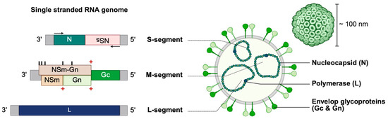
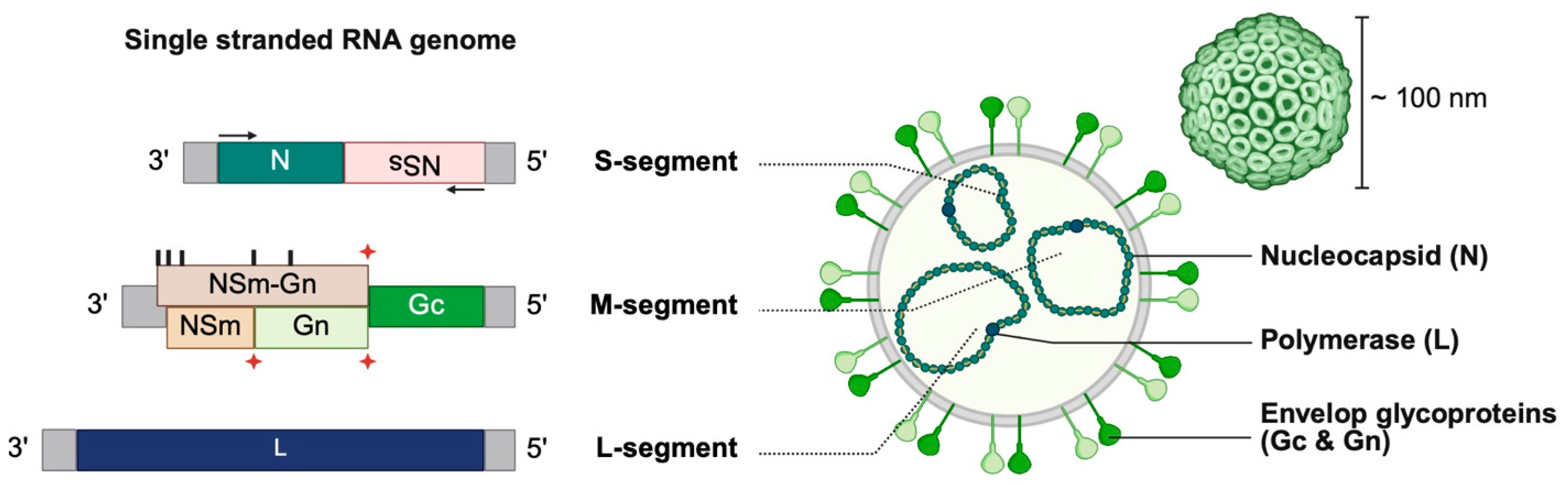
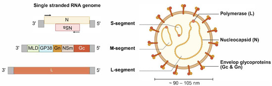
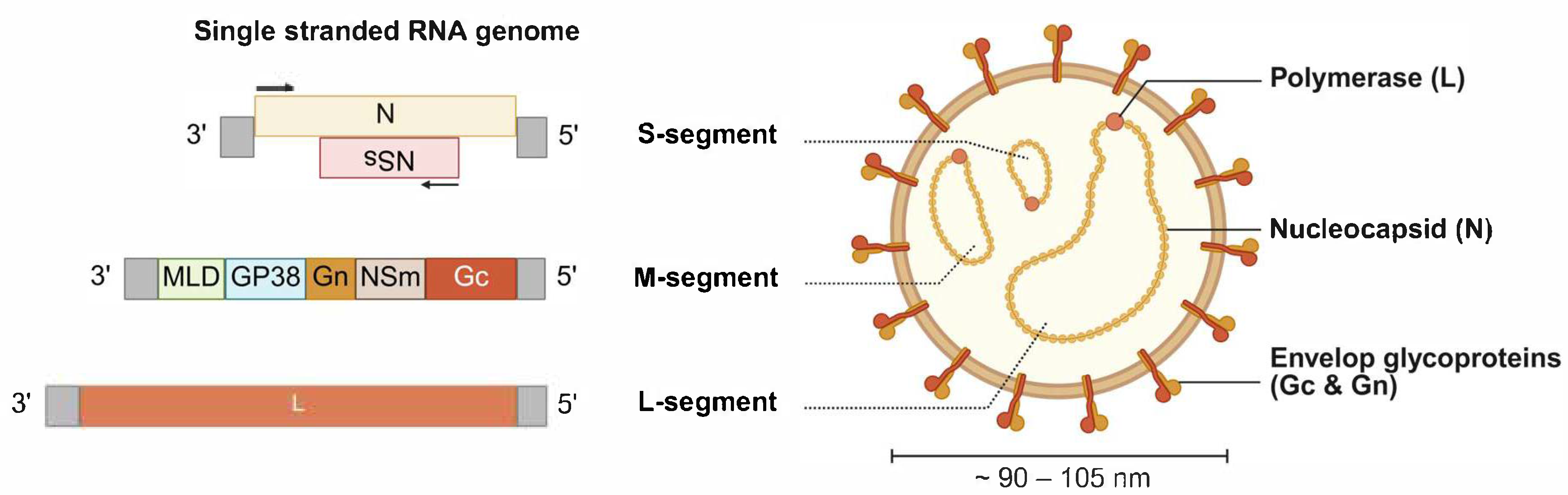

Disclaimer/Publisher’s Note: The statements, opinions and data contained in all publications are solely those of the individual author(s) and contributor(s) and not of MDPI and/or the editor(s). MDPI and/or the editor(s) disclaim responsibility for any injury to people or property resulting from any ideas, methods, instructions or products referred to in the content. |
© 2026 by the authors. Licensee MDPI, Basel, Switzerland. This article is an open access article distributed under the terms and conditions of the Creative Commons Attribution (CC BY) license.