


Abstract
Non-long-terminal-repeat (non-LTR) retrotransposons insert into the genome by priming cDNA synthesis directly from the target site in a process termed target-primed reverse transcription, but the subsequent steps that complete insertion remain poorly understood. In contrast to the relatively uniform products of integrase-mediated LTR retrotransposon integration, non-LTR retrotransposon insertions are structurally heterogeneous, complicating their analysis. Yet this heterogeneity can reveal variation in the underlying insertion reaction when interpreted in an appropriate mechanistic context. Here, we present a framework for mechanism-guided bioinformatic analysis of non-LTR retrotransposon insertions, based on the premise that distinct features of insertion architecture and genomic context report on different biochemical steps of the insertion process. Drawing on historical and recent studies, we identify best practices for sequencing and bioinformatic analysis and consider how extant genomic insertions and experimentally generated de novo insertions provide complementary mechanistic information while imposing different inferential biases. We envision that standardized principles for mechanism-guided analysis will help resolve poorly understood steps of insertion completion, enable comparative investigation across retrotransposons and hosts, and improve evaluation of emerging retrotransposon-based genome-engineering systems.
1. Introduction
Non-long-terminal-repeat (non-LTR) retrotransposons, sometimes called LINEs (long interspersed elements), are a class of eukaryotic transposable elements (TEs) that mobilize through an RNA intermediate. They are among the most ancient extant eukaryotic retrotransposons [1,2] and have achieved widespread evolutionary success, especially in animal genomes, where their activity in extreme cases has generated the majority of genome sequence [3,4].
Non-LTR retrotransposons predominantly initiate insertions through a mechanism called target-primed reverse transcription (TPRT), in which the retrotransposon-encoded catalytic-protein endonuclease (EN) domain nicks one strand of the target site, generating a 3′ hydroxyl that is used to prime reverse transcription of the bound template RNA (Figure 1A, EN-dependent insertion) [5]. Completing an insertion requires nicking the second strand of the target site, forming a 5′ junction, and synthesizing second-strand cDNA. Because the 3′ junction is established during TPRT initiation, whereas the 5′ junction is generated through subsequent and more variable completion steps, non-LTR retrotransposon insertions are structurally asymmetric and heterogeneous. This contrasts with the relatively uniform products of integrase-mediated insertion by long terminal repeat (LTR) retrotransposons, which occurs only after synthesis of full-length double-stranded DNA [6].
Figure 1.
Insertion mechanisms and taxonomic diversity of non-LTR retrotransposons. (A) Schematics of endonuclease (EN)-dependent and EN-independent mechanisms of insertion by non-LTR retrotransposon proteins. “DSB” indicates a double-strand break. (B) Unrooted schematic cladogram of major non-LTR retrotransposon lineages based primarily on RT domain phylogenies [2,7,8]. Branch lengths are arbitrary, and poorly resolved deep relationships are shown as polytomies (multiple descendant branches from a given node). Group II intron RTs are shown as a putative outgroup. In the middle and on the right, the autonomous and nonautonomous elements depicted are representative examples of the respective groups, as indicated. The schematic does not imply that the example nonautonomous element is mobilized by the example autonomous element connected by a dashed line, except for human L1 and Alu. The group nomenclature and clade groupings follow the Repbase classification [2]. The “R2” group includes R2, R4, Hero and NeSL clades. The “L1” group includes Proto1, L1, and Tx1 clades. The “RTE” group includes RTETP, Proto2, RTEX, and RTE clades. The “I” group includes Outcast, Ingi, Vingi, I, Nimb, Tad1, Loa, R1, and Jockey clades. The “CR1” group includes Rex1, CR1, Kiri, L2, L2A, L2B, Crack, and Daphne clades. “APE” indicates apurinic/apyrimidinic endonuclease-like domain, and “RLE” indicates restriction-like endonuclease domain. “DBD” indicates DNA-binding domains. “CTD” indicates C-terminal domain. “RNH” indicates RNase H domain. “TBR” indicates tail-binding region. “SIDE” indicates short internally deleted element. “28S” refers to sequence from the 28S ribosomal RNA gene. “RZ” indicates ribozyme.
Early observations of non-LTR retrotransposon insertions showed precise 3′ ends and variable, often truncated, 5′ ends [9,10,11]. Together with homology of the encoded protein to known reverse transcriptases (RTs), the truncation profile suggested a model in which reverse transcription was primed directly from chromosomal DNA at a characteristic position near the 3′ end of the template RNA, explaining the precise 3′ junction of insertions (see Figure 1A), but used more variable mechanisms to join cDNA to the target site, accounting for 5′ heterogeneity. The central prediction that reverse transcription could be primed directly from the chromosomal target site was experimentally validated in 1993 by Eickbush and colleagues [5], establishing the TPRT mechanism now known to be shared across non-LTR retrotransposons. The genomics-informed discovery of TPRT illustrates the central premise of this review: sequences inserted by non-LTR retrotransposon proteins can serve as molecular records from which transient steps of retrotransposition can be reconstructed.
In the context of renewed interest in studying this class of retroelements [12], including a flurry of innovative retrotransposon-based strategies for transgene insertion [13], here we review historical approaches to and outline best practices for mechanism-guided bioinformatic analysis of inserted sequences. We organize bioinformatic evidence according to the steps of the insertion pathway that are informed by sequence signatures, beginning with the reconstruction of the original target site prior to insertion and continuing to mechanisms of target-site selection, first-strand cDNA synthesis, completion of the insertion, and genome rearrangements. Highlighted examples from the literature primarily focus on animal non-LTR retrotransposons, where most mechanistic work has been performed.
Bioinformatic discovery and characterization of non-LTR retrotransposons themselves—including de novo element discovery, reference/consensus sequence construction, classification and phylogenetic analysis, and prediction of potentially active copies—require a distinct set of approaches and are outside the scope of this review [13,14,15,16,17]. Additionally, recent advances in the use of non-LTR retrotransposon proteins for genome engineering have been reviewed elsewhere [12,13]. We instead focus on the analysis of the insertion events generated by these elements.
2. Structural and Biochemical Diversity of Non-LTR Retrotransposons
Here, we briefly review the structural and biochemical diversity of non-LTR retrotransposons, focusing on experimental models relevant to the subsequent discussion. Although deep phylogenetic relationships remain unresolved, current classifications recognize eight broad groups of known non-LTR retrotransposons, excluding Penelope-like elements (Figure 1B) [1,2,7,8,18]. R2, CRE, and Dualen group elements generally encode a single open reading frame (ORF); L1 and Ambal group elements generally encode two; and RTE, I, and CR1 group elements are more variable [2].
The largest protein, which is encoded by ORF2 of a two-ORF element or by the sole ORF, contains reverse transcriptase (RT) and endonuclease (EN) domains. A third catalytic domain, encoding RNase H activity, is observed in some ORF2 proteins (ORF2p). The presence of other domains and the organization thereof differ substantially among elements even within the same taxonomic group. EN domains are categorized as either apurinic/apyrimidinic endonuclease-like (APE) or restriction-like endonuclease (RLE) according to sequence homology (the presence of either or both EN domains is indicated for each element family in Figure 1B). When present, ORF1p is generally thought to facilitate ribonucleoprotein (RNP) formation and RNA trafficking, but its precise functions remain poorly understood, in part due to substantial variation in sequence and domain architectures across non-LTR retrotransposon lineages [19,20].
Five historically important model systems to highlight are insect R1, animal R2, human LINE-1 (L1), Drosophila I factor, and zebrafish LINE-2 (ZfL2) (Figure 1B). Biochemical, cell culture, and organismal systems for investigating these retrotransposons anchor many of the historical developments in the field. These five representatives differ along major dimensions of non-LTR retrotransposon diversity, including EN domain type, number of ORFs, and insertion-site specificity.
Both R1 and R2 elements insert site-specifically into the sequence encoding the 28S ribosomal RNA in a larger, multicopy repeated gene unit (rDNA), whereas L1, I factor, and ZfL2 elements insert in a dispersed manner. Silk moth Bombyx mori R1 (R1Bm) became an early model when its characteristic 14-bp target-site duplication (TSD) was explained by offset in vitro cleavage of target-site DNA strands, generating 3′ overhangs [21]. B. mori R2 (R2Bm) enabled the foundational biochemical reconstitution of EN cleavage and TPRT [5]. More recently, avian R2 proteins (R2p), particularly zebra finch Taeniopygia guttata R2p, have become models for retrotransposition biochemistry and cell biology [22,23], as well as tools for genome engineering (as reviewed in [12,13]). L1, the sole active autonomous transposable element in humans, is the primary mammalian model, supported by abundant data on somatic and polymorphic insertions and tractable de novo cell culture assays. Here, polymorphic insertions refer to germline-inherited insertions present in some but not all individuals in a population. Drosophila melanogaster I factor provides a genetically inducible in vivo model of germline retrotransposition and associated genome rearrangements through I-R hybrid dysgenesis [24]. Finally, zebrafish Danio rerio L2 elements belong to the CR1 group and variably encode one or two ORFs. ZfL2-1 and ZfL2-2 both stringently recognize their cognate 3′ UTRs [25], and the more extensively studied ZfL2-2 has been shown to mobilize in heterologous cell culture systems [26,27], enabling analysis of host-dependent insertion outcomes [28,29].
In addition to mobilizing their own transcripts, non-LTR retrotransposon proteins can make genome insertions by copying RNAs from nonautonomous elements, including short interspersed elements (SINEs), which lack their own retrotransposition proteins and depend on autonomous elements for mobilization (Figure 1B). RNA sequence or structural features can mediate recognition by the retrotransposon protein. Insertions created by copying cellular mRNAs generate processed pseudogenes.
3. A Framework for Mechanism-Guided Insertion Analysis
We ground this review in the principle that genome sequence analysis is informative for deducing individual biochemical steps across the non-LTR retrotransposon insertion process. As diagrammed in Figure 2, different parts of the insertion or surrounding target site report on different steps of the insertion reaction. Throughout this review, 5′ and 3′ insertion ends are designated relative to the positive-sense RNA template, rather than the initial cDNA strand (Figure 2A).
Figure 2.
Sequence features of non-LTR retrotransposon insertions and related mechanistic interpretations. (A) Schematic representing inference of pre-insertion target site and mobilized template RNA from observed insertion-containing allele. “TSD” indicates target-site duplication. (B–F) Schematics identifying sequence features of retrotransposon insertions, junctions, or reconstructed pre-insertion target sites and relevant mechanistic interpretations for each step. “DSB” indicates double-strand break. “NTA” indicates non-templated addition of nucleotides. “NHEJ” indicates non-homologous end-joining.
Our framework begins with inferring the inputs of the insertion reaction: (1) reconstructing the pre-insertion target site and (2) reconstructing the mobilized RNA and its source (Figure 2A). It continues with inferring the biochemical steps of the insertion reaction: (3) target-site selection and first-strand cleavage (Figure 2B), (4) DNA primer generation and initiation of reverse transcription (Figure 2C), (5) first-strand cDNA synthesis (Figure 2D) and (6) second-strand cleavage (Figure 2E), 5′ junction formation (Figure 2F), and second-strand synthesis. Finally, this framework will address potential off-pathway products of the insertion: (7) insertion-associated genome rearrangements and (8) abortive insertion and collateral genome damage.
In traversing each step of this framework, we will review what evidence can be gathered from the analysis of fixed genomic insertions and from experimentally generated de novo insertions. Here, a fixed genomic insertion refers to an inherited retrotransposon insertion present in essentially all individuals in a population (allele frequency ~1). Where appropriate, we will also consider what evidence is provided by analysis of somatic transposition events or polymorphic insertions. Other kinds of evidence—including in vitro biochemistry and quantifications of retrotransposition frequency—will not be covered by this review except when relevant for context of bioinformatic analyses.
4. Sources of Mechanistic Evidence
The mechanistic conclusions that can be drawn from insertion sequences depend on three linked choices: how insertions arose (extant genomic insertions vs. experimentally generated insertions), how those insertions are sequenced, and how the resulting reads are analyzed bioinformatically. Fixed, somatic, polymorphic, and experimentally generated de novo insertions differ in the biological histories they capture; sequencing methods differ in which events and sequence features they recover; and computational pipelines differ in which aspects of insertion architecture they preserve and interpret. Together, these factors determine both the information available for mechanistic inference and the biases that constrain it.
Section 4.1, Section 4.2 and Section 4.3 consider the distinct strengths and limitations of fixed genomic insertions, somatic insertions, polymorphic insertions, and experimentally generated de novo insertions.
Sequencing method further determines which insertion events and sequence features are recovered. Method choice should be guided by the mechanistic feature of interest, with quantitative recovery of independent insertion events prioritized, where possible, through the use of unique molecular identifiers and minimal PCR amplification. Size selection should be avoided when insertion length or architecture is itself an outcome of interest. Table 1 summarizes representative sequencing approaches, their principal strengths and biases, and example applications.
Table 1.
Assays for sequencing non-LTR retrotransposon insertions. The lists of assays and applications for each category are intended to be representative but non-exhaustive. The “S/L” column identifies whether the method is typically used with short-read (S) and/or long-read (L) sequencing technologies. “PCR bias” refers to sampling and capture biases associated with exponential amplification by PCR. Relevant sources of bias include length bias, sequence composition bias, stochastic dropout and jackpotting, limited or no detection of complex rearrangements, and requirement for insertion to contain primer-binding site for detection. “Insertion sequence requirement” indicates that sequencing method requires insertions to contain a specific sequence and that the associated method is therefore only compatible with de novo insertion assays.
Finally, bioinformatic approaches determine which molecular features can be reconstructed from sequence data. Section 4.4 and Table 2 summarize computational approaches according to their utility for mechanism-guided insertion analysis and provide representative applications.
Table 2.
Computational approaches for analysis of non-LTR retrotransposon insertions. Lists of tools and applications for each category are intended to be representative but non-exhaustive.
4.1. Fixed Genomic Insertions
Fixed non-LTR retrotransposon insertions can be thought of as genomic fossils, and their study as “molecular archaeology” [54]. As with physical fossils, genomic insertions surviving to the present day do not represent a complete record of past insertion events. Insertions are eliminated by neutral genetic drift or purifying selection [64], and mutation, deletion, and recombination obscure information over time. These problems are particularly acute for insertions into repetitive loci such as rDNA or microsatellites, where high recombination rates and concerted evolution are associated with rapid turnover of insertions [65,66,67]. Despite its limitations, analysis of fixed genomic insertions provides the only route to study some non-LTR retrotransposon systems for which biochemical reconstitution or de novo insertion experimental systems are intractable or have not yet been developed.
For analysis of fixed insertions, not all source data are equally informative. When available, haplotype-resolved long-read assemblies enable the most comprehensive analysis of TE insertions [68,69]. However, as non-LTR retrotransposons commonly target multicopy loci encoding conserved non-coding RNAs or microsatellites, telomeres, and other low-complexity sequences [70], analysis of even high-quality, “telomere-to-telomere” genome assemblies may be insufficient for comprehensive analysis of TE insertions. For example, even in the most updated D. melanogaster genome reference (dm6 at the time of this writing), rDNA arrays are not fully assembled [71]. Therefore, a thorough investigation of R1 and R2 insertions would require an analysis of source reads, regardless of assembly status. Analysis of TE insertions is best powered by long reads, which can provide a molecular record of individual insertion events [59,72].
4.2. Somatic and Polymorphic Insertions
Although less common among published studies, analysis of somatic transposition events and of polymorphic insertions can be a powerful tool for generating or testing mechanistic hypotheses. Somatic events and polymorphic insertions avoid some of the bioinformatic challenges associated with fossilized genomic insertions while still reporting on retrotransposition in its native organismal context. These sequences have undergone relatively little decay, and an insertion-less allele of the target site can often provide the pre-insertion target-site sequence.
The primary challenge for analysis of somatic and polymorphic insertions is one of scarcity. Available datasets generally contain fewer of these insertion events than fixed genomic copies or induced de novo insertions. Human tumor samples are an important exception because they are deeply sequenced and broadly sampled by several consortia, providing a wealth of somatic transposition data [73,74,75,76,77].
4.3. De Novo Insertions
De novo sequence insertion assays enable the capture of new genome insertions before evolutionary selection or sequence decay, better preserving the signatures of all the supporting cellular steps. However, as a caveat, these assays do not fully recapitulate the abundance, stoichiometry, localization, or processing of retrotransposon components that occur during endogenous retrotransposition.
We distinguish three primary classes of de novo insertion experiments that differ in how retrotransposition is initiated. The first class involves the delivery of DNA, often episomal, to express non-LTR retrotransposon RNA and proteins (Figure 3A). Traditional DNA-based expression assays, particularly for L1, most often contain an antisense reporter gene (typically conferring drug resistance or fluorescent protein expression) interrupted by a sense intron, thus allowing the selection of cells with bona fide retrotransposition events [78,79]. DNA-based expression constructs typically encode the retrotransposon machinery and template RNA in cis, but separate expression constructs for retrotransposon protein and template RNA have been used for studies of nonautonomous element mobilization and trans complementation [80,81,82,83]. Host-cell machinery determines the level and fate of RNA transcripts in a non-physiological expression context, with potential heterogeneity in the pool of molecules serving as templates for reverse transcription. For downstream sequencing applications, the presence of the donor sequence in a host chromosome or episome can overwhelm signal from RNA-templated genome insertions.
Figure 3.
Experimental systems for generating de novo non-LTR retrotransposon insertions. (A) Schematic of LINE-1 retrotransposition assay involving delivery of episomal construct expressing L1 ORF1p and ORF2p and encoding a sense-orientation intron within an antisense neomycin resistance gene (“neo”). Upon insertion of the RNA encoding the L1 machinery and spliced neo expression cassette (in cis), previously susceptible cells (“G418S”) become drug resistant (“G418R”). “SD” and “SA” indicate splice donor and splice acceptor, respectively. Based on Moran et al. (1996) [78]. (B) Schematic of R2 retrotransposition assay involving delivery of R2p-encoding mRNA and transgene-encoding RNA. Upon insertion of the RNA, cells express GFP. “pro” indicates promoter and “pA” indicates polyadenylation signal sequence. Based on the Precise RNA-mediated INsertion of Transgenes (PRINT) system [23]. (C) Overview of I-R hybrid dysgenesis system for in vivo induction of I factor transposition in D. melanogaster. Schematic depicts genetic cross between female individual from a Reactive strain and male individual from an Inducer strain producing sterile F1 females (“SF”) due to I factor transposition. “×” symbol indicates a lack of transposition events. “✓” symbol indicates active transposition.
The second class involves the delivery of RNA or RNP (Figure 3B). In a cis configuration, an mRNA encoding a non-LTR retrotransposon protein provides both a template for protein translation and a template for TPRT [22,44]. In a trans configuration, an mRNA encodes a retrotransposon protein, while a separate RNA serves as template for TPRT (Figure 3B) [22,23]. A trans format facilitates the separate investigation of the requirements for the RNA template and retrotransposon proteins for insertion. A less common format involves the delivery of a preformed RNP, providing maximal control over the molecular complex responsible for insertion [84]. RNA- and RNP-based modes of delivery restrict insertions to a defined pulse of activity in comparison to plasmid delivery, such that insertion outcomes can be read out within hours of delivery to reduce potentially confounding variables such as cell survival or host silencing of transgenes. Additionally, delivery of the template RNA as a purified transcript has several benefits: it can provide a specific sequence rather than a collection of heterogeneously processed transcripts, and the template RNA can use nucleotide analogs and chemical modifications to engineer a desired RNA fate or half-life.
A third class of de novo insertion experiments entails inducible mobilization of endogenous non-LTR retrotransposons in vivo (Figure 3C). I-R hybrid dysgenesis in D. melanogaster is a model system, in which controlled genetic crosses induce I factor mobilization in the germline of female progeny [85]. Assays relying on mobility of endogenous elements retain physiological context but lack an opportunity to readily alter insertion sequence and to constrain the tempo and molecular details of retrotransposition.
4.4. Bioinformatic Tools and Mechanism-Aware Insertion Analysis
We have evaluated bioinformatic tools for their capacity to analyze insertions in a mechanism-aware manner. Table 2 categorizes computational tools for (1) repeat annotation, (2) de novo, somatic, and polymorphic insertion discovery, (3) amplicon analysis, and (4) mechanism-guided insertion analysis, each with literature references.
Repeat annotation tools such as RepeatMasker identify TE-derived sequence in a host genome assembly using an input library of TE sequences [86]. These annotations are not tailored to features specific to non-LTR retrotransposon insertions but can support custom downstream analysis of truncations, inversions, and junction architectures [54,55]. Insertion-discovery tools—including MELT [58], TLDR [87], and PALMER [59]—infer de novo, somatic, and polymorphic insertion events by comparison of sequencing reads to a host genome assembly. Some insertion-discovery tools, including TLDR and PALMER, additionally reconstruct the insertion architecture, including internal features such as truncations, inversions, and transduced sequence (described below), as well as target-site alterations.
Beyond repeat annotation and insertion-discovery tools, a third category of software analyzes amplicons for genome insertion or editing outcomes. Recent studies adapting non-LTR retrotransposons into genome-engineering tools have often analyzed de novo insertions using genome editing software such as CRISPResso [22,43,44,88,89]. CRISPResso estimates relative insertion efficiency and identifies imprecise junctions or indels within a pool of sequenced amplicons, with the caveat of length-dependent PCR amplification bias; it does not reconstruct the mechanistically informative features of non-LTR insertions.
At the time of writing, there are no widely used bioinformatic tools broadly capable of reconstructing insertion events mediated by non-LTR retrotransposon machinery. However, numerous historical and recent publications provide examples of bespoke analyses that have been applied effectively to mechanistic inquiry [35,38,45,46,47,48,61,62].
5. Reconstructing Inputs, Outputs, and Reaction Steps
5.1. Inputs: Reconstructing the Pre-Insertion Target Site
Reconstruction of the pre-insertion target site (Figure 2A) is a prerequisite for inferring insertion-associated target-site alterations, but this process is challenging for fossilized genomic insertions. Unlike the characteristic 8-bp TSDs associated with the mobility of D. melanogaster P element DNA transposons [90], for example, non-LTR retrotransposon insertions can result in TSDs, target-site truncations, or genomic addition of extra nucleotides, with heterogeneity across insertion loci carrying the same retrotransposon in the same genome. Any attempt to reconstruct an uninserted target-site allele for fossilized insertions is inherently indirect, in that the accumulation of mutations after insertion obscures the ancestral state.
Strategies for target-site inference can be broadly classified as reference-agnostic or alignment-based. The primary reference-agnostic method can be applied only to the subset of insertions that generate TSDs, which can be collapsed to reconstruct the pre-insertion target site. However, even the subset of non-LTR retrotransposons that canonically form TSDs do not always generate TSDs at all insertion loci [54]. For example, when L1 makes EN-independent insertions instead of EN-dependent insertions (Figure 1A), TSDs are absent [28,48,55,91].
Another approach is to align the insertion-containing locus to reference sequence(s) for the same or phylogenetically proximal organisms to infer the pre-insertion target site [92]. This approach has been useful even for insertions in multicopy loci or low-complexity sequence repeats, as demonstrated, for example, for retrotransposons targeting ribosomal RNA genes, U2 small nuclear RNA genes, or other repetitive sequences [16,28,70,93,94,95,96,97,98,99]. However, analysis of insertions into these multicopy target sites poses a unique challenge, in that definitive identification of the specific chromosome position of insertion can be impossible. In these cases, alignment to a consensus reference sequence of a multicopy locus is more accurate, and more appropriate, to report than random coordinate assignment of multi-mapping reads. Anchoring to a consensus reference simplifies downstream analysis, for example in identifying patterns of target-site alterations [23].
In contrast to target-site reconstruction for preexisting mobility events, target sites for de novo insertions can usually be reconstructed by aligning insertion-flanking sequence to the reference genome. Complex rearrangements involving multiple sequence sources, inversions, deletions, or duplications may nevertheless remain difficult to detect and reconstruct [49]. Consequently, such events are often overlooked or not systematically evaluated in insertion sequence analysis. However, recent advances in long-read sequencing, bioinformatic methods, and reporter assays are beginning to enable their detection and mechanistic characterization (see Section 5.7 and Section 5.8).
5.2. Inputs: Reconstructing the Mobilized RNA and Its Source
A parallel effort to target-site reconstruction is inference of the template RNA that gave rise to the inserted sequence (Figure 2A). For endogenous retrotransposition, this can also help identify the source element. In native contexts or de novo insertion assays using plasmid-based expression of a template RNA, transcription and RNA processing can generate heterogeneous templates through variable transcriptional boundaries, splicing, polyadenylation, and self-cleavage. The inserted sequence can diverge further from the actual template RNA sequence due to internal initiation of reverse transcription, non-templated nucleotide additions, premature RT termination, and variable 5′-junction formation by host-cell machineries (Figure 2C,D). RNA ligation and template jumping can further alter the sequence that is ultimately inserted [100,101,102]. Mutations introduced during cDNA synthesis or accumulated after insertion add another source of divergence. Direct delivery of template RNA simplifies reconstruction by defining the input sequence, although heterogeneity can still arise during in vitro transcription or through RNA degradation after delivery [103,104,105].
Inefficient use of the L1-encoded polyadenylation signal can extend transcription into downstream genomic sequence, producing 3′ transductions when this sequence is subsequently inserted [106,107,108,109]. Imprecise transcription initiation or readthrough from upstream promoters can similarly generate 5′ transductions [110,111]. Conversely, inserted sequences do not necessarily reflect the entire transcript sequence: for example, D. melanogaster I factor transcripts include downstream flanking sequence that is not incorporated into insertions [112,113].
Reconstructing the mobilized RNA therefore requires considering alternative RNA-processing products rather than assuming that the inserted sequence derives directly from an annotated retrotransposon transcript. Cryptic splicing of L1 transcripts, for example, generates internal deletions detectable in both RNA and insertion products [114,115]. Conversely, extra sequence can arise through non-templated nucleotide addition, RNA ligation, or template jumping [100,101]. RtcB-mediated ligation, for example, generates U6–L1 chimeric RNAs that can account for chimeric insertions [101,116]. Finally, self-cleaving ribozymes can define template termini, as predicted for the 5′ ends of R2 transcripts [117,118], and observed more broadly across diverse retrotransposon systems [119], making RNA-structure-aware analysis important for accurately predicting template RNA termini.
For extant genomic insertions, care must first be taken to align each insertion to the appropriate family- or subfamily-specific reference, because closely related retrotransposons can share substantial sequence homology. Divergent lineages of the same family can coexist within a genome [120], and use of an incorrect reference can obscure insertion boundaries or lead to misannotation, as demonstrated for primate L1 subfamilies [121,122,123]. Even with accurate family assignment, however, identifying the genomic source locus for an individual insertion is often difficult because non-LTR retrotransposons are typically multicopy. Candidate retrotransposition-competent loci can be identified from genome sequence records by full-length retrotransposon sequence, intact ORFs with preserved catalytic amino acid motifs, and appropriate regulatory sequences, and then tested experimentally [124,125]. More direct source assignment can come from sequence carried into the new insertion: uniquely mappable 3′ or 5′ transduced sequence can identify the source locus [108,109,126], while source-specific single-nucleotide variants or haplotypes within the element can match offspring insertions to candidate genomic copies [63,127].
Trans-mobilized RNAs pose an additional problem because the resulting insertion does not identify the autonomous element that supplied the retrotransposition machinery. If RNP formation is governed by retrotransposon protein recognition of known structural or sequence features at the RNA 3′ end, common sequence features can identify candidate autonomous/nonautonomous element pairs [128,129,130,131]. For non-LTR retrotransposons with relaxed, poly(A)-directed RNP formation [132], autonomous/nonautonomous element pairs can be suggested by similar insertion junction signatures [133,134]. Experimental validation is required to definitively establish these autonomous/nonautonomous pairs [81,82,83,135].
5.3. Insertion Reaction: Target-Site Selection and First-Strand Cleavage
Below, we consider five determinants of target site selection (Figure 2B): (1) retrotransposon protein recognition specificity, (2) template-RNA-guided specificity, (3) host factor interactions, (4) target-site architecture, and (5) regional genomic context. Here, target-site architecture refers to the local physical structure of the DNA at the insertion site, including nicks, overhangs, and replication-associated structures such as Okazaki fragments. Regional genomic context encompasses broader features of the surrounding locus, including chromatin state, transcriptional activity, and replication timing. Importantly, these five determinants of target-site selection are not fully predicted by simple sequence-motif-based identification of target sites. More broadly, non-LTR retrotransposons fall along a spectrum of insertion-site specificity from highly site-specific to broadly dispersed rather than binary categories of site-specific vs. site-non-specific [70,136].
Biochemical mapping of EN–DNA specificity for R1Bm and human L1, together with concordant genomic insertion-site preferences, established endonuclease sequence specificity as the primary model for non-LTR retrotransposon target-site selection [21,133,137,138]. More direct evidence came from EN-domain swapping experiments between SART1 and TRAS1, which transferred target specificity between the two retrotransposon proteins [139]. Additionally, EN-independent L1 insertions lacked the canonical L1 target motif, further supporting a central role for EN sequence specificity [91]. For R2p, off-target mapping implicates nucleotide sequence outside the EN-contacted region as determining specificity [22,23,38], although not fully accounting for its unusually high degree of specificity, which indicates that the biochemical principles of target-site selection remain incompletely resolved.
Beyond protein determinants of specificity, base-pairing between the template RNA and the DNA 3′ end generated by first-strand cleavage can impose additional sequence requirements on TPRT initiation. For R1Bm, the addition of downstream 28S rRNA sequence to the RNA template increased retrotransposition efficiency and 3′ junction precision, consistent with a role for RNA–DNA complementarity [140]. More directly, mutation of the R2 target-site sequence disrupted in vitro TPRT from a prenicked DNA substrate, but activity was restored by compensatory changes to the 3′ tail of the template RNA [23]. Whether RNA sequence or structure contributes to target-site specificity beyond these base-pairing requirements has not been demonstrated.
A clear demonstration of host factor interaction as a determinant of specificity for non-LTR retrotransposons is Dictyostelium discoideum TRE5-A interaction with RNA polymerase III (Pol III) promoter-associated transcription complexes. Despite a high degree of site specificity for Pol III promoters, the exact position of insertion within the promoter sequence varies, suggesting recognition of transcription machinery rather than a specific DNA sequence, as validated through perturbative assays [141,142]. TRE5-A targeting through host factor interaction is analogous to previously characterized recruitment of some Ty LTR retrotransposons to Pol III-transcribed genes by interaction with transcription machinery [143]. As another potential mechanism of host factor influence, the interaction of L1ORF2p with PCNA could bias target-site selection toward lagging-strand templates through repeated PCNA loading during Okazaki fragment synthesis [144,145,146]. However, whether PCNA contributes to L1 target-site selection through this mechanism has not been tested directly.
Local DNA architecture has also been proposed as a determinant of target-site selection, though it has been demonstrated only in the context of L1ORF2p-mediated de novo insertions [45,145]. Mapping of de novo EN-dependent L1 insertions revealed an apparent bias for first-strand nicking in lagging-strand templates at the replication fork, consistent with biochemical preferences for cleavage substrates [35,45,145,147]. In parallel, mapping of de novo EN-independent L1 insertions revealed a bias for the opposite strandedness at replication forks, consistent with priming from Okazaki fragment 3′ ends [45].
The determinants of target-site specificity reviewed so far—those mediated by retrotransposon proteins, retrotransposon RNA, retrotransposon protein interaction with host factors, or target-site DNA architecture—describe predominantly local effects. Regional genomic context can also influence targeting. For human L1, de novo insertion mapping identified no consistent bias for insertion in transcribed regions or open chromatin, in sharp contrast to integration by retroviruses and DNA transposases [35,45]. Also, in contrast to human L1 but more in accord with other mobile genetic elements, de novo D. melanogaster I factor insertions showed a preference for targeting open, transcriptionally active chromatin [148,149].
While fixed genomic insertions provide some information about target-site selection, de novo insertion assays enable more direct and comprehensive identification of target-site preferences. Signatures of target-site sequence preferences can be preserved at fixed genomic insertions [133,150,151], but the contributions of broader genomic context can be more difficult to infer retrospectively. Purifying selection and rapid turnover at repetitive target loci can alter the observed genomic distribution of surviving insertions [35,64,67,149,152]. Host-mediated repression of inserted retrotransposons can also alter local chromatin, complicating inference of the chromatin state at the time of insertion [153]. De novo insertion mapping therefore enables more direct assessment of targeting preferences, both of local target-site sequence and of broader genomic context.
Genome-wide specificity claims require unbiased sampling, explicit detection-sensitivity estimates, retention and reporting of low-frequency events, and quantification of off-target insertions relative to a comprehensive total of all independent insertions. PCR of candidate off-target-site loci alone cannot establish genome-wide specificity. Recent studies adapting non-LTR retrotransposon machinery into gene insertion tools have not uniformly met these standards, in their reliance on candidate-site PCR, exclusion of low-frequency events by bioinformatic filtering, or claims of high specificity without explicit quantification [43,44,89]. See Table 1 for an overview of sequencing methods.
5.4. Insertion Reaction: DNA Primer Generation and Initiation of Reverse Transcription
Generation of the DNA primer for cDNA synthesis can occur by EN-mediated first-strand nicking, or a primer can be co-opted from a preexisting 3′ DNA end (Figure 2C). Early models of the non-LTR retrotransposon insertion mechanism often posited that preexisting DNA breaks could provide 3′ hydroxyl ends to prime reverse transcription [11,154]. Though biochemical studies later demonstrated that EN domain activity is sufficient to generate 3′ hydroxyl ends for TPRT [5,21,137], EN-independent priming from spontaneous DNA breaks and telomere ends is now also established as physiological [42,91,155]. EN-independent priming from Okazaki fragment 3′ ends has also been proposed [45]. The absence of a canonical target-site motif, large-scale target-site rearrangements, and/or internal initiation of reverse transcription can identify candidate EN-independent insertions [45,91]. Because EN-independent insertions exhibit distinct target-site signatures, candidate EN-independent events should be separated from canonical insertions when inferring EN-mediated target-site specificity. Although less deeply characterized than EN-deficient L1ORF2p, EN mutants of avian R2p and R1Bm exhibit diminished but measurable retrotransposition activity [23,140]. Bioinformatic analysis of these insertions suggests altered target-site preferences consistent with RT priming from preexisting 3′ ends [140].
The generation or capture of a DNA primer is only the first requirement for TPRT: productive reverse transcription additionally requires positioning of the RNA template relative to the primer 3′ end. EN-dependent TPRT also requires transfer of the newly generated 3′ end from the EN active site to the RT active site. For some non-LTR retrotransposon proteins, base-pairing of the RNA template to the DNA primer end (terminal complementarity, Figure 2C) is required for initiation [23]. Statistically enriched microhomology at 3′ junctions of extant insertions has suggested base-pairing requirements for RT initiation by taxonomically diverse non-LTR retrotransposon proteins [95,129,131]. In de novo insertion assays, perturbation of template RNA 3′ ends has demonstrated requirements of terminal complementarity for both precision and efficiency of TPRT initiation by R1Bm, SART1Bm, R2Ol, avian R2p, and R7Ag [23,140,156,157,158]. Terminal complementarity requirements are not universal, however: rare human L1 insertions are consistent with internal priming within the RNA template (Figure 2C) rather than initiation at its 3′ terminus [126,159], illustrating flexibility in primer–template positioning. Also, if priming is challenged by inadequate complementarity or insufficient length of single-stranded primer for stable TPRT initiation, extra nucleotides can be added prior to template copying (Figure 2C) [112,160,161].
5.5. Insertion Reaction: First-Strand cDNA Synthesis
Relative to the reaction steps considered thus far, bioinformatically reconstructing first-strand cDNA is particularly challenging. As discussed above (Section 5.2), many different processes before, during, and after first-strand synthesis can alter the final inserted sequence observed. Our analysis will describe historical efforts to disentangle these processes and indicate where future bioinformatic inquiry could provide important contributions.
A central unresolved question is what generates 5′ truncations, which could arise from RNA template degradation, premature termination of reverse transcription, nuclease cleavage of the cDNA, cDNA loss during 5′ junction formation, or a combination of these processes (Figure 2D–F) [162]. Though the hypothesis that 5′ truncation derives from premature termination by an insufficiently processive RT is pervasive in the literature (Figure 2D) [162], biochemical studies of R2 and L1 RTs do not provide support for this conclusion [102,163]. Additional investigation of this hypothesis will be informative for optimization of the genome-engineering applications for non-LTR retrotransposon proteins. Inherently, RNA is prone to hydrolysis and, in cells, to partial degradation (Figure 2D). RNA structural elements or base substitutions designed to improve the biological stability of a template yield notable suppression of 5′ truncation in R2p-mediated insertions, influencing both the percentage of full-length insertions and the amount of sequence lost as a truncation [23,105]. Finally, the cDNA product can be truncated during 5′ junction formation when second-strand synthesis is initiated on an internal region of first-strand cDNA (see Section 5.6 below) [61,62,162,164].
Irregularities in cDNA sequence architecture—including 5′ inversions, internal deletions, and internal duplications—are all candidates for first-strand synthesis-connected processes. Inversions at the 5′ end have been observed in ~20–25% of recently fixed L1 insertions, motivating the twin priming model (Figure 2D), in which reverse transcription primes from both ends of a DNA break at different positions on the same RNA template [165]. Inversion-containing de novo L1 insertions frequently exhibit architectures consistent with twin priming, although the proposed mechanism has not been directly demonstrated [48,49,63]. Similar inversions have not been reported for insertions by other non-LTR retrotransposons, raising the possibility that twin priming may be specific to L1ORF2p because of its tolerance for limited primer–template complementarity [95,159,165]. The occurrence of 5′ inversions in L1ORF2p-mediated de novo insertions varies by host, implicating host-dependent steps in their generation or resolution [61]. Other cDNA irregularities are less mechanistically resolved. Internal deletions and duplications have been observed within non-LTR retrotransposon insertion products [130,166], including de novo insertions [23,48], and therefore could arise from aberrant first- or second-strand synthesis rather than a post-insertion DNA rearrangement.
A final class of noncanonical first-strand synthesis events involves reverse transcription of host RNAs following reverse transcription of the intended template RNA [23,48,116]. While these events are consistent with end-to-end template jumping during first-strand synthesis (Figure 2D) [100], they may also reflect reverse transcription of a pre-ligated chimeric RNA [101].
An informative avenue for future studies would be to make systematic measurements of first-strand cDNA synthesis error rates, which remain poorly characterized for non-LTR retrotransposon RTs in cellular context. Sequencing of de novo L1ORF2p- and avian R2p-mediated insertions has yielded substitution rates on the order of 10−4 per base [23,48], similar to in vitro estimates for L1ORF2p and R2p [167,168]. However, insertion endpoint sequencing convolves errors from template RNA synthesis, first- and second-strand DNA synthesis, and sequencing, precluding direct measurement of first-strand synthesis fidelity in approaches applied to date. In vitro measurements are not a substitute, because they depend on assay conditions including dNTP concentrations and template context. Accurate measurement would require analysis of first-strand cDNA intermediates before second-strand synthesis and should employ low-error consensus sequencing.
5.6. Insertion Reaction: Second-Strand Cleavage, 5′ Junction Formation, and Second-Strand Synthesis
The mechanistic details of insertion completion—second-strand cleavage, 5′ junction formation, and second-strand cDNA synthesis—remain largely unresolved. Sequence-based inference is particularly challenging because endpoint features such as target-site alterations and 5′ truncations may report on multiple steps in the completion process rather than a single biochemical event (Figure 2E,F). De novo insertion studies provide the greatest opportunity to disentangle these processes through experimental perturbation, but new insertion 5′ junctions have often been sequenced far less deeply than 3′ junctions [22,35,38,45]. Mechanistic analysis of completion benefits from the characterization of both insertion junctions, ideally while retaining information about which junctions derive from the same insertion event.
Non-LTR retrotransposon insertions are commonly associated with TSDs, consistent with staggered nicks by the EN domain that generate 3′ overhangs [169,170]. Comparative analyses across host species show that TSD length patterns are more strongly associated with retrotransposon identity than with the host, implicating EN activity as a major determinant of TSD length [61,95]. However, host context can substantially alter the distribution of target-site outcomes even when the modal TSD length is preserved [61,93,97]. As an additional complication, TSDs generated by de novo L1 insertions generally match those observed in fossilized genomic insertions, but there is a much broader distribution of outcomes in the former, for reasons that have not yet been identified [48].
In contrast to most non-LTR retrotransposons, R2 insertions commonly have a 2-bp target-site truncation (TST) instead of a TSD [93]. Early biochemical reconstitutions of nicking by R2Bm protein showed that first- and second-strand cleavage positions matched the predominant junctions of extant B. mori insertions [5], with a 2-bp stagger generating 5′ rather than 3′ overhangs (Figure 2E). More recent studies of avian R2p de novo insertions directly linked R2p-mediated second-strand nicking to 5′-junction architecture and showed that precise cleavage and 5′ junction formation requires the recruitment of a second R2p subunit [46]. R2p variants deficient for second-strand cleavage could still produce full-length gene insertions, but with much more extensive target-site alterations, commonly ~1-kb TSDs [46,171]. Because R2 is the only system in which the retrotransposon EN has been shown to nick both DNA strands in cells, whether typical TSD formation is directly specified by retrotransposon-encoded cleavage activity has not been demonstrated. As an additional complication, both extant and de novo R2p-mediated insertions are often associated with TSTs that are slightly longer than the predicted 2-bp TST [62,97]. This likely occurs through nucleolytic removal of the 3′ overhang generated by 5′-to-3′ strand resection from the target site by host factors (Figure 2E) [62].
Heterogeneity in target-site alterations is accompanied by substantial variation in insertion architecture, particularly at the 5′ end. Full-length and 5′-truncated insertions are associated with different target-site alterations, consistent with distinct completion mechanisms [48,54,55,62]. Across vertebrate hosts, insertions by L1, L2, and Nimb proteins differ in patterns of 5′ truncations and in the use of microhomology for junction formation [28,29,61,95]. Distinct target-site alterations are associated with characteristic 5′-junction signatures, including microhomology and extra nucleotides (Figure 2F), suggesting that different DNA-end structures generated by variable second-strand cleavage may channel insertion intermediates into different completion pathways [28,55,62]. The absence of microhomology at most full-length L1 insertion 5′ junctions led to a model in which the RT jumps from the L1 RNA template to upstream target DNA (Figure 2F) [55], analogous to template jumping characterized for R2 RT [100,102].
Direct perturbation of host DNA repair has provided stronger evidence for multiple alternative completion pathways, although the mechanisms resolved so far may be host-, cell-type-, or element-specific. De novo ZfL2-2 insertions in canonical non-homologous end-joining-deficient DT40 cells were less frequently 5′-truncated [164]. The most detailed mechanistic picture has come from R2p-mediated insertion in human cells, where three repair pathways were associated with distinct 5′-junction outcomes: 53BP1–Shieldin–CST–Polα-primase fill-in with severe 5′ truncation, DNA polymerase theta-mediated end-joining with microhomology use and less severe truncations, and resection-dependent strand annealing with full-length insertions and precise junctions [62]. Unlike ZfL2-2 in DT40 cells, R2p insertion showed no dependency on canonical non-homologous end-joining factors [62], indicating that completion mechanisms can differ across element–host combinations. These findings motivate systematic reevaluation of 5′-junction architectures across extant and de novo non-LTR retrotransposon insertions using updated DNA repair models.
The final step for completing a non-LTR retrotransposon insertion is second-strand synthesis, which is largely invisible in analysis of endpoint insertion outcomes. Consequently, no complete molecular picture of second-strand synthesis has been drawn for any non-LTR retrotransposon insertion system, though involvement of host polymerases has been demonstrated for R2p [62]. Isolated claims of in vitro second-strand synthesis by non-LTR retrotransposon proteins exist [172,173], but relevance of this proposed mechanism to insertion in cells has not been demonstrated. Resolving this final step will likely require approaches that interrogate synthesis directly, such as analysis of reaction intermediates, measurement of host factor recruitment, or detection of polymerase-specific mutational signatures.
5.7. Products: Large-Scale Insertion-Associated Genome Rearrangements
Beyond the local target-site alterations typically associated with non-LTR retrotransposon insertion, larger-scale genome rearrangements can also result, including deletions, duplications, translocations, and inversions. Early studies identified recent D. melanogaster I factor insertions at deletion and inversion breakpoints, suggesting an association between I factor activity and genome rearrangements [24]. However, it could not be determined whether these rearrangements were coincident with insertion or resulted from subsequent ectopic recombination. Later de novo L1 assays recovered frequent deletions, duplications, and inversions at insertion sites [47,48,49], although drug selection over weeks of cell culture likewise prevented definitive assignment of these rearrangements to the insertion event itself.
Comparative genomic analysis subsequently identified rare surviving LINE- and SINE-associated rearrangements ranging from kilobases to candidate megabase-scale events [166,174,175,176], although purifying selection likely removes many such alleles from populations [64]. More direct evidence has come from cancer genomes and de novo insertions. L1 expression and retrotransposition are frequently elevated in cancers, in part through the loss of epigenetic and transcriptional repression [177,178]. Pan-cancer whole-genome sequencing revealed megabase-scale deletions and duplications as well as translocations and inversions associated with L1 retrotransposition [76]. Long-read reconstruction further suggested that some L1-associated translocations and inversions arise through the joining of concurrent retrotransposition intermediates [63], while rearrangement-specific reporter assays demonstrated that such events can occur coincident with L1 insertion [179]. Together, these studies establish L1 retrotransposition as a substantial source of large-scale genome rearrangements in tumors.
Whether comparable rearrangements are common across other non-LTR retrotransposons remains poorly understood. De novo insertion assays and analysis of recent insertions in diverse species will be needed to address this question. For elements such as R2 that target tandemly repeated, difficult-to-assemble loci, detecting rearrangements may require long-read sequencing supplemented by copy-number analysis or cytological methods. I factor-associated rearrangements in D. melanogaster and copy-number loss of PRINTed rDNA arrays in human cells nevertheless suggest that genome rearrangements are not unique to L1 [23,180].
5.8. Products: Abortive Reactions and Collateral Genome Damage
Abortive insertion reactions can occur without leaving a cDNA trace. Such events are therefore invisible to detection by molecular archaeology. For de novo insertion assays as well, standard insertion-mapping approaches generally do not capture collateral genome damage caused by EN activity that does not yield a recognizable TPRT product. Recent work has begun to address this blind spot using reporter-based detection of EN-dependent deletions lacking L1 sequence and genome-wide sequencing of L1-associated rearrangements, including junctions without an inserted L1 product [181,182].
Common methods for detecting or quantifying general genome damage often rely on γH2AX as a marker of DSB formation [183]. Although damage markers are not a direct or comprehensive indicator of DSBs, they provide a sensitive and scalable readout of genome-wide damage without requiring prior knowledge of break sites or the recovery and sequencing of products [184,185]. As non-LTR retrotransposon-based genome supplementation technologies approach clinical development, analysis of collateral damage, including damage not marked by a cDNA insertion, is increasingly of interest. Precedent from CRISPR–Cas9 genome editing illustrates how standard genotyping can substantially underestimate DNA damage: large deletions missed by short-range assays were recognized years after widespread adoption of a supposedly precise method, followed by evidence of chromosome-scale rearrangements and chromothripsis [186,187]. In assays of highly site-specific retrotransposon machinery, with R2 as representative, genomes with recent de novo insertions could be deep-sequenced for target loci lacking insertions as well as for general genome damage. This would reveal any repair signature at double-nicked target sites other than the expected re-ligation of the two-nucleotide overhangs, analogous to detection of DSB repair outcomes at Cas9 target loci [188]. For retrotransposon proteins that make non-site-specific insertions, single-cell or clonal cell line whole-genome sequencing may be necessary to detect global signatures of genome damage; this approach would also allow detection of collateral damage from site-specific insertion machinery.
6. Conclusions
In this review, we outlined historical examples of and best practices for mechanistic inquiry through the analysis of non-LTR retrotransposon insertions and their genomic context. To best enable progress going forward, we propose a standardized set of analyses beginning with a careful reconstruction of the molecular inputs needed to interpret insertion architecture and extending to a quantitative analysis of both junctions and internal sequence features. Reconstruction of the pre-insertion target site, mobilized template, junctions, internal architecture, and associated rearrangements can provide mechanistic information about successive steps of the insertion reaction. Applying this framework across different elements, hosts, and experimental perturbations should help distinguish broadly conserved features of TPRT from element- or host-specific mechanisms, while also improving evaluation of emerging retrotransposon-based genome-engineering systems. A more comprehensive characterization of insertion architecture, beyond identification of the element, target-site motif, and target-site duplications, can substantially expand the mechanistic information obtained from insertion datasets.
Interpretation of insertions as molecular records nevertheless requires caution because endpoint architectures are not necessarily unique to a particular mechanism. Rather than reporting representative examples or modal behavior, studies should report quantitative distributions of outcomes, including complex and difficult-to-resolve events. Conclusions must also account for biases introduced by experimental design, sequencing, and bioinformatic analysis. Robust mechanistic inference often requires recurrent patterns across independent events, experimental perturbation, or orthogonal evidence, and strong causal claims benefit from combining these approaches.
Recent advances provide substantial opportunities to extend mechanism-guided analysis beyond the small number of non-LTR retrotransposons that currently dominate mechanistic understanding. Higher-quality genome assemblies and long-read sequencing increasingly permit reconstruction of complete insertion events and repetitive target loci, while improved de novo insertion systems enable direct perturbation of retrotransposon components and host pathways. Together, these approaches should enable comparative analysis across a broader diversity of element architectures and host backgrounds and help resolve poorly understood aspects of retrotransposition, particularly the mechanisms of insertion completion. More standardized analysis and reporting will, in turn, make these comparisons increasingly informative across studies. As the diversity and scale of insertion datasets continue to grow, treating each insertion as a molecular record of the reaction that generated it should allow sequence analysis to play an increasingly direct role in defining the mechanisms of non-LTR retrotransposition.
Author Contributions
Conceptualization, C.A.H.; writing—original draft preparation, C.A.H.; writing—review and editing, C.A.H. and K.C.; visualization, C.A.H.; supervision, K.C. All authors have read and agreed to the published version of the manuscript.
Funding
This study was supported by the National Institute of General Medical Sciences of the National Institutes of Health under award number R01GM162720.
Institutional Review Board Statement
Not applicable.
Informed Consent Statement
Not applicable.
Data Availability Statement
No new data were created or analyzed in this study. Data sharing is not applicable to this article.
Acknowledgments
We thank members of the Collins lab for thoughtful discussion.
Conflicts of Interest
K.C. is a cofounder, board member, and consultant to Addition Therapeutics, Inc., reviewed and approved by the University of California, Berkeley, in accordance with its conflict-of-interest policies; both she and the company may benefit from commercialization of the PRINT platform. Both authors have patents or patent applications licensed by the University of California.
References
- Malik, H.S.; Burke, W.D.; Eickbush, T.H. The Age and Evolution of Non-LTR Retrotransposable Elements. Mol. Biol. Evol. 1999, 16, 793–805. [Google Scholar] [CrossRef] [Scilit] [PubMed]
- Kojima, K.K. Structural and Sequence Diversity of Eukaryotic Transposable Elements. Genes Genet. Syst. 2019, 94, 233–252. [Google Scholar] [CrossRef] [Scilit] [PubMed]
- Osmanski, A.B.; Paulat, N.S.; Korstian, J.; Grimshaw, J.R.; Halsey, M.; Sullivan, K.A.M.; Moreno-Santillán, D.D.; Crookshanks, C.; Roberts, J.; Garcia, C.; et al. Insights into Mammalian TE Diversity through the Curation of 248 Genome Assemblies. Science 2023, 380, eabn1430. [Google Scholar] [CrossRef] [Scilit] [PubMed]
- Wells, J.N.; Feschotte, C. A Field Guide to Eukaryotic Transposable Elements. Annu. Rev. Genet. 2020, 54, 539–561. [Google Scholar] [CrossRef] [Scilit] [PubMed]
- Luan, D.D.; Korman, M.H.; Jakubczak, J.L.; Eickbush, T.H. Reverse Transcription of R2Bm RNA Is Primed by a Nick at the Chromosomal Target Site: A Mechanism for Non-LTR Retrotransposition. Cell 1993, 72, 595–605. [Google Scholar] [CrossRef] [Scilit] [PubMed]
- Curcio, M.J.; Lutz, S.; Lesage, P. The Ty1 LTR-Retrotransposon of Budding Yeast, Saccharomyces cerevisiae. Microbiol. Spectr. 2015, 3, MDNA3-0053-2014. [Google Scholar] [CrossRef] [Scilit] [PubMed]
- Kojima, K.K.; Fujiwara, H. An Extraordinary Retrotransposon Family Encoding Dual Endonucleases. Genome Res. 2005, 15, 1106–1117. [Google Scholar] [CrossRef] [Scilit] [PubMed]
- Kapitonov, V.V.; Tempel, S.; Jurka, J. Simple and Fast Classification of Non-LTR Retrotransposons Based on Phylogeny of Their RT Domain Protein Sequences. Gene 2009, 448, 207–213. [Google Scholar] [CrossRef] [Scilit] [PubMed]
- Grimaldi, G.; Skowronski, J.; Singer, M.F. Defining the Beginning and End of KpnI Family Segments. EMBO J. 1984, 3, 1753–1759. [Google Scholar] [CrossRef] [Scilit] [PubMed]
- Kazazian, H.H.; Wong, C.; Youssoufian, H.; Scott, A.F.; Phillips, D.G.; Antonarakis, S.E. Haemophilia A Resulting from de Novo Insertion of L1 Sequences Represents a Novel Mechanism for Mutation in Man. Nature 1988, 332, 164–166. [Google Scholar] [CrossRef] [Scilit] [PubMed]
- Schwarz-Sommer, Z.; Leclercq, L.; Göbel, E.; Saedler, H. Cin4, an Insert Altering the Structure of the A1 Gene in Zea Mays, Exhibits Properties of Nonviral Retrotransposons. EMBO J. 1987, 6, 3873–3880. [Google Scholar] [CrossRef] [Scilit] [PubMed]
- Collins, K. Retrotransposon-Based Mechanisms for Transgene Addition to the Human Genome. Curr. Opin. Chem. Biol. 2026, 95, 102762. [Google Scholar] [CrossRef] [Scilit] [PubMed]
- Hassan, N.T.; Sheppard, A.E.; Adelson, D.L.; Collins, K. Diversity and Genome Engineering Applications of R2 Non-LTR Retrotransposons. RNA 2026, 32, 1560–1570. [Google Scholar] [CrossRef] [Scilit] [PubMed]
- Flynn, J.M.; Hubley, R.; Goubert, C.; Rosen, J.; Clark, A.G.; Feschotte, C.; Smit, A.F. RepeatModeler2 for Automated Genomic Discovery of Transposable Element Families. Proc. Natl. Acad. Sci. USA 2020, 117, 9451–9457. [Google Scholar] [CrossRef] [Scilit] [PubMed]
- Goubert, C.; Craig, R.J.; Bilat, A.F.; Peona, V.; Vogan, A.A.; Protasio, A.V. A Beginner’s Guide to Manual Curation of Transposable Elements. Mob. DNA 2022, 13, 7, Erratum in Mob. DNA 2022, 13, 15. [Google Scholar] [CrossRef] [Scilit] [PubMed]
- Hassan, N.T.; Van Treeck, B.; Rodríguez-Vargas, A.; Sheppard, A.E.; Collins, K.; Adelson, D.L. Lineage-Specific Evolution, Structural Diversity, and Activity of R2 Retrotransposons in Animals. Genome Biol. 2026, 27, 174. [Google Scholar] [CrossRef] [Scilit] [PubMed]
- Rodriguez, F.; Arkhipova, I.R. An Overview of Best Practices for Transposable Element Identification, Classification, and Annotation in Eukaryotic Genomes. In Transposable Elements; Branco, M.R., De Mendoza Soler, A., Eds.; Methods in Molecular Biology; Springer: New York, NY, USA, 2023; Volume 2607, pp. 1–23. [Google Scholar]
- Arkhipova, I.R. Using Bioinformatic and Phylogenetic Approaches to Classify Transposable Elements and Understand Their Complex Evolutionary Histories. Mob. DNA 2017, 8, 19. [Google Scholar] [CrossRef] [Scilit] [PubMed]
- Khazina, E.; Weichenrieder, O. Non-LTR Retrotransposons Encode Noncanonical RRM Domains in Their First Open Reading Frame. Proc. Natl. Acad. Sci. USA 2009, 106, 731–736. [Google Scholar] [CrossRef] [Scilit] [PubMed]
- Metcalfe, C.J.; Casane, D. Modular Organization and Reticulate Evolution of the ORF1 of Jockey Superfamily Transposable Elements. Mob. DNA 2014, 5, 19. [Google Scholar] [CrossRef] [Scilit] [PubMed]
- Feng, Q.; Schumann, G.; Boeke, J.D. Retrotransposon R1Bm Endonuclease Cleaves the Target Sequence. Proc. Natl. Acad. Sci. USA 1998, 95, 2083–2088. [Google Scholar] [CrossRef] [Scilit] [PubMed]
- Edmonds, K.K.; Wilkinson, M.E.; Strebinger, D.; Chen, H.; Lash, B.; Schaefer, C.C.; Zhu, S.; Liu, D.; Zilberzwige-Tal, S.; Ladha, A.; et al. Structure and Biochemistry-Guided Engineering of an All-RNA System for DNA Insertion with R2 Retrotransposons. Nat. Commun. 2025, 16, 6079. [Google Scholar] [CrossRef] [Scilit] [PubMed]
- Zhang, X.; Van Treeck, B.; Horton, C.A.; McIntyre, J.J.R.; Palm, S.M.; Shumate, J.L.; Collins, K. Harnessing Eukaryotic Retroelement Proteins for Transgene Insertion into Human Safe-Harbor Loci. Nat. Biotechnol. 2025, 43, 42–51. [Google Scholar] [CrossRef] [Scilit] [PubMed]
- Busseau, I.; Pelisson, A.; Bucheton, A. I Elements of Drosophila Melanogaster Generate Specific Chromosomal Rearrangements during Transposition. Molec. Gen. Genet. 1989, 218, 222–228. [Google Scholar] [CrossRef] [Scilit] [PubMed]
- Hayashi, Y.; Kajikawa, M.; Matsumoto, T.; Okada, N. Mechanism by Which a LINE Protein Recognizes Its 3′ Tail RNA. Nucleic Acids Res. 2014, 42, 10605–10617. [Google Scholar] [CrossRef] [Scilit] [PubMed]
- Honda, H.; Ichiyanagi, K.; Suzuki, J.; Ono, T.; Koyama, H.; Kajikawa, M.; Okada, N. A New System for Analyzing LINE Retrotransposition in the Chicken DT40 Cell Line Widely Used for Reverse Genetics. Gene 2007, 395, 116–124. [Google Scholar] [CrossRef] [Scilit] [PubMed]
- Sugano, T.; Kajikawa, M.; Okada, N. Isolation and Characterization of Retrotransposition-Competent LINEs from Zebrafish. Gene 2006, 365, 74–82. [Google Scholar] [CrossRef] [Scilit] [PubMed]
- Ichiyanagi, K.; Nakajima, R.; Kajikawa, M.; Okada, N. Novel Retrotransposon Analysis Reveals Multiple Mobility Pathways Dictated by Hosts. Genome Res. 2007, 17, 33–41. [Google Scholar] [CrossRef] [Scilit] [PubMed][Green Version]
- Kajikawa, M.; Yamaguchi, K.; Okada, N. A New Mechanism to Ensure Integration during LINE Retrotransposition: A Suggestion from Analyses of the 5′ Extra Nucleotides. Gene 2012, 505, 345–351. [Google Scholar] [CrossRef] [Scilit] [PubMed]
- Richardson, S.R.; Gerdes, P.; Gerhardt, D.J.; Sanchez-Luque, F.J.; Bodea, G.-O.; Muñoz-Lopez, M.; Jesuadian, J.S.; Kempen, M.-J.H.C.; Carreira, P.E.; Jeddeloh, J.A.; et al. Heritable L1 Retrotransposition in the Mouse Primordial Germline and Early Embryo. Genome Res. 2017, 27, 1395–1405. [Google Scholar] [CrossRef] [Scilit] [PubMed]
- Baillie, J.K.; Barnett, M.W.; Upton, K.R.; Gerhardt, D.J.; Richmond, T.A.; De Sapio, F.; Brennan, P.M.; Rizzu, P.; Smith, S.; Fell, M.; et al. Somatic Retrotransposition Alters the Genetic Landscape of the Human Brain. Nature 2011, 479, 534–537. [Google Scholar] [CrossRef] [Scilit] [PubMed]
- Iskow, R.C.; McCabe, M.T.; Mills, R.E.; Torene, S.; Pittard, W.S.; Neuwald, A.F.; Van Meir, E.G.; Vertino, P.M.; Devine, S.E. Natural Mutagenesis of Human Genomes by Endogenous Retrotransposons. Cell 2010, 141, 1253–1261. [Google Scholar] [CrossRef] [Scilit] [PubMed]
- Tang, Z.; Steranka, J.P.; Ma, S.; Grivainis, M.; Rodić, N.; Huang, C.R.L.; Shih, I.-M.; Wang, T.-L.; Boeke, J.D.; Fenyö, D.; et al. Human Transposon Insertion Profiling: Analysis, Visualization and Identification of Somatic LINE-1 Insertions in Ovarian Cancer. Proc. Natl. Acad. Sci. USA 2017, 114, E733–E740. [Google Scholar] [CrossRef] [Scilit] [PubMed]
- Steranka, J.P.; Tang, Z.; Grivainis, M.; Huang, C.R.L.; Payer, L.M.; Rego, F.O.R.; Miller, T.L.A.; Galante, P.A.F.; Ramaswami, S.; Heguy, A.; et al. Transposon Insertion Profiling by Sequencing (TIPseq) for Mapping LINE-1 Insertions in the Human Genome. Mob. DNA 2019, 10, 8. [Google Scholar] [CrossRef] [Scilit] [PubMed]
- Sultana, T.; Van Essen, D.; Siol, O.; Bailly-Bechet, M.; Philippe, C.; Zine El Aabidine, A.; Pioger, L.; Nigumann, P.; Saccani, S.; Andrau, J.-C.; et al. The Landscape of L1 Retrotransposons in the Human Genome Is Shaped by Pre-Insertion Sequence Biases and Post-Insertion Selection. Mol. Cell 2019, 74, 555–570.e7. [Google Scholar] [CrossRef] [Scilit] [PubMed]
- Zhao, B.; Wu, Q.; Ye, A.Y.; Guo, J.; Zheng, X.; Yang, X.; Yan, L.; Liu, Q.-R.; Hyde, T.M.; Wei, L.; et al. Somatic LINE-1 Retrotransposition in Cortical Neurons and Non-Brain Tissues of Rett Patients and Healthy Individuals. PLoS Genet. 2019, 15, e1008043. [Google Scholar] [CrossRef] [Scilit] [PubMed]
- Stern, D.L. Tagmentation-Based Mapping (TagMap) of Mobile DNA Genomic Insertion Sites. bioRxiv 2016. [Google Scholar] [CrossRef] [Scilit]
- Muchenje, K.T.; McCombe, C.L.; Wang, Y.; Oz, T.M.; Li, E.; Saffron, A.; Demirer, G.S. Optimized R2 Retroelement Complexes for DNA Insertion into Plant Genomes. Nat. Biotechnol. 2026. [Google Scholar] [CrossRef] [Scilit] [PubMed]
- An, W.; Han, J.S.; Wheelan, S.J.; Davis, E.S.; Coombes, C.E.; Ye, P.; Triplett, C.; Boeke, J.D. Active Retrotransposition by a Synthetic L1 Element in Mice. Proc. Natl. Acad. Sci. USA 2006, 103, 18662–18667. [Google Scholar] [CrossRef] [Scilit] [PubMed]
- Pradhan, B.; Cajuso, T.; Katainen, R.; Sulo, P.; Tanskanen, T.; Kilpivaara, O.; Pitkänen, E.; Aaltonen, L.A.; Kauppi, L.; Palin, K. Detection of Subclonal L1 Transductions in Colorectal Cancer by Long-Distance Inverse-PCR and Nanopore Sequencing. Sci. Rep. 2017, 7, 14521. [Google Scholar] [CrossRef] [Scilit] [PubMed]
- Ali, Z.; Butt, H.; Alghamdi, R.; Moreno Ramirez, J.L.; Mahfouz, M. Efficient Site-Specific Gene Addition Using R2 Retrotransposons in Tobacco and Rice. Nat. Biotechnol. 2026. [Google Scholar] [CrossRef] [Scilit] [PubMed]
- Tao, J.; Wang, Q.; Mendez-Dorantes, C.; Burns, K.H.; Chiarle, R. Frequency and Mechanisms of LINE-1 Retrotransposon Insertions at CRISPR/Cas9 Sites. Nat. Commun. 2022, 13, 3685. [Google Scholar] [CrossRef] [Scilit] [PubMed]
- Fell, C.W.; Villiger, L.; Lim, J.; Hiraizumi, M.; Tagliaferri, D.; Yarnall, M.T.N.; Lee, A.; Jiang, K.; Kayabolen, A.; Krajeski, R.N.; et al. Reprogramming Site-Specific Retrotransposon Activity to New DNA Sites. Nature 2025, 642, 1080–1089. [Google Scholar] [CrossRef] [Scilit] [PubMed]
- Wang, Y.; Lin, R.-Z.; Harris, M.; Lavayen, B.; Diwanji, N.; McCreedy, B.; Hofmeister, R.; Getts, D. CRISPR-Enabled Autonomous Transposable Element (CREATE) for RNA-Based Gene Editing and Delivery. EMBO Rep. 2025, 26, 1062–1083. [Google Scholar] [CrossRef] [Scilit] [PubMed]
- Flasch, D.A.; Macia, Á.; Sánchez, L.; Ljungman, M.; Heras, S.R.; García-Pérez, J.L.; Wilson, T.E.; Moran, J.V. Genome-Wide de Novo L1 Retrotransposition Connects Endonuclease Activity with Replication. Cell 2019, 177, 837–851.e28. [Google Scholar] [CrossRef] [Scilit] [PubMed]
- Aderounmu, A.M.; Horton, C.A.; Van Treeck, B.; Rodríguez-Vargas, A.; Collins, K. DNA-Bound Avian R2 Non-LTR Retrotransposon Protein Recruits a Second R2 Protein for Genome-Protective Second-Strand Nicking. Proc. Natl. Acad. Sci. USA 2026, 123, e2619612123. [Google Scholar] [CrossRef] [Scilit] [PubMed]
- Gilbert, N.; Lutz-Prigge, S.; Moran, J.V. Genomic Deletions Created upon LINE-1 Retrotransposition. Cell 2002, 110, 315–325. [Google Scholar] [CrossRef] [Scilit] [PubMed]
- Gilbert, N.; Lutz, S.; Morrish, T.A.; Moran, J.V. Multiple Fates of L1 Retrotransposition Intermediates in Cultured Human Cells. Mol. Cell. Biol. 2005, 25, 7780–7795. [Google Scholar] [CrossRef] [Scilit] [PubMed]
- Symer, D.E.; Connelly, C.; Szak, S.T.; Caputo, E.M.; Cost, G.J.; Parmigiani, G.; Boeke, J.D. Human L1 Retrotransposition Is Associated with Genetic Instability In Vivo. Cell 2002, 110, 327–338. [Google Scholar] [CrossRef] [Scilit] [PubMed]
- Wagstaff, B.J.; Hedges, D.J.; Derbes, R.S.; Campos Sanchez, R.; Chiaromonte, F.; Makova, K.D.; Roy-Engel, A.M. Rescuing Alu: Recovery of New Inserts Shows LINE-1 Preserves Alu Activity through A-Tail Expansion. PLoS Genet. 2012, 8, e1002842. [Google Scholar] [CrossRef] [Scilit] [PubMed]
- Gilpatrick, T.; Lee, I.; Graham, J.E.; Raimondeau, E.; Bowen, R.; Heron, A.; Downs, B.; Sukumar, S.; Sedlazeck, F.J.; Timp, W. Targeted Nanopore Sequencing with Cas9-Guided Adapter Ligation. Nat. Biotechnol. 2020, 38, 433–438. [Google Scholar] [CrossRef] [Scilit] [PubMed]
- McDonald, T.L.; Zhou, W.; Castro, C.P.; Mumm, C.; Switzenberg, J.A.; Mills, R.E.; Boyle, A.P. Cas9 Targeted Enrichment of Mobile Elements Using Nanopore Sequencing. Nat. Commun. 2021, 12, 3586. [Google Scholar] [CrossRef] [Scilit] [PubMed]
- van Haasteren, J.; Munis, A.M.; Gill, D.R.; Hyde, S.C. Genome-Wide Integration Site Detection Using Cas9 Enriched Amplification-Free Long-Range Sequencing. Nucleic Acids Res. 2021, 49, e16. [Google Scholar] [CrossRef] [Scilit] [PubMed]
- Szak, S.T.; Pickeral, O.K.; Makalowski, W.; Boguski, M.S.; Landsman, D.; Boeke, J.D. Molecular Archeology of L1 Insertions in the Human Genome. Genome Biol. 2002, 3, research0052.1. [Google Scholar] [CrossRef] [Scilit] [PubMed]
- Zingler, N.; Willhoeft, U.; Brose, H.-P.; Schoder, V.; Jahns, T.; Hanschmann, K.-M.O.; Morrish, T.A.; Löwer, J.; Schumann, G.G. Analysis of 5′ Junctions of Human LINE-1 and Alu Retrotransposons Suggests an Alternative Model for 5′-End Attachment Requiring Microhomology-Mediated End-Joining. Genome Res. 2005, 15, 780–789. [Google Scholar] [CrossRef] [Scilit] [PubMed]
- Chu, C.; Lin, E.W.; Tran, A.; Jin, H.; Ho, N.I.; Veit, A.; Cortes-Ciriano, I.; Burns, K.H.; Ting, D.T.; Park, P.J. The Landscape of Human SVA Retrotransposons. Nucleic Acids Res. 2023, 51, 11453–11465. [Google Scholar] [CrossRef] [Scilit] [PubMed]
- Chen, J.; Wrightsman, T.R.; Wessler, S.R.; Stajich, J.E. RelocaTE2: A High Resolution Transposable Element Insertion Site Mapping Tool for Population Resequencing. PeerJ 2017, 5, e2942. [Google Scholar] [CrossRef] [Scilit] [PubMed]
- Gardner, E.J.; Lam, V.K.; Harris, D.N.; Chuang, N.T.; Scott, E.C.; Pittard, W.S.; Mills, R.E.; The 1000 Genomes Project Consortium; Devine, S.E. The Mobile Element Locator Tool (MELT): Population-Scale Mobile Element Discovery and Biology. Genome Res. 2017, 27, 1916–1929. [Google Scholar] [CrossRef] [Scilit] [PubMed]
- Zhou, W.; Emery, S.B.; Flasch, D.A.; Wang, Y.; Kwan, K.Y.; Kidd, J.M.; Moran, J.V.; Mills, R.E. Identification and Characterization of Occult Human-Specific LINE-1 Insertions Using Long-Read Sequencing Technology. Nucleic Acids Res. 2020, 48, 1146–1163. [Google Scholar] [CrossRef] [Scilit] [PubMed]
- Chu, C.; Borges-Monroy, R.; Viswanadham, V.V.; Lee, S.; Li, H.; Lee, E.A.; Park, P.J. Comprehensive Identification of Transposable Element Insertions Using Multiple Sequencing Technologies. Nat. Commun. 2021, 12, 3836. [Google Scholar] [CrossRef] [Scilit] [PubMed]
- Yamaguchi, K.; Kajikawa, M.; Okada, N. Integrated Mechanism for the Generation of the 5′ Junctions of LINE Inserts. Nucleic Acids Res. 2014, 42, 13269–13279, Erratum in Nucleic Acids Res. 2015, 43, 2487–2488. [Google Scholar] [CrossRef] [Scilit] [PubMed]
- McIntyre, J.J.R.; Horton, C.A.; Collins, K. Different DNA Repair Pathways Support Intact or Truncated Insertions by R2 Retrotransposon Protein. Science 2026, 391, eadz3121. [Google Scholar] [CrossRef] [Scilit] [PubMed]
- Zumalave, S.; Santamarina, M.; Espasandín, N.P.; Zamora, J.; Garcia-Souto, D.; Temes, J.; Baker, T.M.; Rodríguez-Castro, J.; Otero, P.; Pequeño-Valtierra, A.; et al. Concurrent L1 Retrotransposition Events Promote Reciprocal Translocations in Human Tumorigenesis. Science 2026, 392, eaee4513. [Google Scholar] [CrossRef] [Scilit] [PubMed]
- Boissinot, S.; Entezam, A.; Furano, A.V. Selection Against Deleterious LINE-1-Containing Loci in the Human Lineage. Mol. Biol. Evol. 2001, 18, 926–935. [Google Scholar] [CrossRef] [Scilit] [PubMed]
- Eickbush, T.H.; Eickbush, D.G. Finely Orchestrated Movements: Evolution of the Ribosomal RNA Genes. Genetics 2007, 175, 477–485. [Google Scholar] [CrossRef] [Scilit] [PubMed]
- Ellegren, H. Microsatellites: Simple Sequences with Complex Evolution. Nat. Rev. Genet. 2004, 5, 435–445. [Google Scholar] [CrossRef] [Scilit] [PubMed]
- Jakubczak, J.L.; Zenni, M.K.; Woodruff, R.C.; Eickbush, T.H. Turnover of R1 (Type I) and R2 (Type II) Retrotransposable Elements in the Ribosomal DNA of Drosophila Melanogaster. Genetics 1992, 131, 129–142. [Google Scholar] [CrossRef] [Scilit] [PubMed]
- Hoyt, S.J.; Storer, J.M.; Hartley, G.A.; Grady, P.G.S.; Gershman, A.; De Lima, L.G.; Limouse, C.; Halabian, R.; Wojenski, L.; Rodriguez, M.; et al. From Telomere to Telomere: The Transcriptional and Epigenetic State of Human Repeat Elements. Science 2022, 376, eabk3112. [Google Scholar] [CrossRef] [Scilit] [PubMed]
- Yoo, D.; Rhie, A.; Hebbar, P.; Antonacci, F.; Logsdon, G.A.; Solar, S.J.; Antipov, D.; Pickett, B.D.; Safonova, Y.; Montinaro, F.; et al. Complete Sequencing of Ape Genomes. Nature 2025, 641, 401–418. [Google Scholar] [CrossRef] [Scilit] [PubMed]
- Kojima, K.K.; Fujiwara, H. Cross-Genome Screening of Novel Sequence-Specific Non-LTR Retrotransposons: Various Multicopy RNA Genes and Microsatellites Are Selected as Targets. Mol. Biol. Evol. 2004, 21, 207–217. [Google Scholar] [CrossRef] [Scilit] [PubMed]
- Shukla, H.G.; Chakraborty, M.; Emerson, J.J. Genetic Variation in Recalcitrant Repetitive Regions of the Drosophila Melanogaster Genome. Genome Res. 2025, 35, 2023–2040. [Google Scholar] [CrossRef] [Scilit] [PubMed]
- Chang, C.-H.; Chavan, A.; Palladino, J.; Wei, X.; Martins, N.M.C.; Santinello, B.; Chen, C.-C.; Erceg, J.; Beliveau, B.J.; Wu, C.-T.; et al. Islands of Retroelements Are Major Components of Drosophila Centromeres. PLoS Biol. 2019, 17, e3000241. [Google Scholar] [CrossRef] [Scilit] [PubMed]
- Lee, E.; Iskow, R.; Yang, L.; Gokcumen, O.; Haseley, P.; Luquette, L.J.; Lohr, J.G.; Harris, C.C.; Ding, L.; Wilson, R.K.; et al. Landscape of Somatic Retrotransposition in Human Cancers. Science 2012, 337, 967–971. [Google Scholar] [CrossRef] [Scilit] [PubMed]
- Helman, E.; Lawrence, M.S.; Stewart, C.; Sougnez, C.; Getz, G.; Meyerson, M. Somatic Retrotransposition in Human Cancer Revealed by Whole-Genome and Exome Sequencing. Genome Res. 2014, 24, 1053–1063. [Google Scholar] [CrossRef] [Scilit] [PubMed]
- Tubio, J.M.C.; Li, Y.; Ju, Y.S.; Martincorena, I.; Cooke, S.L.; Tojo, M.; Gundem, G.; Pipinikas, C.P.; Zamora, J.; Raine, K.; et al. Extensive Transduction of Nonrepetitive DNA Mediated by L1 Retrotransposition in Cancer Genomes. Science 2014, 345, 1251343. [Google Scholar] [CrossRef] [Scilit] [PubMed]
- Rodriguez-Martin, B.; Alvarez, E.G.; Baez-Ortega, A.; Zamora, J.; Supek, F.; Demeulemeester, J.; Santamarina, M.; Ju, Y.S.; Temes, J.; Garcia-Souto, D.; et al. Pan-Cancer Analysis of Whole Genomes Identifies Driver Rearrangements Promoted by LINE-1 Retrotransposition. Nat. Genet. 2020, 52, 306–319, Erratum in Nat. Genet. 2023, 55, 1080. [Google Scholar] [CrossRef] [Scilit] [PubMed]
- Solovyov, A.; Behr, J.M.; Hoyos, D.; Banks, E.; Drong, A.W.; Thornlow, B.; Zhong, J.Z.; Garcia-Rivera, E.; McKerrow, W.; Chu, C.; et al. Pan-Cancer Multi-Omic Model of LINE-1 Activity Reveals Locus Heterogeneity of Retrotransposition Efficiency. Nat. Commun. 2025, 16, 2049, Erratum in Nat. Commun 2025, 16, 2870. [Google Scholar] [CrossRef] [Scilit] [PubMed]
- Moran, J.V.; Holmes, S.E.; Naas, T.P.; DeBerardinis, R.J.; Boeke, J.D.; Kazazian, H.H. High Frequency Retrotransposition in Cultured Mammalian Cells. Cell 1996, 87, 917–927. [Google Scholar] [CrossRef] [Scilit] [PubMed]
- Ostertag, E.M.; Prak, E.T.; DeBerardinis, R.J.; Moran, J.V.; Kazazian, H.H. Determination of L1 Retrotransposition Kinetics in Cultured Cells. Nucleic Acids Res. 2000, 28, 1418–1423. [Google Scholar] [CrossRef] [Scilit] [PubMed]
- Wei, W.; Gilbert, N.; Ooi, S.L.; Lawler, J.F.; Ostertag, E.M.; Kazazian, H.H.; Boeke, J.D.; Moran, J.V. Human L1 Retrotransposition: cis Preference versus trans Complementation. Mol. Cell. Biol. 2001, 21, 1429–1439. [Google Scholar] [CrossRef] [Scilit] [PubMed]
- Dewannieux, M.; Esnault, C.; Heidmann, T. LINE-Mediated Retrotransposition of Marked Alu Sequences. Nat. Genet. 2003, 35, 41–48. [Google Scholar] [CrossRef] [Scilit] [PubMed]
- Hancks, D.C.; Goodier, J.L.; Mandal, P.K.; Cheung, L.E.; Kazazian, H.H. Retrotransposition of Marked SVA Elements by Human L1s in Cultured Cells. Hum. Mol. Genet. 2011, 20, 3386–3400. [Google Scholar] [CrossRef] [Scilit] [PubMed]
- Raiz, J.; Damert, A.; Chira, S.; Held, U.; Klawitter, S.; Hamdorf, M.; Löwer, J.; Strätling, W.H.; Löwer, R.; Schumann, G.G. The Non-Autonomous Retrotransposon SVA Is Trans -Mobilized by the Human LINE-1 Protein Machinery. Nucleic Acids Res. 2012, 40, 1666–1683. [Google Scholar] [CrossRef] [Scilit] [PubMed]
- Eickbush, D.G.; Luan, D.D.; Eickbush, T.H. Integration of Bombyx mori R2 Sequences into the 28S Ribosomal RNA Genes of Drosophila melanogaster. Mol. Cell. Biol. 2000, 20, 213–223. [Google Scholar] [CrossRef] [Scilit] [PubMed]
- Bucheton, A.; Paro, R.; Sang, H.M.; Pelisson, A.; Finnegan, D.J. The Molecular Basis of I-R Hybrid Dysgenesis in Drosophila Melanogaster: Identification, Cloning, and Properties of the I Factor. Cell 1984, 38, 153–163. [Google Scholar] [CrossRef] [Scilit] [PubMed]
- Smit, A.F.A.; Hubley, R.; Green, P. RepeatMasker, Open-4.0; Institute for Systems Biology: Seattle, WA, USA, 2013–2015; Available online: https://www.repeatmasker.org/ (accessed on 30 August 2026).
- Ewing, A.D.; Smits, N.; Sanchez-Luque, F.J.; Faivre, J.; Brennan, P.M.; Richardson, S.R.; Cheetham, S.W.; Faulkner, G.J. Nanopore Sequencing Enables Comprehensive Transposable Element Epigenomic Profiling. Mol. Cell 2020, 80, 915–928.e5. [Google Scholar] [CrossRef] [Scilit] [PubMed]
- Clement, K.; Rees, H.; Canver, M.C.; Gehrke, J.M.; Farouni, R.; Hsu, J.Y.; Cole, M.A.; Liu, D.R.; Joung, J.K.; Bauer, D.E.; et al. CRISPResso2 Provides Accurate and Rapid Genome Editing Sequence Analysis. Nat. Biotechnol. 2019, 37, 224–226. [Google Scholar] [CrossRef] [Scilit] [PubMed]
- Chen, Y.; Luo, S.; Hu, Y.; Mao, B.; Wang, X.; Lu, Z.; Shan, Q.; Zhang, J.; Wang, S.; Feng, G.; et al. All-RNA-Mediated Targeted Gene Integration in Mammalian Cells with Rationally Engineered R2 Retrotransposons. Cell 2024, 187, 4674–4689.e18. [Google Scholar] [CrossRef] [Scilit] [PubMed]
- O’Hare, K.; Rubin, G.M. Structures of P Transposable Elements and Their Sites of Insertion and Excision in the Drosophila Melanogaster Genome. Cell 1983, 34, 25–35. [Google Scholar] [CrossRef] [Scilit] [PubMed]
- Morrish, T.A.; Gilbert, N.; Myers, J.S.; Vincent, B.J.; Stamato, T.D.; Taccioli, G.E.; Batzer, M.A.; Moran, J.V. DNA Repair Mediated by Endonuclease-Independent LINE-1 Retrotransposition. Nat. Genet. 2002, 31, 159–165. [Google Scholar] [CrossRef] [Scilit] [PubMed]
- Srikanta, D.; Sen, S.K.; Huang, C.T.; Conlin, E.M.; Rhodes, R.M.; Batzer, M.A. An Alternative Pathway for Alu Retrotransposition Suggests a Role in DNA Double-Strand Break Repair. Genomics 2009, 93, 205–212. [Google Scholar] [CrossRef] [Scilit] [PubMed][Green Version]
- George, J.A.; Burke, W.D.; Eickbush, T.H. Analysis of the 5′ Junctions of R2 Insertions with the 28S Gene: Implications for Non-LTR Retrotransposition. Genetics 1996, 142, 853–863. [Google Scholar] [CrossRef] [Scilit] [PubMed]
- Ichiyanagi, K.; Okada, N. Genomic Alterations upon Integration of Zebrafish L1 Elements Revealed by the TANT Method. Gene 2006, 383, 108–116. [Google Scholar] [CrossRef] [Scilit] [PubMed]
- Ichiyanagi, K.; Okada, N. Mobility Pathways for Vertebrate L1, L2, CR1, and RTE Clade Retrotransposons. Mol. Biol. Evol. 2008, 25, 1148–1157. [Google Scholar] [CrossRef] [Scilit] [PubMed]
- Stage, D.E.; Eickbush, T.H. Sequence Variation within the rRNA Gene Loci of 12 Drosophila Species. Genome Res. 2007, 17, 1888–1897. [Google Scholar] [CrossRef] [Scilit] [PubMed]
- Stage, D.E.; Eickbush, T.H. Origin of Nascent Lineages and the Mechanisms Used to Prime Second-Strand DNA Synthesis in the R1 and R2 Retrotransposons of Drosophila. Genome Biol. 2009, 10, R49. [Google Scholar] [CrossRef] [Scilit] [PubMed]
- Gladyshev, E.A.; Arkhipova, I.R. Rotifer rDNA-Specific R9 Retrotransposable Elements Generate an Exceptionally Long Target Site Duplication upon Insertion. Gene 2009, 448, 145–150. [Google Scholar] [CrossRef] [Scilit] [PubMed][Green Version]
- Kojima, K.K.; Jurka, J. Ancient Origin of the U2 Small Nuclear RNA Gene-Targeting Non-LTR Retrotransposons Utopia. PLoS ONE 2015, 10, e0140084. [Google Scholar] [CrossRef] [Scilit] [PubMed]
- Bibillo, A.; Eickbush, T.H. End-to-End Template Jumping by the Reverse Transcriptase Encoded by the R2 Retrotransposon. J. Biol. Chem. 2004, 279, 14945–14953. [Google Scholar] [CrossRef] [Scilit] [PubMed]
- Moldovan, J.B.; Wang, Y.; Shuman, S.; Mills, R.E.; Moran, J.V. RNA Ligation Precedes the Retrotransposition of U6/LINE-1 Chimeric RNA. Proc. Natl. Acad. Sci. USA 2019, 116, 20612–20622. [Google Scholar] [CrossRef] [Scilit] [PubMed]
- Bibillo, A.; Eickbush, T.H. High Processivity of the Reverse Transcriptase from a Non-Long Terminal Repeat Retrotransposon. J. Biol. Chem. 2002, 277, 34836–34845. [Google Scholar] [CrossRef] [Scilit] [PubMed]
- Pleiss, J.A.; Derrick, M.L.; Uhlenbeck, O.C. T7 RNA Polymerase Produces 5′ End Heterogeneity during in Vitro Transcription from Certain Templates. RNA 1998, 4, 1313–1317. [Google Scholar] [CrossRef] [Scilit] [PubMed]
- Gholamalipour, Y.; Karunanayake Mudiyanselage, A.; Martin, C.T. 3′ End Additions by T7 RNA Polymerase Are RNA Self-Templated, Distributive and Diverse in Character—RNA-Seq Analyses. Nucleic Acids Res. 2018, 46, 9253–9263. [Google Scholar] [CrossRef] [Scilit] [PubMed]
- Palm, S.M.; Horton, C.A.; Zhang, X.; Collins, K. Structure and Sequence at an RNA Template 5′ End Influence Insertion of Transgenes by an R2 Retrotransposon Protein. RNA 2024, 30, 1227–1245. [Google Scholar] [CrossRef] [Scilit] [PubMed]
- Holmes, S.E.; Dombroski, B.A.; Krebs, C.M.; Boehm, C.D.; Kazazian, H.H. A New Retrotransposable Human L1 Element from the LRE2 Locus on Chromosome 1q Produces a Chimaeric Insertion. Nat. Genet. 1994, 7, 143–148. [Google Scholar] [CrossRef] [Scilit] [PubMed]
- Moran, J.V.; DeBerardinis, R.J.; Kazazian, H.H. Exon Shuffling by L1 Retrotransposition. Science 1999, 283, 1530–1534. [Google Scholar] [CrossRef] [Scilit] [PubMed]
- Goodier, J.L.; Ostertag, E.M.; Kazazian, H.H. Transduction of 3′-Flanking Sequences Is Common in L1 Retrotransposition. Hum. Mol. Genet. 2000, 9, 653–657. [Google Scholar] [CrossRef] [Scilit] [PubMed]
- Pickeral, O.K.; Makałowski, W.; Boguski, M.S.; Boeke, J.D. Frequent Human Genomic DNA Transduction Driven by LINE-1 Retrotransposition. Genome Res. 2000, 10, 411–415. [Google Scholar] [CrossRef] [Scilit] [PubMed]
- Lavie, L.; Maldener, E.; Brouha, B.; Meese, E.U.; Mayer, J. The Human L1 Promoter: Variable Transcription Initiation Sites and a Major Impact of Upstream Flanking Sequence on Promoter Activity. Genome Res. 2004, 14, 2253–2260. [Google Scholar] [CrossRef] [Scilit] [PubMed]
- Salvador-Palomeque, C.; Sanchez-Luque, F.J.; Fortuna, P.R.J.; Ewing, A.D.; Wolvetang, E.J.; Richardson, S.R.; Faulkner, G.J. Dynamic Methylation of an L1 Transduction Family during Reprogramming and Neurodifferentiation. Mol. Cell. Biol. 2019, 39, e00499-18. [Google Scholar] [CrossRef] [Scilit] [PubMed]
- Chaboissier, M.-C.; Finnegan, D.; Bucheton, A. Retrotransposition of the I Factor, a Non-Long Terminal Repeat Retrotransposon of Drosophila, Generates Tandem Repeats at the 3′ End. Nucleic Acids Res. 2000, 28, 2467–2472. [Google Scholar] [CrossRef] [Scilit] [PubMed][Green Version]
- Chambeyron, S.; Bucheton, A.; Busseau, I. Tandem UAA Repeats at the 3′-End of the Transcript Are Essential for the Precise Initiation of Reverse Transcription of the I Factor in Drosophila Melanogaster. J. Biol. Chem. 2002, 277, 17877–17882. [Google Scholar] [CrossRef] [Scilit] [PubMed]
- Belancio, V.P.; Hedges, D.J.; Deininger, P. LINE-1 RNA Splicing and Influences on Mammalian Gene Expression. Nucleic Acids Res. 2006, 34, 1512–1521. [Google Scholar] [CrossRef] [Scilit] [PubMed]
- Larson, P.A.; Moldovan, J.B.; Jasti, N.; Kidd, J.M.; Beck, C.R.; Moran, J.V. Spliced Integrated Retrotransposed Element (SpIRE) Formation in the Human Genome. PLoS Biol. 2018, 16, e2003067. [Google Scholar] [CrossRef] [Scilit] [PubMed]
- Law, C.-T.; Burns, K.H. Comparative Genomics Reveals LINE-1 Recombination with Diverse RNAs. Cell Genom. 2026, 6, 101165. [Google Scholar] [CrossRef] [Scilit] [PubMed]
- Eickbush, D.G.; Burke, W.D.; Eickbush, T.H. Evolution of the R2 Retrotransposon Ribozyme and Its Self-Cleavage Site. PLoS ONE 2013, 8, e66441. [Google Scholar] [CrossRef] [Scilit] [PubMed]
- Eickbush, D.G.; Eickbush, T.H. R2 Retrotransposons Encode a Self-Cleaving Ribozyme for Processing from an rRNA Cotranscript. Mol. Cell. Biol. 2010, 30, 3142–3150. [Google Scholar] [CrossRef] [Scilit] [PubMed]
- Ruminski, D.J.; Webb, C.-H.T.; Riccitelli, N.J.; Lupták, A. Processing and Translation Initiation of Non-Long Terminal Repeat Retrotransposons by Hepatitis Delta Virus (HDV)-like Self-Cleaving Ribozymes. J. Biol. Chem. 2011, 286, 41286–41295. [Google Scholar] [CrossRef] [Scilit] [PubMed]
- Burke, W.D.; Eickbush, D.G.; Xiong, Y.; Jakubczak, J.; Eickbush, T.H. Sequence Relationship of Retrotransposable Elements R1 and R2 within and between Divergent Insect Species. Mol. Biol. Evol. 1993, 10, 163–185. [Google Scholar] [CrossRef] [Scilit] [PubMed][Green Version]
- Smit, A.F.A.; Tóth, G.; Riggs, A.D.; Jurka, J. Ancestral, Mammalian-Wide Subfamilies of LINE-1 Repetitive Sequences. J. Mol. Biol. 1995, 246, 401–417. [Google Scholar] [CrossRef] [Scilit] [PubMed]
- Campitelli, L.F.; Yellan, I.; Albu, M.; Barazandeh, M.; Patel, Z.M.; Blanchette, M.; Hughes, T.R. Reconstruction of Full-Length LINE-1 Progenitors from Ancestral Genomes. Genetics 2022, 221, iyac074. [Google Scholar] [CrossRef] [Scilit] [PubMed]
- Khan, H.; Smit, A.; Boissinot, S. Molecular Evolution and Tempo of Amplification of Human LINE-1 Retrotransposons since the Origin of Primates. Genome Res. 2006, 16, 78–87. [Google Scholar] [CrossRef] [Scilit] [PubMed]
- Brouha, B.; Schustak, J.; Badge, R.M.; Lutz-Prigge, S.; Farley, A.H.; Moran, J.V.; Kazazian, H.H. Hot L1s Account for the Bulk of Retrotransposition in the Human Population. Proc. Natl. Acad. Sci. USA 2003, 100, 5280–5285. [Google Scholar] [CrossRef] [Scilit] [PubMed]
- Beck, C.R.; Collier, P.; Macfarlane, C.; Malig, M.; Kidd, J.M.; Eichler, E.E.; Badge, R.M.; Moran, J.V. LINE-1 Retrotransposition Activity in Human Genomes. Cell 2010, 141, 1159–1170. [Google Scholar] [CrossRef] [Scilit] [PubMed]
- Ovchinnikov, I.; Troxel, A.B.; Swergold, G.D. Genomic Characterization of Recent Human LINE-1 Insertions: Evidence Supporting Random Insertion. Genome Res. 2001, 11, 2050–2058. [Google Scholar] [CrossRef] [Scilit] [PubMed]
- Scott, E.C.; Gardner, E.J.; Masood, A.; Chuang, N.T.; Vertino, P.M.; Devine, S.E. A Hot L1 Retrotransposon Evades Somatic Repression and Initiates Human Colorectal Cancer. Genome Res. 2016, 26, 745–755. [Google Scholar] [CrossRef] [Scilit] [PubMed]
- Kajikawa, M.; Okada, N. LINEs Mobilize SINEs in the Eel through a Shared 3′ Sequence. Cell 2002, 111, 433–444. [Google Scholar] [CrossRef] [Scilit] [PubMed]
- Gilbert, C.; Pace, J.K.; Waters, P.D. Target Site Analysis of RTE1_LA and Its AfroSINE Partner in the Elephant Genome. Gene 2008, 425, 1–8. [Google Scholar] [CrossRef] [Scilit] [PubMed]
- Eickbush, D.G.; Eickbush, T.H. R2 and R2/R1 Hybrid Non-Autonomous Retrotransposons Derived by Internal Deletions of Full-Length Elements. Mob. DNA 2012, 3, 10. [Google Scholar] [CrossRef] [Scilit] [PubMed]
- Nishiyama, E.; Ohshima, K. Cross-Kingdom Commonality of a Novel Insertion Signature of RTE-Related Short Retroposons. Genome Biol. Evol. 2018, 10, 1471–1483. [Google Scholar] [CrossRef] [Scilit] [PubMed]
- Doucet, A.J.; Wilusz, J.E.; Miyoshi, T.; Liu, Y.; Moran, J.V. A 3′ Poly(A) Tract Is Required for LINE-1 Retrotransposition. Mol. Cell 2015, 60, 728–741. [Google Scholar] [CrossRef] [Scilit] [PubMed]
- Jurka, J. Sequence Patterns Indicate an Enzymatic Involvement in Integration of Mammalian Retroposons. Proc. Natl. Acad. Sci. USA 1997, 94, 1872–1877. [Google Scholar] [CrossRef] [Scilit] [PubMed]
- Ostertag, E.M.; Goodier, J.L.; Zhang, Y.; Kazazian, H.H. SVA Elements Are Nonautonomous Retrotransposons That Cause Disease in Humans. Am. J. Hum. Genet. 2003, 73, 1444–1451. [Google Scholar] [CrossRef] [Scilit] [PubMed]
- Esnault, C.; Maestre, J.; Heidmann, T. Human LINE Retrotransposons Generate Processed Pseudogenes. Nat. Genet. 2000, 24, 363–367. [Google Scholar] [CrossRef] [Scilit] [PubMed]
- Bringaud, F.; Biteau, N.; Zuiderwijk, E.; Berriman, M.; El-Sayed, N.M.; Ghedin, E.; Melville, S.E.; Hall, N.; Baltz, T. The Ingi and RIME Non-LTR Retrotransposons Are Not Randomly Distributed in the Genome of Trypanosoma Brucei. Mol. Biol. Evol. 2004, 21, 520–528. [Google Scholar] [CrossRef] [Scilit] [PubMed]
- Feng, Q.; Moran, J.V.; Kazazian, H.H.; Boeke, J.D. Human L1 Retrotransposon Encodes a Conserved Endonuclease Required for Retrotransposition. Cell 1996, 87, 905–916. [Google Scholar] [CrossRef] [Scilit] [PubMed]
- Cost, G.J.; Boeke, J.D. Targeting of Human Retrotransposon Integration Is Directed by the Specificity of the L1 Endonuclease for Regions of Unusual DNA Structure. Biochemistry 1998, 37, 18081–18093. [Google Scholar] [CrossRef] [Scilit] [PubMed]
- Takahashi, H.; Fujiwara, H. Transplantation of Target Site Specificity by Swapping the Endonuclease Domains of Two LINEs. EMBO J. 2002, 21, 408–417. [Google Scholar] [CrossRef] [Scilit] [PubMed]
- Anzai, T.; Osanai, M.; Hamada, M.; Fujiwara, H. Functional Roles of 3′-Terminal Structures of Template RNA during in Vivo Retrotransposition of Non-LTR Retrotransposon, R1Bm. Nucleic Acids Res. 2005, 33, 1993–2002. [Google Scholar] [CrossRef] [Scilit] [PubMed]
- Siol, O.; Boutliliss, M.; Chung, T.; Glöckner, G.; Dingermann, T.; Winckler, T. Role of RNA Polymerase III Transcription Factors in the Selection of Integration Sites by the Dictyostelium Non-Long Terminal Repeat Retrotransposon TRE5-A. Mol. Cell. Biol. 2006, 26, 8242–8251. [Google Scholar] [CrossRef] [Scilit] [PubMed]
- Spaller, T.; Groth, M.; Glöckner, G.; Winckler, T. TRE5-A Retrotransposition Profiling Reveals Putative RNA Polymerase III Transcription Complex Binding Sites on the Dictyostelium Extrachromosomal rDNA Element. PLoS ONE 2017, 12, e0175729. [Google Scholar] [CrossRef] [Scilit] [PubMed]
- Cheung, S.; Manhas, S.; Measday, V. Retrotransposon Targeting to RNA Polymerase III-Transcribed Genes. Mob. DNA 2018, 9, 14. [Google Scholar] [CrossRef] [Scilit] [PubMed]
- Taylor, M.S.; LaCava, J.; Mita, P.; Molloy, K.R.; Huang, C.R.L.; Li, D.; Adney, E.M.; Jiang, H.; Burns, K.H.; Chait, B.T.; et al. Affinity Proteomics Reveals Human Host Factors Implicated in Discrete Stages of LINE-1 Retrotransposition. Cell 2013, 155, 1034–1048. [Google Scholar] [CrossRef] [Scilit] [PubMed]
- Thawani, A.; Florez Ariza, A.J.; Nogales, E.; Collins, K. Template and Target-Site Recognition by Human LINE-1 in Retrotransposition. Nature 2024, 626, 186–193. [Google Scholar] [CrossRef] [Scilit] [PubMed]
- Yu, C.; Gan, H.; Han, J.; Zhou, Z.-X.; Jia, S.; Chabes, A.; Farrugia, G.; Ordog, T.; Zhang, Z. Strand-Specific Analysis Shows Protein Binding at Replication Forks and PCNA Unloading from Lagging Strands When Forks Stall. Mol. Cell 2014, 56, 551–563, Erratum in Mol. Cell 2014, 56, P609. [Google Scholar] [CrossRef] [Scilit] [PubMed]
- Miller, B.D.; Smail, B.A.; Van Eeuwen, T.; Kodama, H.; Kondo, K.; Jiang, H.; O’Brien, A.; Dai, N.; Trachman, R.J.; Guan, S.; et al. Rapid DNA Cleavage by the LINE-1 Endonuclease Proximal to DNA Ends and at Mismatches. J. Biol. Chem. 2026, 302, 110994. [Google Scholar] [CrossRef] [Scilit] [PubMed]
- Siudeja, K.; Van Den Beek, M.; Riddiford, N.; Boumard, B.; Wurmser, A.; Stefanutti, M.; Lameiras, S.; Bardin, A.J. Unraveling the Features of Somatic Transposition in the Drosophila Intestine. EMBO J. 2021, 40, e106388. [Google Scholar] [CrossRef] [Scilit] [PubMed]
- Cao, J.; Yu, T.; Xu, B.; Hu, Z.; Zhang, X.; Theurkauf, W.E.; Weng, Z. Epigenetic and Chromosomal Features Drive Transposon Insertion in Drosophila melanogaster. Nucleic Acids Res. 2023, 51, 2066–2086. [Google Scholar] [CrossRef] [Scilit] [PubMed]
- Roiha, H.; Miller, J.R.; Woods, L.C.; Glover, D.M. Arrangements and Rearrangements of Sequences Flanking the Two Types of rDNA Insertion in D. Melanogaster. Nature 1981, 290, 749–754. [Google Scholar] [CrossRef] [Scilit] [PubMed]
- Fujiwara, H.; Ogura, T.; Takada, N.; Miyajima, N.; Ishikawa, H.; Maekawa, H. Introns and Their Flanking Sequences of Bombyx Mori rDNA. Nucleic Acids Res. 1984, 12, 6861–6869. [Google Scholar] [CrossRef] [Scilit] [PubMed]
- Pérez-González, C.E.; Eickbush, T.H. Rates of R1 and R2 Retrotransposition and Elimination from the rDNA Locus of Drosophila Melanogaster. Genetics 2002, 162, 799–811. [Google Scholar] [CrossRef] [Scilit] [PubMed]
- Lee, Y.C.G.; Karpen, G.H. Pervasive Epigenetic Effects of Drosophila Euchromatic Transposable Elements Impact Their Evolution. eLife 2017, 6, e25762. [Google Scholar] [CrossRef] [Scilit] [PubMed]
- Weiner, A.M.; Deininger, P.L.; Efstratiadis, A. Nonviral Retroposons: Genes, Pseudogenes, and Transposable Elements Generated by the Reverse Flow of Genetic Information. Annu. Rev. Biochem. 1986, 55, 631–661. [Google Scholar] [CrossRef] [PubMed]
- Morrish, T.A.; Garcia-Perez, J.L.; Stamato, T.D.; Taccioli, G.E.; Sekiguchi, J.; Moran, J.V. Endonuclease-Independent LINE-1 Retrotransposition at Mammalian Telomeres. Nature 2007, 446, 208–212. [Google Scholar] [CrossRef] [Scilit] [PubMed]
- Osanai, M.; Takahashi, H.; Kojima, K.K.; Hamada, M.; Fujiwara, H. Essential Motifs in the 3′ Untranslated Region Required for Retrotransposition and the Precise Start of Reverse Transcription in Non-Long-Terminal-Repeat Retrotransposon SART1. Mol. Cell. Biol. 2004, 24, 7902–7913. [Google Scholar] [CrossRef] [Scilit] [PubMed]
- Nichuguti, N.; Hayase, M.; Fujiwara, H. Both the Exact Target Site Sequence and a Long Poly(A) Tail Are Required for Precise Insertion of the 18S Ribosomal DNA-Specific Non-Long Terminal Repeat Retrotransposon R7Ag. Mol. Cell. Biol. 2016, 36, 1494–1508. [Google Scholar] [CrossRef] [Scilit] [PubMed]
- Kuroki-Kami, A.; Nichuguti, N.; Yatabe, H.; Mizuno, S.; Kawamura, S.; Fujiwara, H. Targeted Gene Knockin in Zebrafish Using the 28S rDNA-Specific Non-LTR-Retrotransposon R2Ol. Mob. DNA 2019, 10, 23. [Google Scholar] [CrossRef] [Scilit] [PubMed]
- Srikanta, D.; Sen, S.K.; Conlin, E.M.; Batzer, M.A. Internal Priming: An Opportunistic Pathway for L1 and Alu Retrotransposition in Hominins. Gene 2009, 448, 233–241. [Google Scholar] [CrossRef] [Scilit] [PubMed][Green Version]
- Luan, D.D.; Eickbush, T.H. RNA Template Requirements for Target DNA-Primed Reverse Transcription by the R2 Retrotransposable Element. Mol. Cell. Biol. 1995, 15, 3882–3891. [Google Scholar] [CrossRef] [Scilit] [PubMed]
- Luan, D.D.; Eickbush, T.H. Downstream 28S Gene Sequences on the RNA Template Affect the Choice of Primer and the Accuracy of Initiation by the R2 Reverse Transcriptase. Mol. Cell. Biol. 1996, 16, 4726–4734. [Google Scholar] [CrossRef] [Scilit] [PubMed]
- Levin, H.L.; Lee, S.; Anand, A. The 5′ Truncation of Retrotransposon L1: A Process of Genome Integrity. Genetics 2025, 231, iyaf202. [Google Scholar] [CrossRef] [Scilit] [PubMed]
- Piskareva, O.; Schmatchenko, V. DNA Polymerization by the Reverse Transcriptase of the Human L1 Retrotransposon on Its Own Template in Vitro. FEBS Lett. 2006, 580, 661–668. [Google Scholar] [CrossRef] [Scilit] [PubMed]
- Suzuki, J.; Yamaguchi, K.; Kajikawa, M.; Ichiyanagi, K.; Adachi, N.; Koyama, H.; Takeda, S.; Okada, N. Genetic Evidence That the Non-Homologous End-Joining Repair Pathway Is Involved in LINE Retrotransposition. PLoS Genet. 2009, 5, e1000461. [Google Scholar] [CrossRef] [Scilit] [PubMed]
- Ostertag, E.M.; Kazazian, H.H. Twin Priming: A Proposed Mechanism for the Creation of Inversions in L1 Retrotransposition. Genome Res. 2001, 11, 2059–2065. [Google Scholar] [CrossRef] [Scilit] [PubMed]
- Han, K.; Sen, S.K.; Wang, J.; Callinan, P.A.; Lee, J.; Cordaux, R.; Liang, P.; Batzer, M.A. Genomic Rearrangements by LINE-1 Insertion-Mediated Deletion in the Human and Chimpanzee Lineages. Nucleic Acids Res. 2005, 33, 4040–4052. [Google Scholar] [CrossRef] [Scilit] [PubMed]
- Jamburuthugoda, V.K.; Eickbush, T.H. The Reverse Transcriptase Encoded by the Non-LTR Retrotransposon R2 Is as Error-Prone as That Encoded by HIV-1. J. Mol. Biol. 2011, 407, 661–672. [Google Scholar] [CrossRef] [Scilit] [PubMed]
- Potapov, V.; Krudup, S.; Maguire, S.; Unlu, I.; Guan, S.; Buss, J.A.; Smail, B.A.; Van Eeuwen, T.; Taylor, M.S.; Burns, K.H.; et al. Discrete Measurements of RNA Polymerase and Reverse Transcriptase Fidelity Reveal Evolutionary Tuning. RNA 2024, 30, 1246–1258. [Google Scholar] [CrossRef] [Scilit] [PubMed]
- Eickbush, T.H.; Jamburuthugoda, V.K. The Diversity of Retrotransposons and the Properties of Their Reverse Transcriptases. Virus Res. 2008, 134, 221–234. [Google Scholar] [CrossRef] [Scilit] [PubMed]
- Han, J.S. Non-Long Terminal Repeat (Non-LTR) Retrotransposons: Mechanisms, Recent Developments, and Unanswered Questions. Mob. DNA 2010, 1, 15. [Google Scholar] [CrossRef] [Scilit] [PubMed]
- Lee, R.J.; Horton, C.A.; Van Treeck, B.; McIntyre, J.J.R.; Collins, K. Conserved and Divergent DNA Recognition Specificities and Functions of R2 Retrotransposon N-Terminal Domains. Cell Rep. 2024, 43, 114239. [Google Scholar] [CrossRef] [Scilit] [PubMed]
- Cost, G.J.; Feng, Q.; Jacquier, A.; Boeke, J.D. Human L1 Element Target-primed Reverse Transcription in Vitro. EMBO J. 2002, 21, 5899–5910. [Google Scholar] [CrossRef] [Scilit] [PubMed]
- Dangerfield, T.L.; Zhou, J.; Neumeyer, J.; Eickbush, T.; Johnson, K.A. Single-Turnover Kinetic Analysis of Non-Long Terminal Repeat Retrotransposition Defines the Pathway and Rate Constants Leading to Second-Strand Synthesis. Nucleic Acids Res. 2026, 54, gkag223. [Google Scholar] [CrossRef] [Scilit] [PubMed]
- Callinan, P.A.; Wang, J.; Herke, S.W.; Garber, R.K.; Liang, P.; Batzer, M.A. Alu Retrotransposition-Mediated Deletion. J. Mol. Biol. 2005, 348, 791–800. [Google Scholar] [CrossRef] [Scilit] [PubMed]
- Sen, S.K.; Huang, C.T.; Han, K.; Batzer, M.A. Endonuclease-Independent Insertion Provides an Alternative Pathway for L1 Retrotransposition in the Human Genome. Nucleic Acids Res. 2007, 35, 3741–3751. [Google Scholar] [CrossRef] [Scilit] [PubMed]
- Lee, J.; Ha, J.; Son, S.-Y.; Han, K. Human Genomic Deletions Generated by SVA-Associated Events. Comp. Funct. Genom. 2012, 2012, 807270. [Google Scholar] [CrossRef] [Scilit] [PubMed]
- Burns, K.H. Transposable Elements in Cancer. Nat. Rev. Cancer 2017, 17, 415–424. [Google Scholar] [CrossRef] [Scilit] [PubMed]
- Mendez-Dorantes, C.; Burns, K.H. LINE-1 Retrotransposition and Its Deregulation in Cancers: Implications for Therapeutic Opportunities. Genes Dev. 2023, 37, 948–967. [Google Scholar] [CrossRef] [Scilit] [PubMed]
- Mendez-Dorantes, C.; Kalinowski, J.C.; Burn, A.; Schofield, P.; Law, C.-T.; Isik, E.; Burns, K.H. LINE-1 Insertion Intermediates Recombine with One Another or with DNA Breaks to Form Genome Rearrangements. Nat. Commun. 2026, 17, 9302. [Google Scholar] [CrossRef] [Scilit] [PubMed]
- Proust, J.; Prudhommeau, C.; Ladevèze, V.; Gotteland, M.; Fontyne-Branchard, M.C. I-R Hybrid Dysgenesis in Drosophila Melanogaster. Use of in Situ Hybridization to Show the Association of I Factor DNA with Induced Sex-Linked Recessive Lethals. Mutat. Res.-Fundam. Mol. Mech. Mutagen. 1992, 268, 265–285. [Google Scholar] [CrossRef] [Scilit] [PubMed]
- Kines, K.J.; Sokolowski, M.; DeFreece, C.; Shareef, A.; deHaro, D.L.; Belancio, V.P. Large Deletions, Cleavage of the Telomeric Repeat Sequence, and Reverse Transcriptase-Mediated DNA Damage Response Associated with Long Interspersed Element-1 ORF2p Enzymatic Activities. Genes 2024, 15, 143. [Google Scholar] [CrossRef] [Scilit] [PubMed]
- Mendez-Dorantes, C.; Zeng, X.; Karlow, J.A.; Schofield, P.; Turner, S.; Leventhal, M.; Kalinowski, J.; Wang, J.; Zumalave, S.; Tubio, J.M.C.; et al. Chromosomal Rearrangements and Instability Caused by LINE-1 Retrotransposition. bioRxiv 2024. [Google Scholar] [CrossRef] [Scilit] [PubMed]
- Bonner, W.M.; Redon, C.E.; Dickey, J.S.; Nakamura, A.J.; Sedelnikova, O.A.; Solier, S.; Pommier, Y. γH2AX and Cancer. Nat. Rev. Cancer 2008, 8, 957–967. [Google Scholar] [CrossRef] [Scilit] [PubMed]
- Gasior, S.L.; Wakeman, T.P.; Xu, B.; Deininger, P.L. The Human LINE-1 Retrotransposon Creates DNA Double-Strand Breaks. J. Mol. Biol. 2006, 357, 1383–1393. [Google Scholar] [CrossRef] [Scilit] [PubMed]
- Belgnaoui, S.M.; Gosden, R.G.; Semmes, O.J.; Haoudi, A. Human LINE-1 Retrotransposon Induces DNA Damage and Apoptosis in Cancer Cells. Cancer Cell. Int. 2006, 6, 13. [Google Scholar] [CrossRef] [Scilit] [PubMed]
- Mou, H.; Smith, J.L.; Peng, L.; Yin, H.; Moore, J.; Zhang, X.-O.; Song, C.-Q.; Sheel, A.; Wu, Q.; Ozata, D.M.; et al. CRISPR/Cas9-Mediated Genome Editing Induces Exon Skipping by Alternative Splicing or Exon Deletion. Genome Biol. 2017, 18, 108. [Google Scholar] [CrossRef] [Scilit] [PubMed]
- Leibowitz, M.L.; Papathanasiou, S.; Doerfler, P.A.; Blaine, L.J.; Sun, L.; Yao, Y.; Zhang, C.-Z.; Weiss, M.J.; Pellman, D. Chromothripsis as an On-Target Consequence of CRISPR–Cas9 Genome Editing. Nat. Genet. 2021, 53, 895–905. [Google Scholar] [CrossRef] [Scilit] [PubMed]
- van Overbeek, M.; Capurso, D.; Carter, M.M.; Thompson, M.S.; Frias, E.; Russ, C.; Reece-Hoyes, J.S.; Nye, C.; Gradia, S.; Vidal, B.; et al. DNA Repair Profiling Reveals Nonrandom Outcomes at Cas9-Mediated Breaks. Mol. Cell 2016, 63, 633–646. [Google Scholar] [CrossRef] [Scilit] [PubMed]
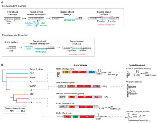
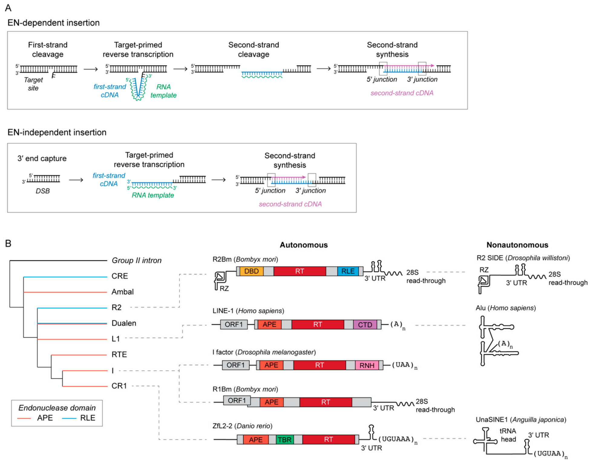
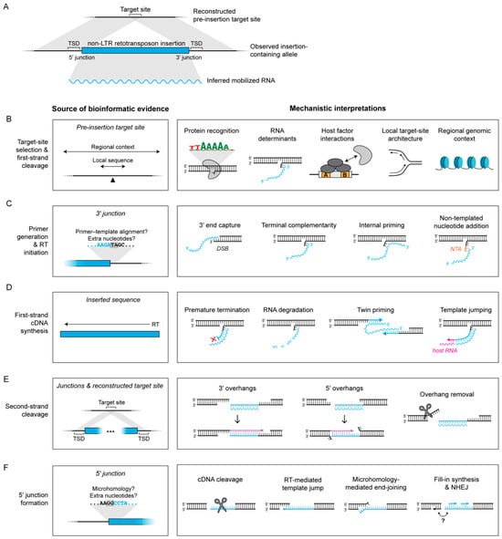
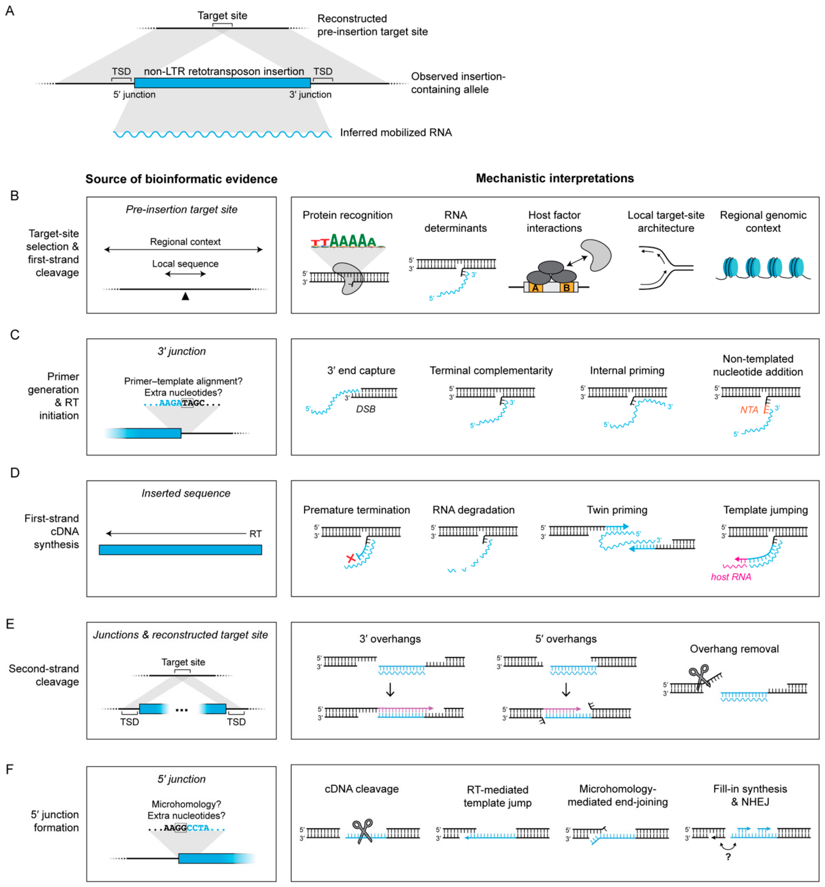
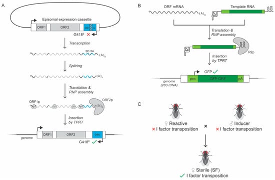
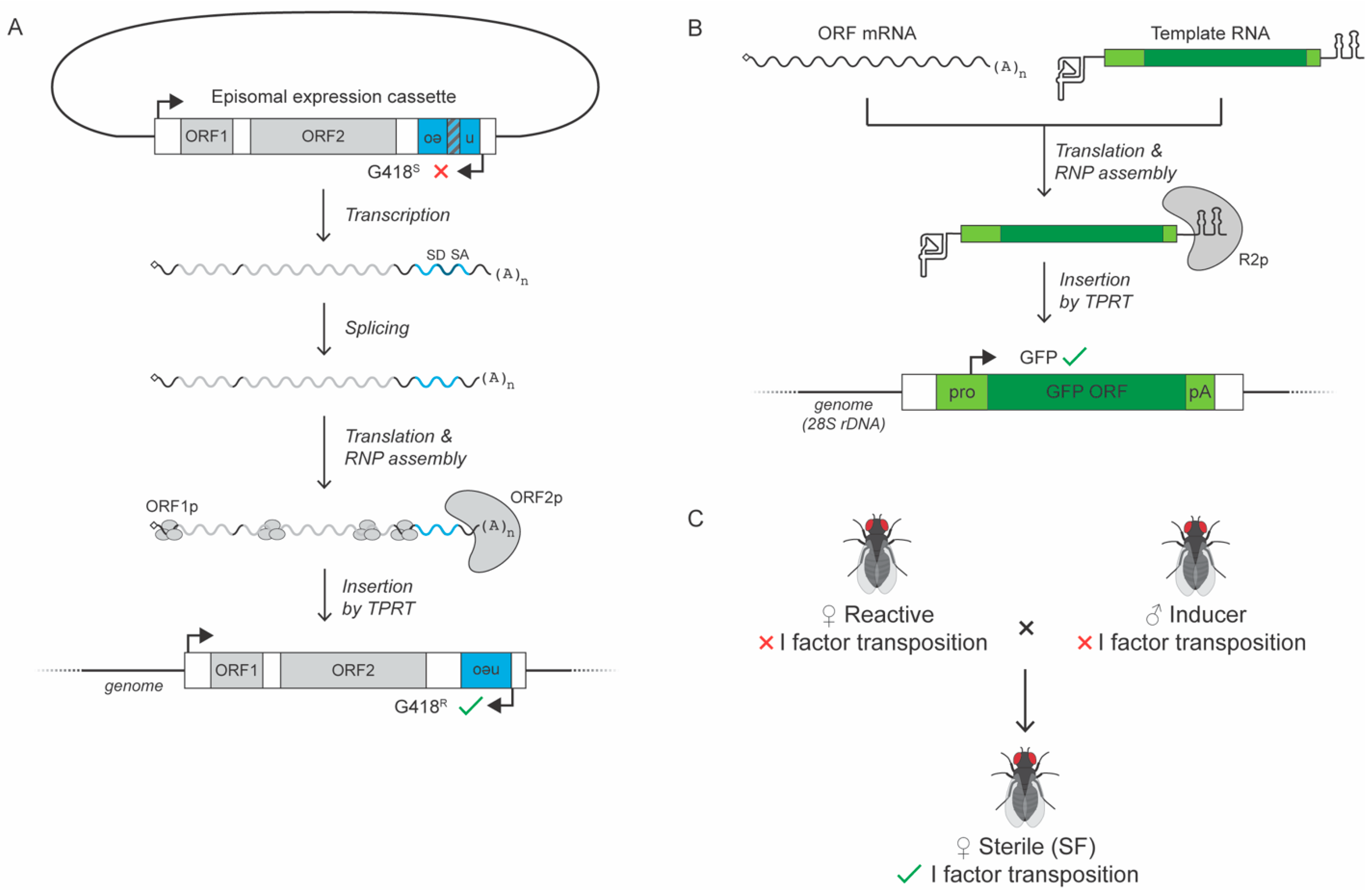

Disclaimer/Publisher’s Note: The statements, opinions and data contained in all publications are solely those of the individual author(s) and contributor(s) and not of MDPI and/or the editor(s). MDPI and/or the editor(s) disclaim responsibility for any injury to people or property resulting from any ideas, methods, instructions or products referred to in the content. |
© 2026 by the authors. Licensee MDPI, Basel, Switzerland. This article is an open access article distributed under the terms and conditions of the Creative Commons Attribution (CC BY) license.