


Abstract
‘Candidatus Phytoplasma rubi’ (elm yellows group, 16SrV-E phytoplasma subgroup) is the causal agent of rubus stunt disease, a disorder affecting economically important plants—raspberries and blackberries. Although this phytoplasma has been extensively studied in cultivated Rubus crops, its occurrence and molecular identity in wild Rubus species populations in North-Eastern Europe remain poorly documented. In this study, phytoplasmas associated with symptomatic wild Rubus idaeus L. and Rubus nessensis Hall plants were investigated in natural forest ecosystems of the Curonian Spit, Lithuania. A total of 65 symptomatic plants were surveyed, and phytoplasma infection was detected in 30 samples by nested PCR targeting the 16S rRNA gene. Positive samples were characterized using a multilocus molecular approach based on sequence analysis of the additional cpn60 and secA genes. All strains showed high nucleotide sequence similarity across the analysed loci and consistently clustered with reference strains of ‘Candidatus Phytoplasma rubi’. Virtual RFLP profiles derived from the 16S rRNA and cpn60 genes were nearly identical to those of established 16SrV-E phytoplasma subgroup reference strains and clearly distinct from other 16SrV phytoplasma subgroups. These results provide not only the first detailed multilocus molecular characterization of ‘Candidatus Phytoplasma rubi’-related strains infecting wild Rubus species in Lithuania but also represent the first report of this phytoplasma in naturally occurring R. idaeus and R. nessensis plants in the country, thereby extending the known geographical occurrence of this pathogen and documenting its presence in wild Rubus hosts from unmanaged forest habitats in the Eastern Baltic region of Northern Europe.
1. Introduction
Phytoplasmas are phloem-limited, cell wall-less plant pathogenic bacteria that infect a wide range of herbaceous and woody plant species. These pathogens are transmitted primarily by phloem-feeding insects [1,2]. They cause many economically important plant diseases worldwide [1,3]. In Rubus species, phytoplasma infection causes rubus stunt (RS or RuS) disease, a disorder associated with stunting, witches’ broom, floral abnormalities, and severe yield losses [4]. In addition to ‘Candidatus (Ca.) Phytoplasma rubi’, other phytoplasmas have also been reported in Rubus species. Aster yellows phytoplasmas (16SrI-B) [5,6,7] have been detected in raspberry and blackberry, X-disease phytoplasmas (16SrIII) [8,9] in loganberry and black raspberry, stolbur phytoplasmas (16SrXII-A) in raspberry and blackberry [10,11,12], and the blackPort phytoplasma (16SrV-I) in blackberry [13]. Rubus stunt is widely distributed across Europe and has been reported from numerous countries in Central, Western and Southern Europe, as well as from other regions worldwide [7,14,15,16]. Molecular studies have shown that this disease is associated with ‘Ca. Phytoplasma rubi’ strains. This phytoplasma species belongs to the 16SrV (elm yellows) phytoplasma group (16SrV-E subgroup) and is regarded as the main causal agent of rubus stunt in Europe [4,17].
The studies on RS disease caused by ‘Ca. Phytoplasma rubi’ and related strains have focused on cultivated raspberry and blackberry plantations [4,18]. However, the epidemiology of phytoplasmas is linked to complex ecological systems involving insect vectors and alternative plant hosts. Several studies have shown that ‘Ca. Phytoplasma rubi’-related strains can occur in several plant species (Rubus caesius L., Rubus fruticosus L., Malva sylvestris L., Rosa canina L. and Fragaria × ananassa) [4,19,20]. In phytoplasma pathosystems, non-crop hosts in surrounding vegetation may contribute to pathogen persistence and spread, depending on local vector ecology and host competence [18,21]. However, the occurrence of ‘Ca. Phytoplasma rubi’ in wild Rubus species and their possible role in the ecology of this pathogen remain insufficiently studied. Data from natural forests and semi-natural habitats remain scarce.
To date, most molecular studies on ‘Ca. Phytoplasma rubi’ have been conducted in West-Southern and Central Europe, particularly in countries such as the Czech Republic, Germany, Poland, France, Italy, and Portugal [7,13,18,20,22,23]. These studies have focused mainly on cultivated Rubus crops. In contrast, information from Northern and Northeastern Europe remains limited. The Baltic region is a particularly underexplored area with respect to the diversity, distribution, and ecological context of phytoplasmas affecting Rubus species [16]. Lithuania lies near the northern margin of the known European distribution of rubus stunt disease. Previous studies have documented the presence of ‘Ca. Phytoplasma rubi’ in cultivated Rubus idaeus L. in the country [16]. However, to date, there have been no reports of this phytoplasma infecting wild Rubus species, and no multilocus molecular characterization of ‘Ca. Phytoplasma rubi’ from natural forest ecosystems in Lithuania has been available. Rubus nessensis Hall, a common wild species in northern European forests and semi-natural habitats, has not previously been investigated as a potential host of phytoplasmas in Eastern Baltic region of Northern Europe.
In this study, phytoplasma infections were investigated in symptomatic wild R. idaeus and R. nessensis plants collected from natural forest ecosystems in Lithuania. The detected phytoplasmas were characterized using a multilocus molecular approach. This approach was based on sequence analysis of the 16S rRNA, cpn60, and secA genes. Phylogenetic analysis and virtual RFLP profiling were used to support the classification. This study documents the occurrence of ‘Ca. Phytoplasma rubi’-related strains in wild Rubus species at the northern edge of its known European range. It provides new insights into the geographical distribution of this pathogen. It also improves our understanding of its ecological context. Finally, it expands current knowledge of the ecological context of ‘Ca. Phytoplasma rubi’ and provides a basis for future studies on the epidemiological significance of wild Rubus populations.
2. Materials and Methods
2.1. Field Survey and Sample Collection
Field surveys were conducted in September 2025 in natural forest ecosystems of the Curonian Spit, western Lithuania (55°32′28″ N, 21°58′15″ E). The surveyed sites consisted of unmanaged or semi-natural mixed forest stands dominated by pine (Pinus sylvestris L. and Pinus mugo Turra) with a heterogeneous understory composed primarily of herbaceous vegetation and including naturally occurring Rubus shrub species.
Wild Rubus idaeus L. and Rubus nessensis Hall plants exhibiting symptoms commonly associated with phytoplasma infection were identified during visual inspection. Observed symptoms included shortened internodes, excessive shoot proliferation, leaf size reduction, leaf chlorosis or reddening, phyllody of flowers, and general growth decline. Symptomatic plants occurred either as isolated individuals or in small spatial clusters.
A total of 65 symptomatic Rubus plants were sampled, including 21 R. idaeus and 44 R. nessensis individuals (Figure S1). From each plant, leaves at different developmental stages (from young to fully expanded), including their petioles, were collected preferentially from actively growing shoots. Samples were placed in sterile polyethylene bags, transported to the laboratory in insulated containers, and stored at −80 °C until DNA extraction.
2.2. DNA Extraction
Total genomic DNA was extracted from approximately 100 mg of plant tissue per sample. Plant material was homogenized under liquid nitrogen using sterile mortars and pestles. Total genomic DNA was extracted from leaf tissues using a Genomic DNA Purification Kit (Thermo Fisher Scientific Baltics, Vilnius, Lithuania) according to the manufacturer’s instructions [16].
The quality and concentration of extracted DNA were assessed spectrophotometrically using a NanoDrop™ spectrophotometer (Thermo Fisher Scientific, Vilnius, Lithuania). DNA extracts were diluted with sterile nuclease-free water and stored at −20 °C until further analysis.
2.3. Amplification of Phytoplasmas Genetic Loci
Phytoplasma detection and multilocus molecular characterization were performed by PCR-based amplification of the 16S rRNA, cpn60, and secA gene loci. Initial phytoplasma detection was carried out using direct and nested PCR assays targeting the 16S rRNA gene. The primer pairs P1/P7 [24,25] were used for direct PCR amplification, followed by nested PCR using the internal primer pair R16F2n/R16R2 [26] (Table S1). All samples that tested positive in the 16S rRNA nested PCR assay were subsequently subjected to multilocus molecular characterization. Partial fragments of the cpn60 gene were amplified using a nested PCR approach with the primer pairs H279p/H280p [27] and D0317/D0318 [27], whereas the secA gene was amplified using a direct and semi-nested PCR with the primer pairs SecAfor1/SecArev3 [28] and SecAfor2/SecArev3 [28] (Table S1).
All PCR reactions were performed in a final volume of 25 μL containing 125 μM of each dNTP, 0.5 μM of each primer, 1 U of AmpliTaq Gold DNA polymerase with 1× reaction buffer (Thermo Fisher Scientific, Vilnius, Lithuania), and 2 μL of template DNA (100–200 ng). For nested and semi-nested PCR assays, 2 μL of a 1:50 dilution of the first-round PCR product was used as template. Thermal cycling conditions and annealing temperatures were selected according to the respective primer pairs and PCR strategies that are summarized in Table S1 [24,25,26,27,28].
Amplified products were separated by electrophoresis on 1.0%–1.5% agarose gels, stained with SYBR Green dye (Thermo Fisher Scientific, Vilnius, Lithuania), and visualized under UV illumination. Samples producing amplicons of the expected size were considered phytoplasma-positive. Only samples yielding clear and single bands were selected for subsequent sequencing. Positive and negative controls were included in all PCR runs. DNA extracted from a known phytoplasma-infected plant served as a positive control, whereas nuclease-free water and DNA extracted from asymptomatic Rubus plants, including both R. idaeus and R. nessensis, were used as negative controls. All samples that tested positive in the 16S rRNA nested PCR assay were sequenced. Due to the high sequence similarity observed among the obtained sequences, a representative subset of strains was selected for multilocus characterization (cpn60 and secA).
2.4. DNA Sequencing, Phylogenetic and Virtual and Enzymatic RFLP Analyses
PCR products of the 16S rRNA, cpn60, and secA genes were purified using a GeneJET PCR purification kit (Thermo Fisher Scientific, Vilnius, Lithuania) according to the manufacturer’s instructions. Purified PCR products were sequenced by a commercial service provider (SeqVision, Vilnius, Lithuania) using Oxford Nanopore Technologies. Library preparation, sequencing, and primary data processing were performed by the provider according to standard protocols. Consensus sequences were generated from multiple reads per amplicon. Sequence quality was assessed through NCBI BLASTn web server (National Center for Biotechnology Information, Bethesda, MD, USA) comparison and by evaluating consistency across independently amplified loci (16S rRNA, cpn60, and secA). The high concordance observed among loci supports the reliability of the obtained sequences despite the high overall similarity.
Sequence data were assembled and edited, and consensus sequences were compared with reference sequences available in the GenBank database using BLAST. Multiple sequence alignments were performed using standard alignment software MEGA software version 11.0.13 [29], and sequence similarities were calculated by pairwise comparison. The nucleotide sequences generated in this study were deposited in the GenBank database under accession numbers: PX970422 to PX970431 for 16S rDNA, PX984888 to PX984897 for cpn60, PX984898 to PX984907 for secA (Table S2). Consensus sequences of the 16S rRNA, cpn60, and secA genes obtained in this study were compared with reference phytoplasma sequences retrieved from GenBank (NCBI). A detailed overview of all sampled plants, including PCR results, sequenced loci, and accession numbers, is provided in Supplementary Table S2.
Phylogenetic trees were reconstructed using the neighbor-joining method, which is suitable for the analysis of highly similar sequences, implemented in MEGA11.0.13. Evolutionary distances were calculated according to a suitable nucleotide substitution model. Bootstrap analysis was performed with 1000 replicates to assess branch support. Sequence alignments were generated using the MUSCLE algorithm and manually inspected prior to analysis. Gaps and ambiguous positions were treated using pairwise deletion. The same set of reference sequences was used for each locus to ensure comparability of tree topology. Resulting phylogenies were examined to determine the taxonomic placement and subgroup affiliation of the Lithuanian phytoplasma strains. No evidence of mixed sequence signal was observed in the obtained reads, and consensus sequences were generated to represent the dominant haplotype in each sample. Ambiguous nucleotide positions were not detected or were resolved conservatively during sequence assembly.
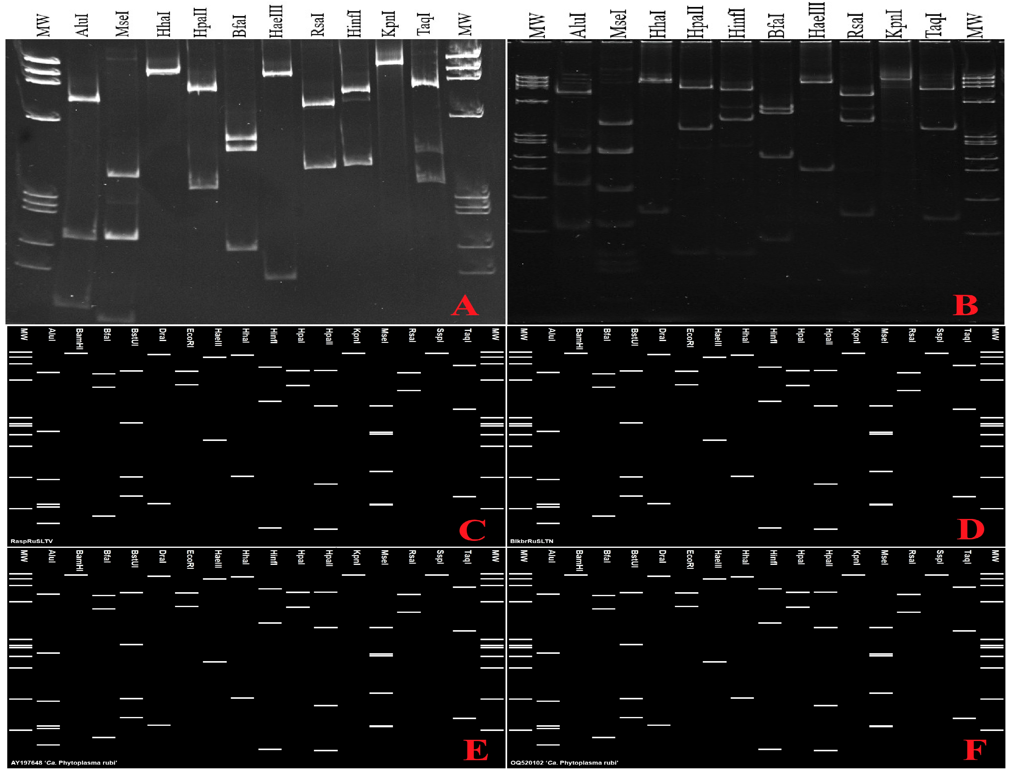
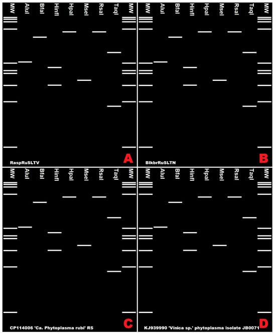
Actual enzymatic restriction fragment length polymorphism (RFLP) analysis was performed on the 16S rRNA gene (R16F2n/R16R2 fragments) using the restriction enzymes AluI, MseI, HhaI, HpaII, BfaI, HaeIII, RsaI, HinfI, KpnI, and TaqI. The analysis was conducted on all samples that tested positive in the 16S rRNA nested PCR assay. Restriction digestions were performed according to standard protocols, and resulting fragments were separated by 5% bis-acrylamide gel electrophoresis and visualized under UV illumination. The obtained restriction profiles were consistent across all analysed samples. Representative profiles from one Rubus idaeus strain (RaspRuSLTV01) and one Rubus nessensis strain (BlkbrRuSLTN01) are shown in Results Section 3.2. The obtained profiles were compared with virtual RFLP patterns and reference strains to confirm subgroup affiliation [18,20]. In addition, virtual RFLP analyses were performed in silico on the 16S rRNA and cpn60 gene sequences to determine phytoplasma group and subgroup affiliations. Virtual digestions with selected restriction enzymes were carried out using the pDRAW32 software (AcaClone 1.1.154 Software), the online iPhyClassifier tool (https://plantpathology.ba.ars.usda.gov/cgi-bin/resource/iphyclassifier.cgi accessed on 10 January 2026) and CpnClassiPhyR online tool (http://cpnclassiphyr.ca/CpnClassiPhyR.php accessed on 11 February 2026). Virtual restriction profiles obtained for the sequences generated in this study were compared with those of reference phytoplasma strains representing the recognized 16SrV subgroups to assign subgroup affiliation.
3. Results
3.1. Field Survey and Symptom Description
During field surveys conducted in natural forest ecosystems of the Curonian Spit (Lithuania), wild populations of Rubus idaeus and Rubus nessensis were examined for the presence of symptoms commonly associated with phytoplasma infection. Symptomatic plants were observed across multiple locations within the surveyed area, occurring either as isolated individuals or as small, spatially localized groups. No regular spatial pattern indicative of anthropogenic planting or management was evident.
Both Rubus species exhibited a comparable range of symptoms. Infected plants were generally characterized by reduced and stunted growth compared with neighbouring asymptomatic individuals. Internodes were frequently shortened, resulting in a compact plant shape appearance. Shoot proliferation was commonly observed, particularly in the upper portions of the plants stem, with the formation of multiple thin, weak shoots. Leaf symptoms varied among plants and included reduced leaf size, leaf deformation, and chlorosis or reddish discoloration, especially along the veins. In some plants, phyllody was observed, in which floral parts were partially or completely transformed into leaf-like structures. In some cases, leaves appeared brittle and exhibited abnormal orientation. Symptom severity differed markedly among individual plants, even within proximity, and ranged from mild growth suppression to pronounced morphological alterations.
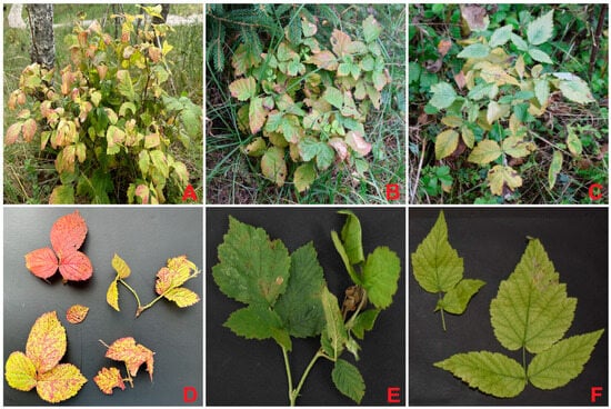
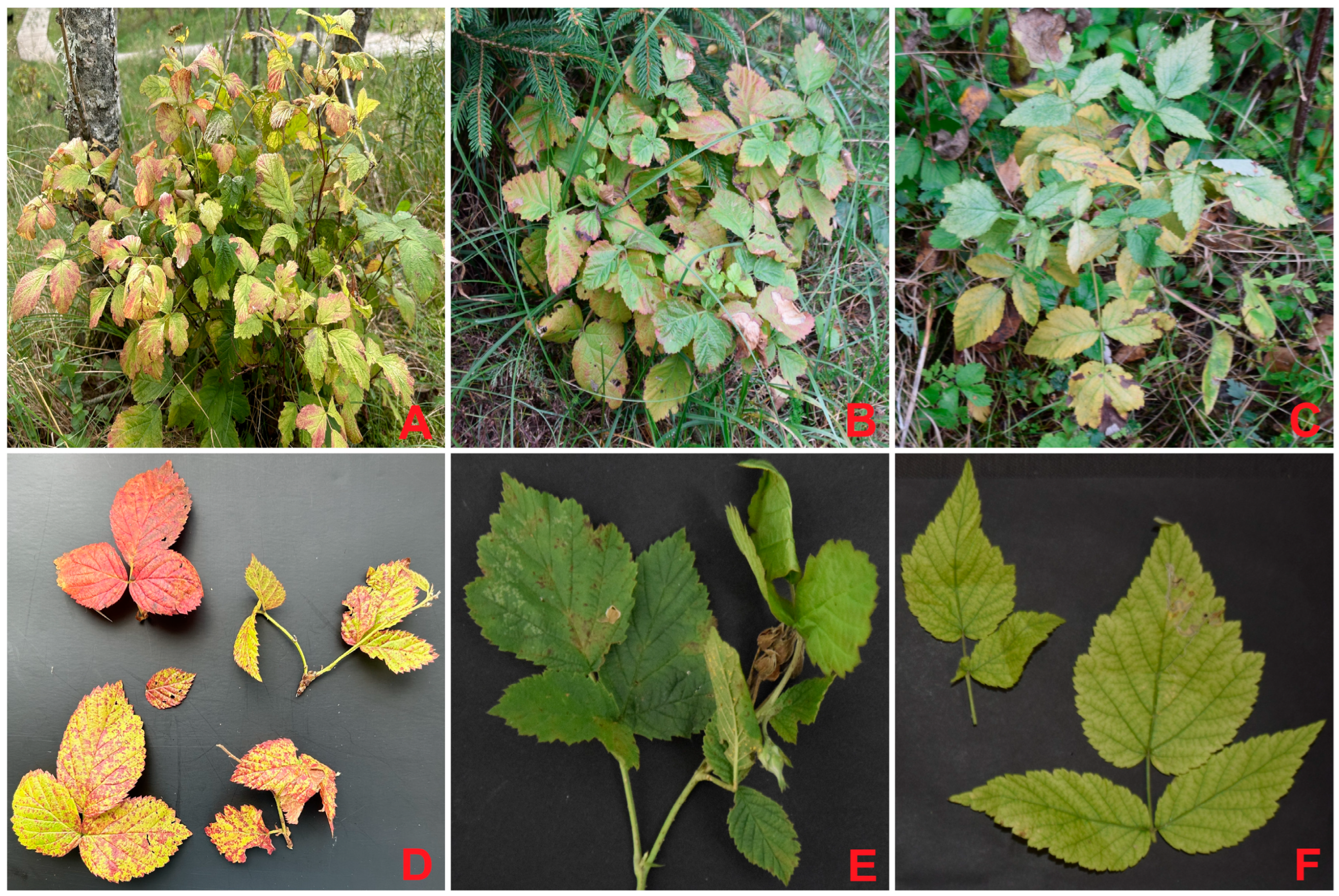
In R. idaeus, the most common symptoms included dwarfism, reduced leaf size, and leaf discoloration manifested as yellowing and reddening. Diseased plants were clearly weaker than asymptomatic individuals and showed suppressed vegetative development (Figure 1).
Figure 1.
Rubus stunt (RaspRuSLTV) affected wild raspberry (Rubus idaeus) plants, showing typical disease symptoms. Raspberries shown in panels: (A,D) exhibited strong leaf yellowing and reddening, reduced growth and dieback; (B,E) showed leaf yellowing and reddening, dwarfing, phyllody, reduced leaf size and dieback; (C,F) were severely stunted, with pronounced leaf reddening, reduction in leaf size, and chlorosis.
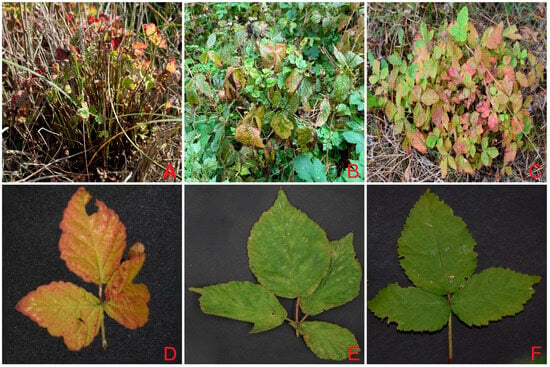
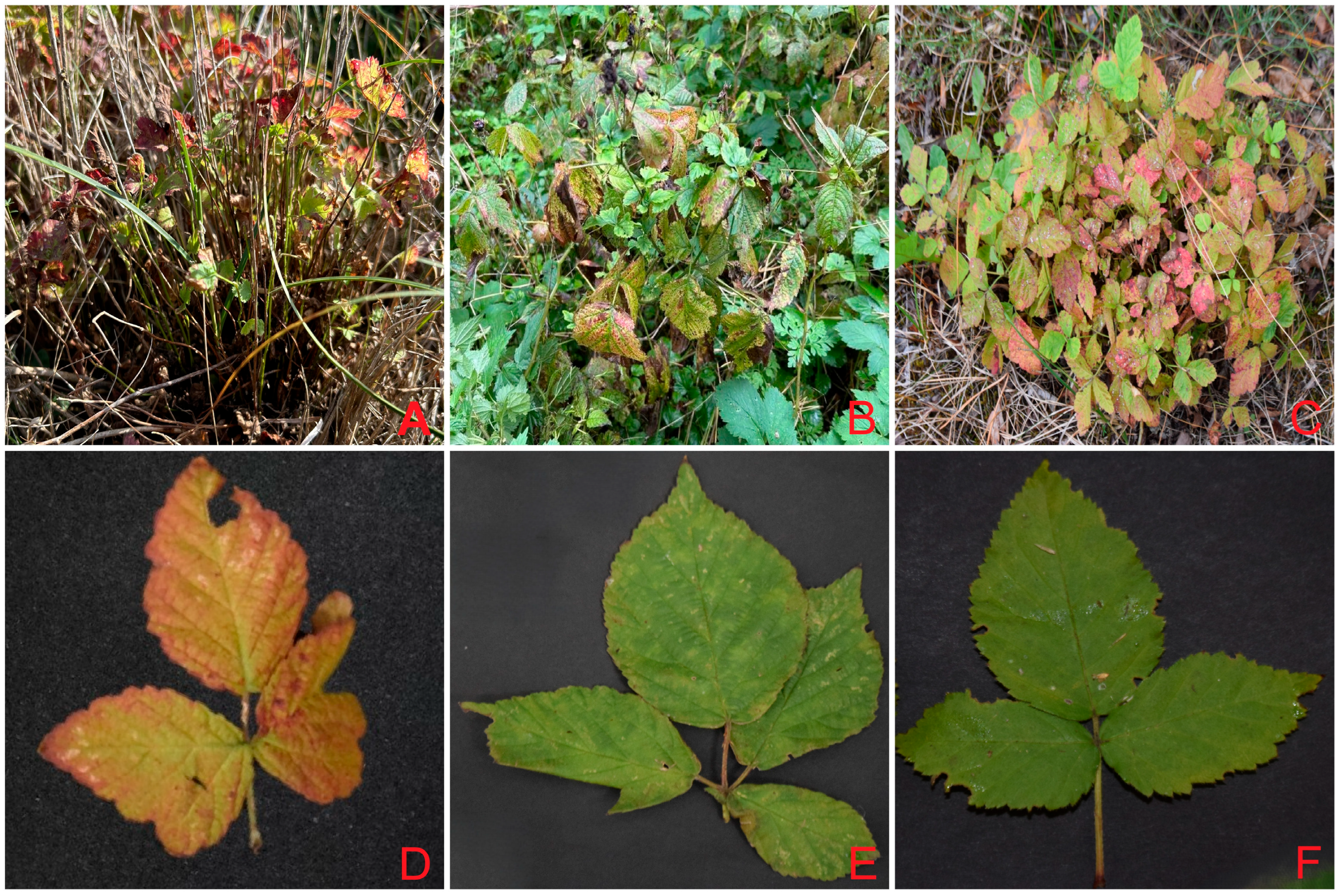
Symptoms observed in R. nessensis were generally more severe and included strong growth suppression, reduced leaf size, shortened internodes, and intensive shoot proliferation resulting in witches’-broom-like structures. Leaf discoloration, including chlorosis and reddening was also observed. These symptoms were associated with pronounced distortion of plant architecture (Figure 2).
Figure 2.
Rubus stunt (BlkbrRuSLTN) affected wild blackberry (Rubus nessensis) plants showing typical disease symptoms. Blackberries shown in panels: (A,D) were severely stunted and exhibited irregular and dense shoot branching, shortened internodes, and strong leaf yellowing and reddening with reduced leaf size. (B,E) showed leaf yellowing and reddening, localized chlorosis, reduced leaf size, and abnormal shoot branching. (C,F) were strongly stunted, with pronounced shoot proliferation, reduced leaf size, chlorosis, and intensive leaf yellowing and reddening.
No obvious signs of other biotic stress factors, such as fungal infections, insect feeding damage, or mechanical injury, were associated with the observed symptoms. The combination of growth abnormalities and foliar symptoms was considered indicative of possible phytoplasma infection and served as the basis for selecting plants for subsequent molecular analyses. These symptoms were used only as a field-screening criterion and were not considered diagnostic, as similar symptom expression may result from other biotic or abiotic factors.
3.2. Detection and Identification of Phytoplasmas Based on 16S rRNA Gene Sequences
Total genomic DNAs extracted from 65 symptomatic Rubus sp. plants were screened for the presence of phytoplasmas using a nested PCR assay targeting the 16S rRNA gene. Amplifications were performed with the universal primer pairs P1/P7 in the first PCR round and R16F2n/R16R2 in the nested reaction. A phytoplasma-specific amplicon of approximately 1.2 kb was obtained from 30 samples, including 10 R. idaeus and 20 R. nessensis plants. When expressed relative to the number of symptomatic plants sampled, this corresponds to detection rates of approximately 47.6% (10/21) in R. idaeus and 45.5% (20/44) in R. nessensis. All positive amplicons were sequenced, and high-quality partial 16S rRNA gene sequences were obtained for each strain. The Lithuanian strains were designated RaspRuSLTV (from R. idaeus) and BlkbrRuSLTN (from R. nessensis). Comparative sequence analysis showed that the 16S rDNA sequences of raspberry and blackberry phytoplasma strains were highly similar to each other, sharing 99.9%–100% nucleotide identity across the analysed region.
All 30 samples positive in the 16S rRNA nested PCR were sequenced. Because the sequences were highly similar, only 10 representative strains were selected for multilocus (cpn60 and secA) characterization and downstream phylogenetic analyses. Because most sequences were identical or nearly identical, a representative subset of strains was selected for further analyses: five RaspRuSLTV and five BlkbrRuSLTN strains. BLAST analysis against sequences deposited in the GenBank database revealed that the detected strains shared 99.84%–100% nucleotide identity with ‘Candidatus Phytoplasma rubi’ strain RS (CP114006) [30], Fragaria × ananassa witches’-broom phytoplasma strain StraWB3230 (PX122036) [20], and ‘Ca. Phytoplasma rubi’ strain RuS-L from cultivated raspberry in eastern Lithuania (HM118514), previously generated by the authors and sequence data were unpublished at the time of submission. According to the current phytoplasma species delineation guidelines, a novel ‘Candidatus Phytoplasma’ species should be recognized only when the 16S rRNA gene sequence identity is below 98.65% or when genome- and ecology-based criteria are fulfilled. In the present study, the high sequence similarity observed supports the assignment of the detected phytoplasmas to ‘Candidatus Phytoplasma rubi’-related strains [31].
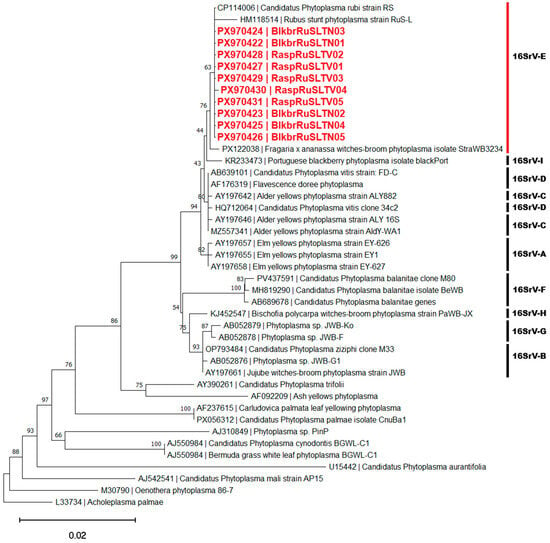
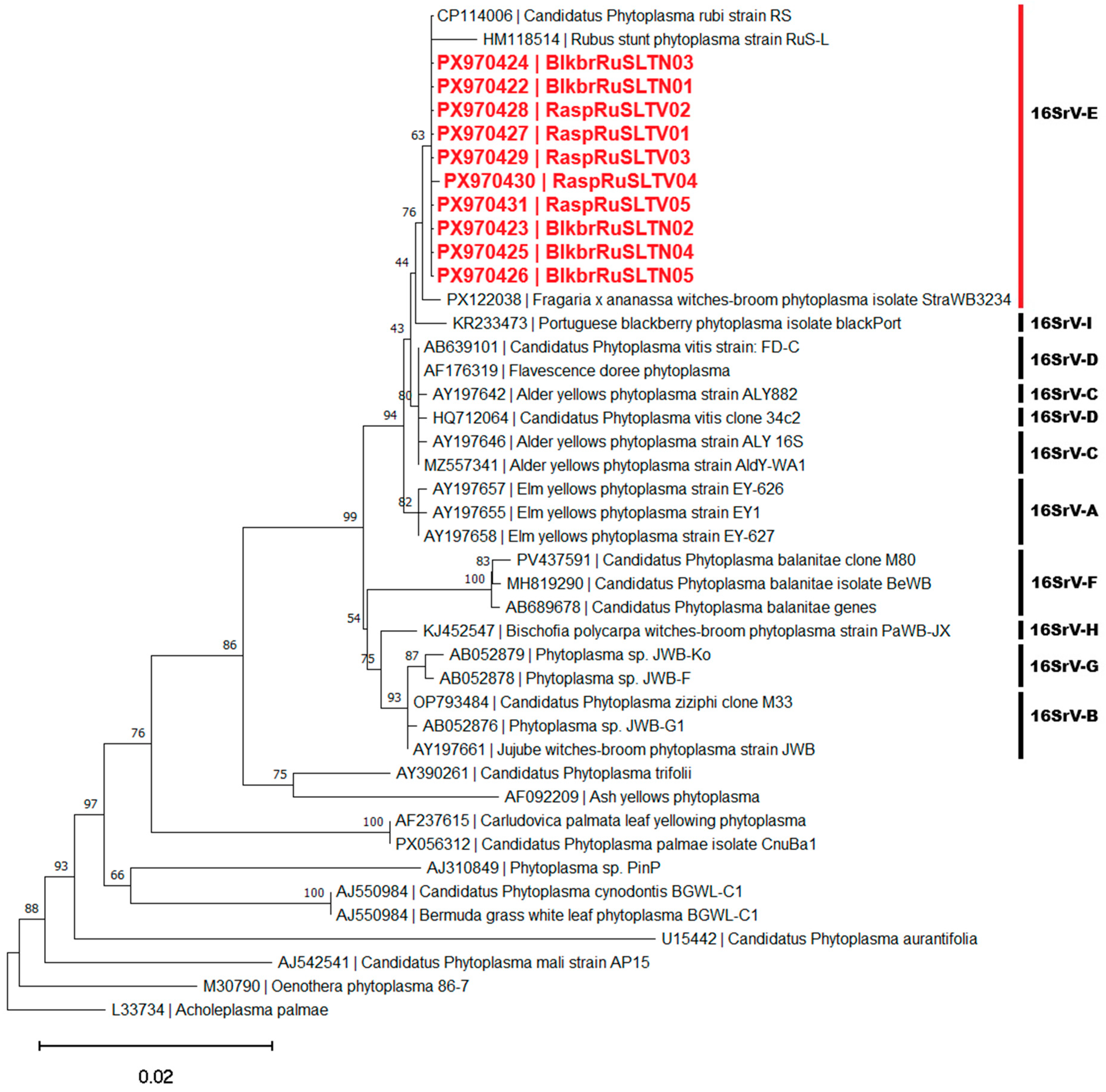
Phylogenetic analysis based on partial 16S rRNA gene sequences placed RaspRuSLTV and BlkbrRuSLTN strains within the 16SrV (elm yellows) phytoplasma group. Because most sequences were identical or nearly identical, a representative subset of strains was selected for further analyses: five RaspRuSLTV and five BlkbrRuSLTN strains. In the reconstructed phylogenetic tree, the Lithuanian strains clustered closely with established reference strains of ‘Ca. Phytoplasma rubi’ and formed a monophyletic group (Figure 3). Within the 16SrV group, the Lithuanian strains grouped specifically with representatives of the 16SrV-E subgroup (including HM118514, CP114006, and PX122038), while reference strains representing other 16SrV subgroups formed distinct and more distant branches.
Figure 3.
Phylogenetic tree constructed using the neighbor-joining method (1000 bootstrap repetitions) with 16S rDNA sequences of RaspRuSLTV and BlkbrRuSLTN strains detected in Lithuania (highlighted in red), and other 16SrV phytoplasma group (Elm yellows) members and ‘Ca. Phytoplasma’ representatives. Acholeplasma palmae was used as an outgroup. The bar represents a phylogenetic distance of 0.02 nucleotide substitutions per site. The GenBank accession number is provided for each phytoplasma strain. Bootstrap values are shown on branches of the phylogenetic tree.
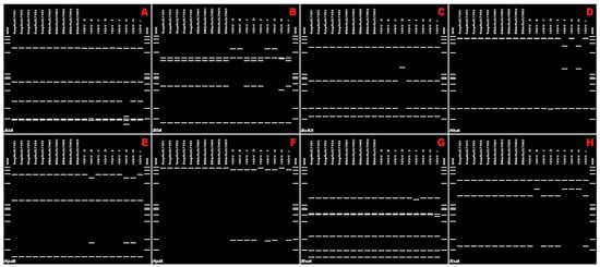
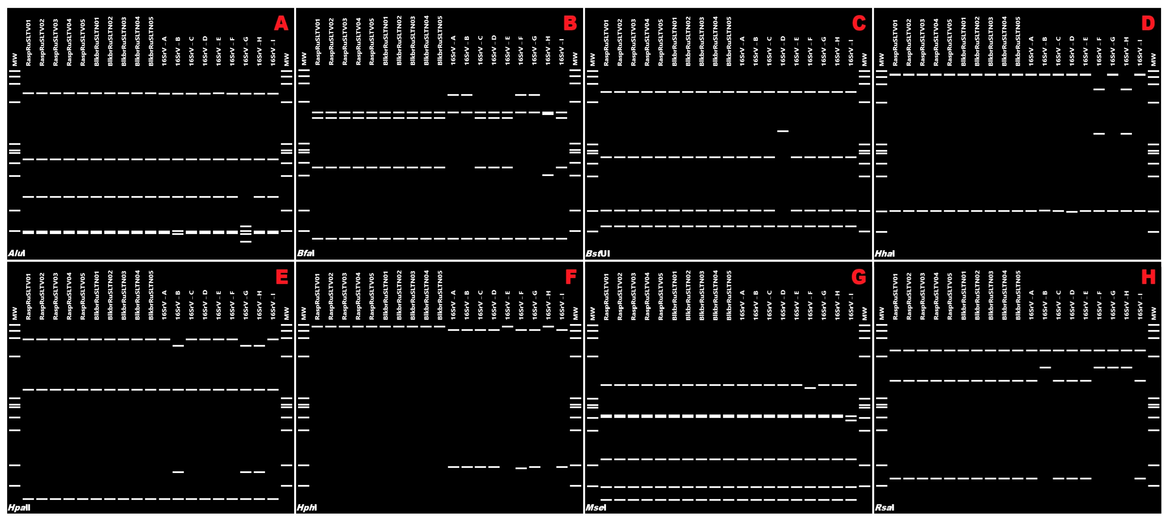
To further support subgroup assignment, virtual RFLP analysis using AcaClone pDRAW32 was performed on the R16F2n/R16R2-amplified 16S rRNA gene fragments of the RaspRuSLTV and BlkbrRuSLTN phytoplasma strains. Several restriction enzymes (BamHI, DraI, EcoRI, HaeIII, HinfI, HpaII, KpnI, SspI, TaqI) generated identical profiles among all reference strains representing the 16SrV subgroups and were therefore not informative for subgroup differentiation. In contrast, virtual digestions with AluI, BfaI, BstUI, HhaI, HpaII, HphI, MseI, and RsaI produced distinct restriction profiles among the 16SrV subgroups, enabling reliable subgroup-level discrimination (Figure 4). Among these enzymes, AluI, BfaI, BstUI, HhaI, HpaII, HphI, MseI, and RsaI showed clear discriminatory power and resolved the 16SrV group into characteristic restriction pattern clusters or uniquely distinguished specific subgroups (Figure 4). Notably, digestion with HphI allowed clear separation of the 16SrV-E subgroup from other members of the 16SrV (elm yellows) phytoplasma group (Figure 4F).
Figure 4.
Virtual restriction profiles generated with AluI (A), BfaI (B), BstUI (C), HhaI (D), HpaII (E), HphI (F), MseI (G), and RsaI (H) using 16S rDNA R16F2n/R2 fragments sequences of phytoplasmas detected in raspberry (R. idaeus) (RaspRuSLTV01–RaspRuSLTV05) and blackberry (R. nessensis) (BlkbrRuSLTN01–BlkbrRuSLTN05), and of representative 16SrV group phytoplasmas. Virtual digestions were generated using pDraw32 Software (http://www.acaclone.com). Other phytoplasmas are: 16SrV-A, AC: AY197655, elm yellows from USA; 16SrV-B, AC: AB052876, jujube witches’ broom from Japan; 16SrV-C, AC: AY197642, alder yellows from southern Italy; 16SrV-D, AC: AY197644, ‘flavescence dore’e’ from northern Italy; 16SrV-E, AC: AY197648, rubus stunt strain RuS from USA; 16SrV-F, AC: AB689678, balanites witches’ broom from South Korea; 16SrV-G, AC: AB052879, jujube witches’ broom from Korea; 16SrV-H, AC: KJ452547 Bischofia polycarpa witches’ broom phytoplasma from China; 16SrV-I, AC: KR233473 blackberry phytoplasma isolate blackPort from Portugal. MW, marker phiX174 HaeIII-digested with fragment sizes, from top to bottom: 1353, 1078, 872, 603, 310, 281, 271, 234, 194, 118 and 72 bp.
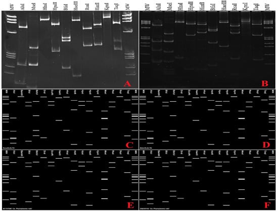
Comparison of the virtual and actual enzymatic restriction profiles showed that the patterns obtained for the RaspRuSLTV and BlkbrRuSLTN strains were indistinguishable from those of the reference ‘Candidatus Phytoplasma rubi’ strains RS (CP114006) [30] and StraWB3230 (PX122036) [20], as implemented in the iPhyClassifier platform, both assigned to the 16SrV-E subgroup. These profiles were clearly distinct from those of other 16SrV subgroup representatives (Figure 5). Experimental RFLP analysis was performed on representative strains RaspRuSLTV01 and BlkbrRuSLTN01, and the resulting profiles correspond to those shown in Figure 5A,B. These results confirm that the phytoplasmas detected in Rubus idaeus and Rubus nessensis in Lithuania belong to the 16SrV-E subgroup related to ‘Candidatus Phytoplasma rubi’.
Figure 5.
Comparison of actual and virtual RFLP profiles of 16S rRNA gene (R16F2n/R2 fragment) phytoplasma strains. Actual RFLP profiles are shown for RaspRuSLTV01 (A) and BlkbrRuSLTN01 (B). Virtual RFLP profiles generated using the iPhyClassifier online tool are shown for RaspRuSLTV01 (C), BlkbrRuSLTN01 (D), Rubus stunt phytoplasma strain RUS, acc. no. AY197648 (E) and ‘Candidatus Phytoplasma rubi’ strain RS, acc. no. CP114006 (F). Actual RFLP analysis was performed using restriction enzymes AluI, MseI, HhaI, HpaII, BfaI, HaeIII, RsaI, HinfI, KpnI and TaqI. Lanes correspond to restriction digestions with the listed enzymes from left to right. Identical restriction profiles were observed across all analysed samples. MW, φX174 HaeIII DNA ladder with fragment sizes (bp): 1353, 1078, 872, 603, 310, 281, 271, 234, 194, 118 and 72.
3.3. Confirmation of Phytoplasma Identity Based on the cpn60 Gene
To further confirm the taxonomic placement of the phytoplasmas detected in wild Rubus species, amplification and sequence analysis of the chaperonin-60 (cpn60) gene were performed. PCR amplification using the universal phytoplasma primer cocktail H279p/H280p and D0317/D0318 (applied at a 1:7 molar ratio) successfully generated an amplicon of approximately 600 bp from 10 phytoplasma-positive samples, derived from 5 R. idaeus and 5 R. nessensis plants. All amplicons were successfully sequenced and subjected to comparative sequence analyses. Pairwise comparison of the obtained cpn60 sequences revealed a high degree of sequence conservation among the Lithuanian strains. Nucleotide identity values between RaspRuSLTV and BlkbrRuSLTN strains were 100% for all analysed gene fragments, indicating complete sequence identity.
BLAST analysis of the cpn60 sequences against the GenBank database showed 99.84%–100% nucleotide identity with ‘Candidatus Phytoplasma rubi’ strain RS (CP114006) [30], as well as with the Vinca sp. phytoplasma strain JB0071_B02_CFIA1-1 (KJ939990) [27], both strains are assigned to the 16SrV-E subgroup.
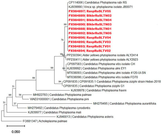
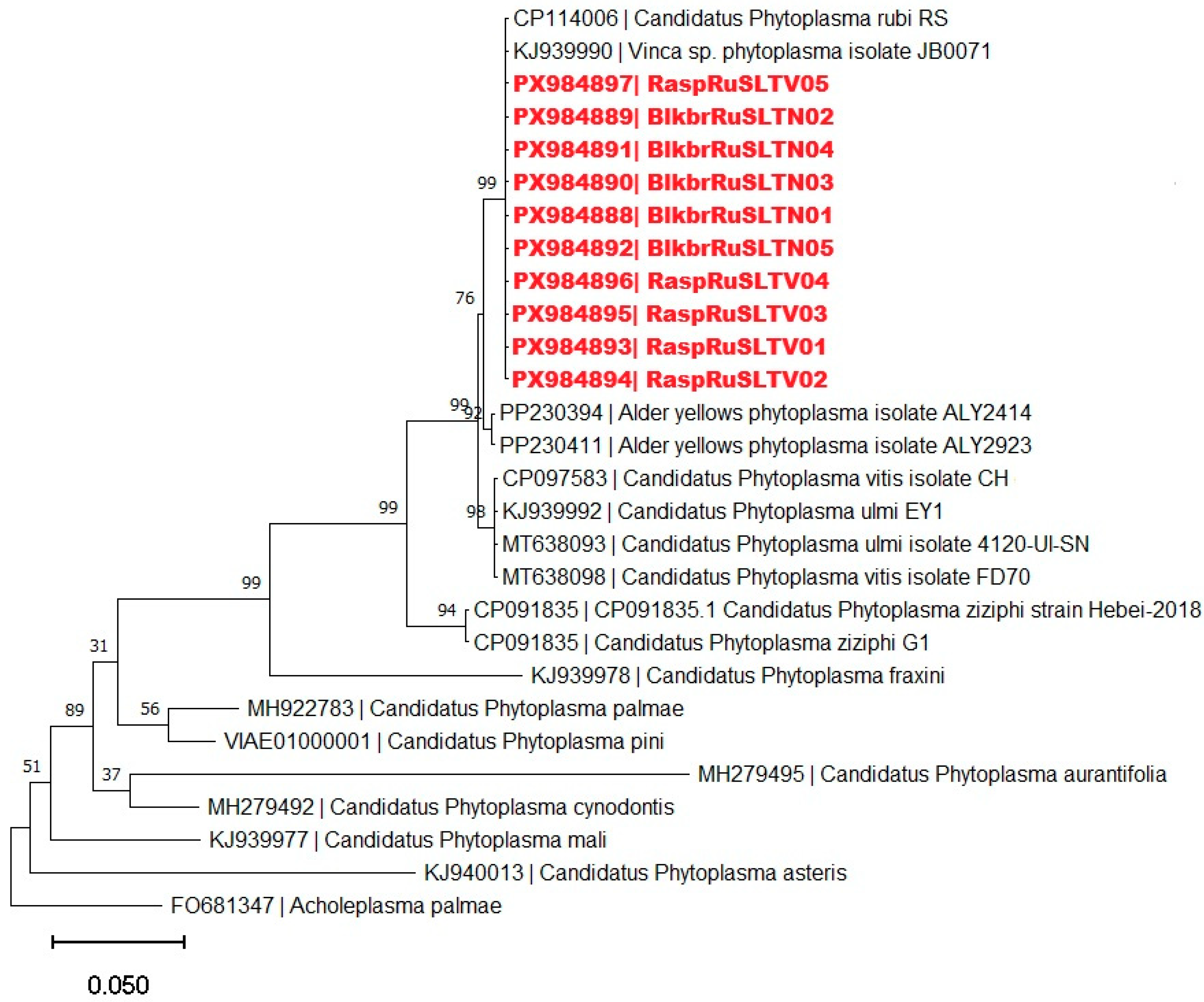
Phylogenetic analysis based on cpn60 gene sequences placed all RaspRuSLTV and BlkbrRuSLTN strains within a clade comprised of reference strain of ‘Ca. Phytoplasma rubi’ and other members of the 16SrV-E phytoplasma subgroup (Figure 6). Phytoplasma strains representing other 16SrV subgroups formed distinct and clearly separated branches, confirming the close phylogenetic relationship between the Lithuanian strains and the rubus stunt phytoplasma lineage.
Figure 6.
Phylogenetic tree constructed using the neighbor-joining method with cpn60 sequences of RaspRuSLTV and BlkbrRuSLTN strains detected in Lithuania (highlighted in red), 16SrV phytoplasma group members and ‘Ca. Phytoplasma’ representatives. Acholeplasma palmae was used as an outgroup. The bar represents a phylogenetic distance of 0.05 nucleotide substitutions per site. The GenBank accession number is provided for each phytoplasma. Bootstrap values are shown on branches of the phylogenetic tree.
To further evaluate subgroup affiliation, virtual RFLP analysis was performed on the cpn60 gene fragments amplified with the primer pairs H279p/H280p and D0317/D0318 from the RaspRuSLTV and BlkbrRuSLTN phytoplasma strains. Several restriction enzymes (such as AluI, BfaI, HpaII and RsaI) produced identical profiles across all reference strains representing the 16SrV subgroups and were therefore not informative for subgroup discrimination. In contrast, a subset of enzymes (such as DraI, HaeIII, HpaII, MboI, MseI and TaqI) generated distinct and subgroup-specific restriction profiles, enabling reliable differentiation among members of the 16SrV group (Figure S2). Notably, digestion with HaeIII separated the 16SrV-E and 16SrV-C subgroups from the remaining members of the 16SrV group, whereas HpaII further differentiated the 16SrV-E subgroup from the 16SrV-C subgroup.
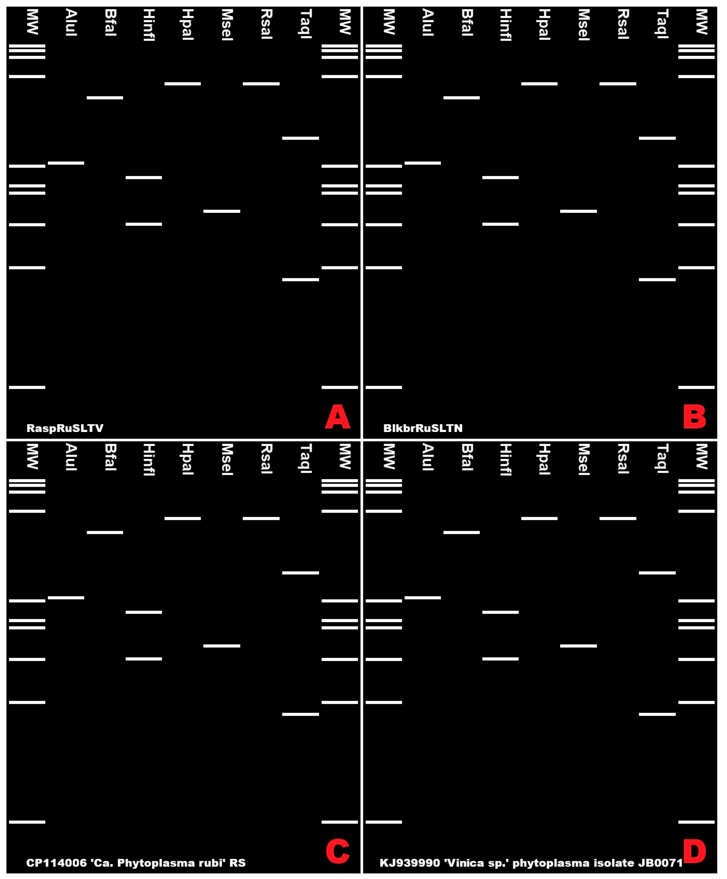
Comparison of the resulting virtual restriction profiles demonstrated that the patterns obtained for the RaspRuSLTV and BlkbrRuSLTN strains were identical to those of ‘Candidatus Phytoplasma rubi’ strain RS (CP114006) [30] and ‘Vinca sp.’ phytoplasma strain JB0071 (KJ939990) [27], which were retrieved and analysed using the CpnClassiPhyR database and assigned to the 16SrV-E subgroup, and were clearly distinct from the profiles of other 16SrV subgroup representatives (Figure 7). These results independently corroborate the 16SrV-E subgroup assignment inferred from analyses based on the 16S rRNA gene.
Figure 7.
Virtual RFLP analysis performed using the CpnClassiPhyR online tool at http://cpnclassiphyr.ca/CpnClassiPhyR.php (accessed on 11 February 2026) of the cpn60 fragments from raspberry (R. idaeus) (RaspRuSLTV01) (A), blackberry (R. nessensis) (BlkbrRuSLTN01) (B) and the ‘Ca. Phytoplasma rubi’ strain RS (C) and ‘Vinica sp.’ phytoplasma strain JB0071 (D). MW, marker phiX174 HaeIII-digested with fragment sizes, from top to bottom: 1353, 1078, 872, 603, 310, 281, 271, 234, 194, 118 and 72 bp.
3.4. Confirmation of Phytoplasma Identity Based on the secA Gene
To further validate the taxonomic assignment of the phytoplasmas detected in wild Rubus species, a fragment of the protein translocase subunit A (secA) gene was amplified using a semi-nested PCR approach. Amplifications were performed with the primer pairs SecAfor1/SecArev3 in the first PCR round and SecAfor2/SecArev3 in the semi-nested reaction. The amplicons of approximately 480 bp were successfully obtained from all 10 selected phytoplasma-positive samples, including 5 Rubus idaeus and 5 Rubus nessensis plants. All amplicons were sequenced and subjected to comparative sequence analyses. Pairwise comparison of the obtained secA sequences revealed a very high level of sequence conservation among the Lithuanian strains. Nucleotide identity values between RaspRuSLTV and BlkbrRuSLTN strains ranged from 99.84% to 100%, indicating minimal sequence divergence within the analysed fragment.
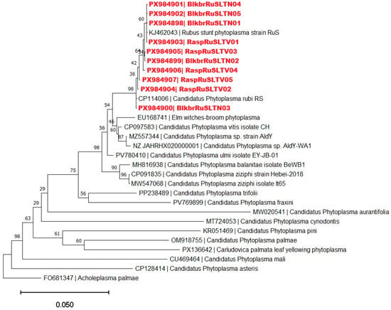
BLAST analysis against the GenBank database showed that the secA sequences of the Lithuanian strains shared 99.84%–100% nucleotide identity with reference sequences of ‘Ca. Phytoplasma rubi’, including strains RS (CP114006) [30] and RuS (KJ462043) [28], both assigned to the 16SrV-E subgroup.
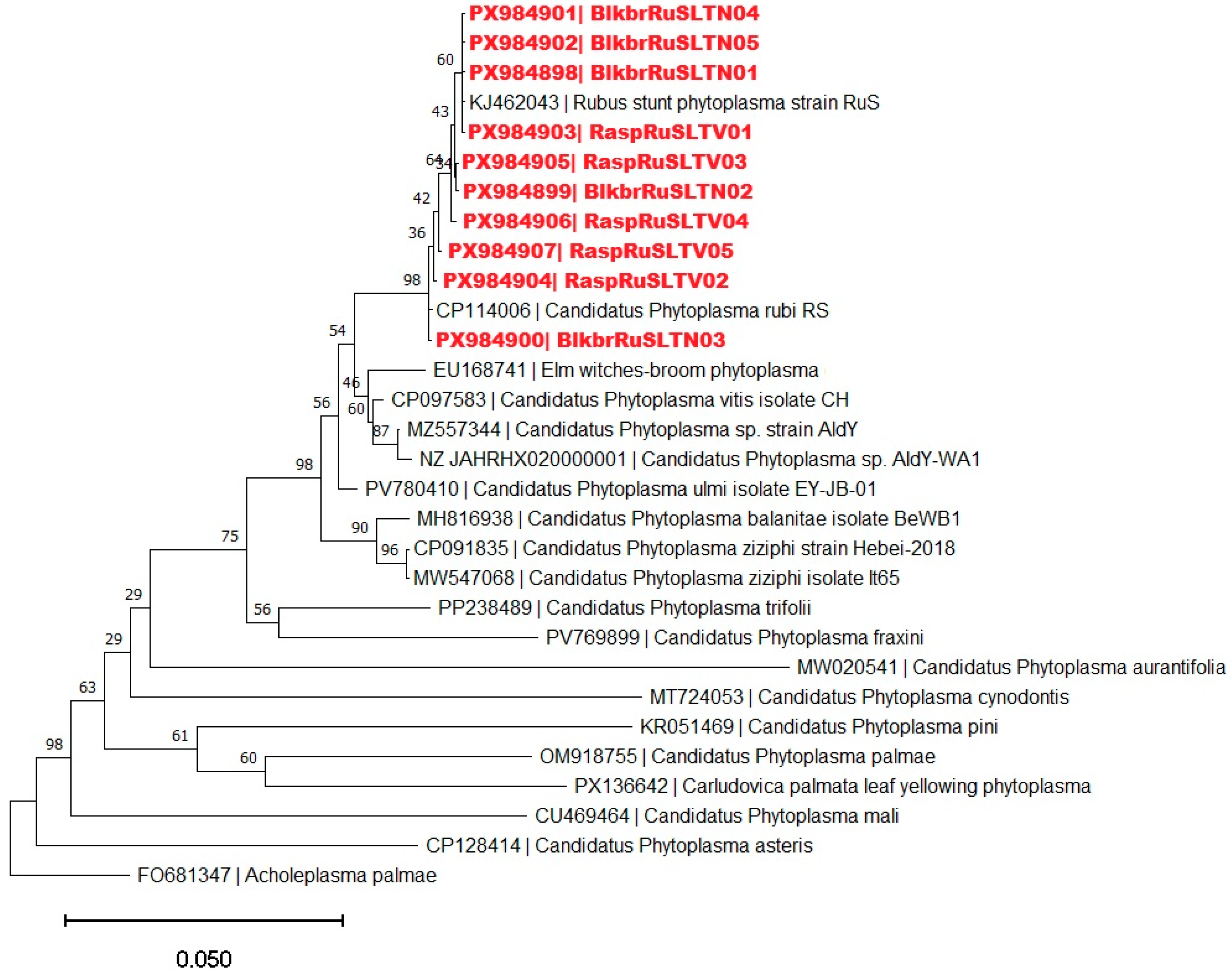
Phylogenetic analysis based on secA gene sequences consistently placed all RaspRuSLTV and BlkbrRuSLTN strains within a strongly supported monophyletic group comprising reference strains of ‘Ca. Phytoplasma rubi’ (Figure 8). Within the broader 16SrV phytoplasma clade, the Lithuanian strains clustered specifically with representatives of the 16SrV-E subgroup, whereas phytoplasmas belonging to other 16SrV subgroups formed distinct and more distant lineages. These results corroborate the 16SrV-E subgroup assignment inferred from analyses of the 16S rRNA and cpn60 gene sequences.
Figure 8.
Phylogenetic tree constructed using the neighbor-joining method with secA sequences of RaspRuSLTV and BlkbrRuSLTN strains detected in Lithuania (highlighted in red), 16SrV phytoplasma group members and other ‘Ca. Phytoplasma’ representatives. Acholeplasma palmae was used as an outgroup. The bar represents a phylogenetic distance of 0.05 nucleotide substitutions per site. The GenBank accession number is provided for each phytoplasma. Bootstrap values are shown on branches of the phylogenetic tree.
4. Discussion
The present study provides the first detailed multilocus molecular characterization of ‘Candidatus Phytoplasma rubi’-related strains infecting wild Rubus idaeus and Rubus nessensis populations in Lithuania. All phytoplasma strains detected in symptomatic plants from the Curonian Spit national park of unique ecosystems were assigned to the 16SrV-E subgroup based on multilocus sequence analysis and virtual RFLP profiling. To our knowledge, this represents not only the first such study conducted in Lithuania but also one of the few studies documenting ‘Ca. Phytoplasma rubi’ in wild Rubus populations in the Eastern Baltic region of Europe. The consistent classification of all Lithuanian strains as members of the 16SrV-E subgroup across three independent genetic markers provides strong evidence for their taxonomic identity. In this study, phytoplasma presence was confirmed exclusively by molecular methods, and symptom observations were not used for diagnostic purposes. Although the 16S rRNA gene remains the primary marker for phytoplasma detection and group-level classification, its limited resolving power within closely related lineages of the 16SrV group is well recognized [32,33,34,35]. In this study, consistent results were obtained from 16S rRNA, cpn60, and secA gene sequence analyses. Together with phylogenetic inference and virtual RFLP profiling, these data provide a robust basis for precise subgroup-level identification. The three genetic markers gave consistent results and clearly distinguished the detected phytoplasmas from other 16SrV phytoplasmas, including alder yellows, flavescence dorée and Portuguese blackberry phytoplasma related strains.
Across all analysed loci, the Lithuanian strains exhibited a high level of nucleotide sequence identity (>99.8%), irrespective of host species (R. idaeus or R. nessensis). No host- or site-specific sequence variation was detected, indicating that both Rubus species were infected by the same phytoplasma. Moreover, the sequences obtained from wild Rubus plants were nearly identical to each other and very similar to previously reported ‘Candidatus Phytoplasma rubi’ sequences from cultivated raspberry in Lithuania and other European agricultural systems, indicating that these strains belong to closely related, or potentially the same, genetic lineage. However, the present data does not allow conclusions regarding transmission pathways, movement, or exchange between natural and cultivated systems. This observation is consistent with the narrow host range of ‘Candidatus Phytoplasma rubi’ within the genus Rubus and suggests limited host-driven diversification in the genomic regions examined [18,36,37]. The variability in symptom expression observed in the field likely reflects the combined influence of infection age, phytoplasma titre, host physiological status, and environmental conditions rather than genetic differentiation among strains. These symptoms were used only as a field-screening criterion for sample selection and are not specific to phytoplasma infection, as similar symptom expressions may result from other biotic or abiotic factors.
Rubus stunt disease, caused by ‘Ca. Phytoplasma rubi’, has been reported from numerous European countries and is recognized as an economically important disease of cultivated raspberry and blackberry [7,13,14]. Molecular studies from Central and Western Europe have consistently shown that rubus stunt disease in cultivated Rubus species is associated with phytoplasmas belonging to the 16SrV-E phytoplasma subgroup [13,18,20,22,23]. The present findings show that this taxonomic lineage is present in wild raspberries and blackberries in Lithuania. This extends the known geographical range of ‘Ca. Phytoplasma rubi’ toward Northern Europe [14]. Importantly, the detection of this phytoplasma in unmanaged forest habitats indicates that its ecological niche is not restricted to agricultural systems.
The detection of ‘Ca. Phytoplasma rubi’ in wild Rubus species broadens the known ecological context of this pathogen in Lithuania. Because infected plants were identified in unmanaged forest habitats, these findings indicate that the pathogen is not confined to cultivated Rubus systems. However, the present study was not designed to test reservoir function, inoculum dynamics, or epidemiological connectivity between wild and cultivated hosts. No insect vector sampling, transmission assays, systematic screening of asymptomatic plants, or temporal monitoring of clonal patches were performed. Therefore, any epidemiological role of wild Rubus populations, including possible reservoir function, should at present be regarded as a plausible hypothesis rather than a demonstrated conclusion, as wild Rubus hosts have been suggested to act as long-term reservoirs in other regions [4,21,35,38,39,40,41]. In this context, insect vectors associated with rubus stunt phytoplasmas have also been reported elsewhere [21,42], although their role in the studied area remains unknown. In unmanaged habitats, infected wild Rubus plants may represent a potential source of inoculum for nearby cultivated systems, although this has not been directly assessed in the present study [4,38,39,40,41].
Phytoplasmas are transmitted not only by insect vectors but can also be disseminated through vegetative propagation of infected plants. Wild Rubus species are characterized by intensive clonal growth and vegetative reproduction, including the formation of root suckers, rhizomes and rooting of arching shoots [43,44,45]. Because wild Rubus species reproduce clonally, infected genotypes could in principle support local persistence of phytoplasma infection within natural stands. However, the present study did not include temporal or within-patch monitoring, and this possibility remains to be tested.
Several limitations should be considered when interpreting the ecological significance of these findings. Because asymptomatic plants were not systematically analyzed, the present study does not allow assessment of symptom specificity in wild Rubus hosts. The survey was restricted to symptomatic plants and did not include systematic sampling of asymptomatic wild Rubus individuals. In addition, no insect vectors were collected, no transmission assays were conducted, no quantitative assessment of phytoplasma titre was performed, and no temporal monitoring of infected clonal patches was carried out. Accordingly, the present study cannot determine whether wild Rubus populations function as confirmed reservoirs, how infection is maintained locally, or whether transmission occurs between natural and cultivated systems. Rather, the study provides a molecular baseline for future work addressing these questions.
5. Conclusions
This study provides the first multilocus molecular characterization of ‘Candidatus Phytoplasma rubi’-related strains in Lithuania. This is the first report of the phytoplasma in wild Rubus idaeus and Rubus nessensis in Lithuania, contributing to the current knowledge of its occurrence in Northern Europe. Using a robust diagnostic framework based on 16S rRNA, cpn60, and secA gene sequence analyses, supported by phylogenetic reconstruction and virtual RFLP profiling, all detected phytoplasma strains were consistently assigned as the ‘Ca. Phytoplasma rubi’-related and belonging to 16SrV-E phytoplasma subgroup. The congruent results obtained across three independent genetic markers demonstrate a high degree of genetic uniformity among Lithuanian strains and provide strong evidence for their taxonomic identity. The detection of ‘Ca. Phytoplasma rubi’ in unmanaged forest ecosystems extends the known geographical and ecological range of this pathogen in Northern Europe. It also shows that wild raspberries and blackberries in Lithuania can host phytoplasma strains closely related to those previously reported from cultivated Rubus crops.
These findings identify wild Rubus populations as an ecologically relevant component of the host range of ‘Ca. Phytoplasma rubi’ in Lithuania. However, their epidemiological significance, including possible reservoir function or involvement in transmission between natural and cultivated habitats, was not directly addressed in this study and remains to be investigated. The molecular baseline established here provides a foundation for future studies incorporating vector analysis, asymptomatic host screening, and spatial or temporal comparisons of wild and cultivated Rubus populations.
Supplementary Materials
The following supporting information can be downloaded at: https://www.mdpi.com/article/10.3390/f17040493/s1, Figure S1: Map with location of collected red raspberry (Rubus idaeus) and blackberry (Rubus nessensis) plant samples from the Curonian Spit (55°32′28″ N, 21°58′15″ E). Figure S2: Virtual restriction profiles generated with DraI (A), HaeIII (B), HpaII (C), MboI (D), MseI (E) and TaqI (F) from cpn60 D0317/D0318 fragments sequences of phytoplasmas detected in raspberry (R. idaeus) (RaspRuSLTV01–RaspRuSLTV05) and blackberry (R. nessensis) (BlkubrRuSLTN01–BlkubRuSLTN05), and of representative 16SrV group phytoplasmas. Table S1: used primers. Table S2: Summary of sampled plants, host species, PCR results, sequenced loci and GenBank accession numbers for the 16S rRNA, cpn60 and secA gene sequences generated in this study.
Author Contributions
Conceptualization, D.V. and A.I.; methodology, D.V.; software, M.D.; validation, D.V., M.D., M.Ž.-E. and A.I.; formal analysis, M.D.; investigation, M.D. and A.I.; resources, M.D., A.I., M.Ž.-E. and D.V.; data curation, D.V.; writing—original draft preparation, M.D.; writing—review and editing, A.I., M.Ž.-E. and D.V.; visualization, M.D. and M.Ž.-E.; supervision, D.V. All authors have read and agreed to the published version of the manuscript.
Funding
This research received no external funding.
Data Availability Statement
The nucleotide sequence data generated in this study have been deposited in the NCBI GenBank database under accession numbers: PX970422 to PX970431 for 16S rDNA, PX984888 to PX984897 for cpn60, PX984898 to PX984907 for secA. All other data supporting the findings of this study are available within the article and its Supplementary Materials.
Acknowledgments
During the preparation of this manuscript, the authors used ChatGPT-5.2 (OpenAI) for the purposes of improving grammar and clarity. The authors have reviewed and edited the output and take full responsibility for the content of this publication.
Conflicts of Interest
The authors declare no conflicts of interest.
Abbreviations
The following abbreviations are used in this manuscript:
| RS | Rubus stunt (disease) |
| PCR | Polymerase chain reaction |
| DNA | Deoxyribonucleic acid |
| dNTPs | Deoxynucleotide triphosphates |
| 16S rRNA | 16S ribosomal RNA |
| cpn60 | chaperonin-60 gene |
| secA | protein translocase subunit A gene |
| RFLP | restriction fragment length polymorphism |
| BLAST | Basic Local Alignment Search Tool |
| NCBI | National Center for Biotechnology Information |
| MEGA | Molecular Evolutionary Genetics Analysis software |
| bp | Base pairs |
| kb | Kilobase pairs |
References
- Wang, R.; Bai, B.; Li, D.; Wang, J.; Huang, W.; Wu, Y.; Zhao, L. Phytoplasma: A plant pathogen that cannot be ignored in agricultural production—Research progress and outlook. Mol. Plant Pathol. 2024, 25, e13437. [Google Scholar] [CrossRef] [Scilit]
- Kumari, S.; Nagendran, K.; Rai, A.B.; Singh, B.; Rao, G.P.; Bertaccini, A. Global status of phytoplasma diseases in vegetable crops. Front. Microbiol. 2019, 10, 1349. [Google Scholar] [CrossRef] [Scilit] [PubMed]
- Weintraub, P.G.; Beanland, L. Insect vectors of phytoplasmas. Annu. Rev. Entomol. 2006, 51, 91–111. [Google Scholar] [CrossRef] [Scilit] [PubMed]
- Linck, H.; Reineke, A. Rubus stunt: A review of an important phytoplasma disease in Rubus spp. J. Plant Dis. Prot. 2019, 126, 393–399. [Google Scholar] [CrossRef] [Scilit]
- Fahmeed, F.; Rosete, Y.A.; Pérez, K.A.; Boa, E.; Lucas, J. First report of ‘Candidatus Phytoplasma asteris’ (group 16SrI) infecting fruits and vegetables in Islamabad, Pakistan. J. Phytopathol. 2009, 157, 639–641. [Google Scholar] [CrossRef] [Scilit]
- Reeder, R.; Kelly, P.L.; Arocha, Y. ‘Candidatus Phytoplasma asteris’ identified in blackberry (Rubus fruticosus agg.) in the United Kingdom. Plant Pathol. 2010, 59, 394. [Google Scholar] [CrossRef] [Scilit]
- Cieślińska, M. Detection and characterization of phytoplasmas associated with diseases of Rubus spp. in Poland. J. Plant Pathol. 2011, 93, 51–56. [Google Scholar]
- Davies, D.L. The occurrence of two phytoplasmas associated with stunted Rubus species in the UK. Plant Pathol. 2000, 49, 86–88. [Google Scholar] [CrossRef] [Scilit]
- Davis, R.E.; Dally, E.L. Revised subgroup classification of group 16SrV phytoplasmas and placement of flavescence dorée–associated phytoplasmas in two distinct subgroups. Plant Dis. 2001, 85, 790–797. [Google Scholar] [CrossRef] [Scilit]
- Borroto Fernández, E.G.; Calari, A.; Hanzer, V.; Katinger, H.; Bertaccini, A.; Laimer, M. Phytoplasma-infected plants in Austrian forests: Role as a reservoir? Bull. Insectol. 2007, 60, 391–392. [Google Scholar]
- Bobev, S.G.; De Jonghe, K.; Maes, M. First report of ‘Candidatus Phytoplasma solani’ on blackberry (Rubus fruticosus) in Bulgaria. Plant Dis. 2013, 97, 282. [Google Scholar] [CrossRef] [Scilit] [PubMed]
- Kuzmanovic, S.; Starovic, M.; Pavlovic, S.; Gavrilovic, V.; Aleksic, G.; Stojanovic, S.; Josic, D. Detection of stolbur phytoplasma on blackberry: A new natural host in Serbia. Genetika 2011, 43, 559–568. [Google Scholar] [CrossRef] [Scilit]
- Fránová, J.; De Sousa, E.; Koloniuk, I.; Mimoso, C.; Matos, J.; Cardoso, F.; Contaldo, N.; Paltrinieri, S.; Bertaccini, A. Multi-gene characterization of a new ‘Candidatus Phytoplasma rubi’-related strain associated with blackberry witches’ broom. Int. J. Syst. Evol. Microbiol. 2016, 66, 1438–1446. [Google Scholar] [CrossRef] [Scilit] [PubMed]
- Mäurer, R.; Seemüller, E. Nature and genetic relatedness of the mycoplasma-like organism causing rubus stunt in Europe. Plant Pathol. 1995, 44, 244–249. [Google Scholar] [CrossRef] [Scilit]
- Sertkaya, G.; Osler, R.; Musetti, R.; Ermacora, P.; Martini, M. Detection of phytoplasmas in Rubus spp. by microscopy and molecular techniques in Turkey. Acta Hortic. 2004, 656, 181–186. [Google Scholar] [CrossRef] [Scilit]
- Valiūnas, D.; Jomantienė, R.; Davis, R.E. Phytoplasmas detected in cultivated fruit plants in Lithuania. Bull. Insectol. 2007, 60, 139–140. [Google Scholar]
- Davis, R.E.; Dally, E.L.; Converse, R.H. Molecular identification of a phytoplasma associated with witches’-broom disease of black raspberry in Oregon and its classification in group 16SrIII, new subgroup Q. Plant Dis. 2001, 85, 1121. [Google Scholar] [CrossRef] [Scilit]
- Fránová, J.; Přibylová, J.; Zemek, R.; Tan, J.L.; Hamborg, Z.; Blystad, D.-R.; Lenz, O.; Koloniuk, I. First report of ‘Candidatus Phytoplasma rubi’ associated with rubus stunt disease of raspberry and blackberry in the Czech Republic. Plant Dis. 2023, 107, 2510. [Google Scholar] [CrossRef] [Scilit]
- Jarausch, W.; Jarausch-Wehrheim, B.; Danet, J.L.; Broquaire, J.M.; Dosba, F.; Saillard, C.; Garnier, M. Detection and indentification of european stone fruit yellows and other phytoplasmas in wild plants in the surroundings of apricot chlorotic leaf roll-affected orchards in Southern France. Eur. J. Plant Pathol. 2001, 107, 209–217. [Google Scholar] [CrossRef] [Scilit]
- Marcone, C.; Palmieri, C.; Sellitto, A. A witches’-broom disease of cultivated strawberry associated with ‘Candidatus Phytoplasma rubi’-related strains in Southern Italy. Plants 2025, 14, 2914. [Google Scholar] [CrossRef] [Scilit] [PubMed]
- Linck, H.; Reineke, A. Preliminary survey on putative insect vectors for rubus stunt phytoplasmas. J. Appl. Entomol. 2019, 143, 328–332. [Google Scholar] [CrossRef] [Scilit]
- Böhm, J.W.; Duckeck, D.; Duduk, B.; Schneider, B.; Kube, M. Genome comparison of ‘Candidatus Phytoplasma rubi’ with genomes of other 16SrV phytoplasmas highlights special group features. Appl. Microbiol. 2023, 3, 1083–1100. [Google Scholar] [CrossRef] [Scilit]
- Rufo, R.; Batlle, A.; Camprubi, A.; Montesinos, E.; Calvet, C. Control of rubus stunt and stolbur diseases in Madagascar periwinkle. Plant Pathol. 2016, 66, 551–558. [Google Scholar] [CrossRef] [Scilit]
- Deng, S.; Hiruki, C. Genetic relatedness between two nonculturable mycoplasmalike organisms revealed by nucleic acid hybridization and polymerase chain reaction. Phytopathology 1991, 81, 1475–1479. [Google Scholar] [CrossRef] [Scilit]
- Schneider, B.; Seemüller, E.; Smart, C.D.; Kirkpatrick, B.C. Phylogenetic classification of plant pathogenic mycoplasma-like organisms or phytoplasmas. In Molecular and Diagnostic Procedures in Mycoplasmology; Academic Press: San Diego, CA, USA, 1995; pp. 369–380. [Google Scholar] [CrossRef] [Scilit]
- Gundersen, D.E.; Lee, I.-M. Ultrasensitive detection of phytoplasmas by nested PCR assays using two universal primer pairs. Phytopathol. Mediterr. 1996, 35, 114–151. [Google Scholar]
- Dumonceaux, T.J.; Green, M.; Hammond, C.; Perez, E.; Olivier, C. Molecular diagnostic tools for detection and differentiation of phytoplasmas based on chaperonin-60 reveal differences in host plant infection patterns. PLoS ONE 2014, 9, e116039. [Google Scholar] [CrossRef] [Scilit] [PubMed]
- Hodgetts, J.; Boonham, N.; Mumford, R.; Harrison, N.; Dickinson, M. Phytoplasma phylogenetics based on analysis of secA and 23S rRNA gene sequences for improved resolution of candidate species of ‘Candidatus Phytoplasma’. Int. J. Syst. Evol. Microbiol. 2008, 58, 1826–1837. [Google Scholar] [CrossRef] [Scilit] [PubMed]
- Tamura, K.; Stecher, G.; Kumar, S. MEGA11: Molecular evolutionary genetics analysis version 11. Mol. Biol. Evol. 2021, 38, 3022–3027. [Google Scholar] [CrossRef] [Scilit]
- Duckeck, D.; Zübert, C.; Böhm, J.W.; Carminati, G.; Schneider, B.; Kube, M. Complete genome of ‘Candidatus Phytoplasma rubi’ RS, a phytopathogenic bacterium associated with rubus stunt disease. Microbiol. Resour. Announc. 2023, 12, e01303-22. [Google Scholar] [CrossRef] [Scilit] [PubMed]
- Wei, W.; Zhao, Y. Phytoplasma taxonomy: Nomenclature, classification, and identification. Biology 2022, 11, 1119. [Google Scholar] [CrossRef] [Scilit]
- Zhao, Y.; Wei, W.; Lee, I.-M.; Shao, J.; Suo, X.; Davis, R.E. The iPhyClassifier, an interactive online tool for phytoplasma classification and taxonomic assignment. Methods Mol. Biol. 2012, 938, 329–338. [Google Scholar] [CrossRef] [Scilit]
- Muirhead, K.; Pérez-López, E.; Bahder, B.W.; Hill, J.E.; Dumonceaux, T. The CpnClassiPhyR is a resource for cpn60 universal target-based classification of phytoplasmas. Plant Dis. 2019, 103, 2494–2497. [Google Scholar] [CrossRef] [Scilit]
- Zhao, Y.; Davis, R.E. Criteria for phytoplasma 16Sr group/subgroup delineation and the need of a platform for proper registration of new groups and subgroups. Int. J. Syst. Evol. Microbiol. 2016, 66, 2121–2123. [Google Scholar] [CrossRef] [Scilit] [PubMed]
- Dickinson, M.; Hodgetts, J. PCR analysis of phytoplasmas based on the secA gene. Methods Mol. Biol. 2012, 938, 205–215. [Google Scholar] [CrossRef] [Scilit]
- Malembic-Maher, S.; Salar, P.; Filippin, L.; Carle, P.; Angelini, E.; Foissac, X. Genetic diversity of European phytoplasmas of the 16SrV taxonomic group and proposal of ‘Candidatus Phytoplasma rubi’. Int. J. Syst. Evol. Microbiol. 2011, 61, 2129–2134. [Google Scholar] [CrossRef] [Scilit] [PubMed]
- Bertaccini, A.; Arocha-Rosete, Y.; Contaldo, N.; Duduk, B.; Fiore, N.; Montano, H.G.; Kube, M.; Kuo, C.-H.; Martini, M.; Oshima, K.; et al. Revision of the ‘Candidatus Phytoplasma’ species description guidelines. Int. J. Syst. Evol. Microbiol. 2022, 72, 005353. [Google Scholar] [CrossRef] [Scilit]
- Marie-Jeanne, V.; Bonnot, F.; Thébaud, G.; Peccoud, J.; Labonne, G.; Sauvion, N. Multi-scale spatial genetic structure of the vector-borne pathogen ‘Candidatus Phytoplasma prunorum’ in orchards and in wild habitats. Sci. Rep. 2020, 10, 5002. [Google Scholar] [CrossRef] [Scilit]
- Yu, S.-S.; Wei, W. Beyond single-pathogen models: Understanding mixed infections involving phytoplasmas and other plant pathogens. Plants 2025, 14, 2049. [Google Scholar] [CrossRef] [Scilit]
- Boyle, J.A.; Yang, Y.; Caballero, A.; Hector, E.N.; Ellett, A.; Glasgow, E.; Stinchcombe, J.R.; Bontrager, M. The ecological dynamics and consequences of phytoplasma in a spring ephemeral. bioRxiv 2025. [Google Scholar] [CrossRef] [Scilit]
- Sundararaj, R.; Rishi, R.R.; Manjula, K.N. Emerging phytoplasma diseases in forestry and the role of determining the insect vectors for the management of the diseases. Indian J. Entomol. 2023, 85, 1105–1114. [Google Scholar] [CrossRef] [Scilit]
- Linck, H.; Krüger, E.; Reineke, A. A multiplex TaqMan qPCR assay for sensitive and rapid detection of phytoplasmas infecting Rubus species. PLoS ONE 2017, 12, e0177808. [Google Scholar] [CrossRef] [Scilit]
- Lambrecht-McDowell, S.C.; Radosevich, S.R. Population demographics and trade-offs to reproduction of an invasive and noninvasive species of Rubus. Biol. Invasions 2005, 7, 281–295. [Google Scholar] [CrossRef] [Scilit]
- Makarov, S.; Upadyshev, M.; Sungurova, N.; Tyukavina, O.; Kulikova, E.; Kuznetsova, I. Clonal micropropagation of wild berry plants of the genus Rubus. Food Process. Tech. Technol. 2024, 54, 60–70. [Google Scholar] [CrossRef] [Scilit]
- Barrett, S.C.H. Influences of clonality on plant sexual reproduction. Proc. Natl. Acad. Sci. USA 2015, 112, 8859–8866. [Google Scholar] [CrossRef] [Scilit] [PubMed]
Disclaimer/Publisher’s Note: The statements, opinions and data contained in all publications are solely those of the individual author(s) and contributor(s) and not of MDPI and/or the editor(s). MDPI and/or the editor(s) disclaim responsibility for any injury to people or property resulting from any ideas, methods, instructions or products referred to in the content. |
© 2026 by the authors. Licensee MDPI, Basel, Switzerland. This article is an open access article distributed under the terms and conditions of the Creative Commons Attribution (CC BY) license.