


Abstract
Artificial intelligence (AI) technologies, which are being actively developed in modern medicine today, increase the speed and quality of patient care. This article mainly seeks to demonstrate the use of various options of computer analysis of clinical images to solve practical problems of increasing the efficiency of routine diagnostics using retrospective analysis, as well as show the potential for its widespread implementation (due to the scalability of the architecture) in practical healthcare, exemplified by ultrasound (US) and magnetic resonance imaging (MRI) data analysis. This is an interuniversity study, its research protocol was conducted in accordance with the Declaration of Helsinki, and the protocol was approved by the local Ethics Committee of Orel State University named after I. S. Turgenev (Protocol No. 25 dated 16 November 2022). The software was developed using Python 3.7 and open neural network models. Statistical processing included an efficiency assessment for which IBM SPSS Statistics 20.0 was used. Detection errors in the analysis of 550 US cases did not exceed 6–8% and were associated with technical difficulties due to image quality. When studying 1030 MRI studies, only 0.19% of cases failed to obtain reliable image analysis results. The differences in the average values for the dimensional characteristics of the studied vessels were 0.11–0.12 mm. The effectiveness of AI in clinical tasks is presented. The improvement in segmentation accuracy was achieved through the use of step-by-step image optimization during the AI training stage. The evolution of technologies in medicine, aimed at digitalization and personalization, is intended to improve the quality and speed of studying images in practical work.
1. Introduction
The widespread introduction of computer technology and an increase in computing power have made it possible to actively introduce AI technologies into everyday life [1,2,3].
The dissemination of these software tools and their improvement are facilitated by a diverse set of available tools that allow using ready-made models, further training them, and creating arbitrary original combinations. The unification of software products is complemented by the development of programming technologies introducing more flexible tools for data interpretation and visualization [4,5].
Currently, computer technologies are being actively introduced into education of specialists in various specialties, including the training of medical workers [6]. The use of AI tools makes it possible to improve educational and methodological materials and introduce diversity into demonstration and control tasks. In recent years, students have been mastering computer tools to prepare for classes, which reflects the impact of new educational technologies with the introduction of AI on the image of the education system as a whole [7,8,9].
The development of informatization in the domestic healthcare system has served as an important factor for the introduction of AI systems into the work of specialized medical institutions. The COVID-19 pandemic period coincided with the technological maturity and availability of AI tools, which was reflected in its successful application to reduce the workload among radiologists [10]. The peculiarity of the clinical work of healthcare professionals requires constant analysis of large amounts of diagnostic information, identification and observation of certain patterns that make it possible to assess the condition of patients and predict the further course of diseases and their outcome. The most striking examples of the use of AI in medicine are systems for analyzing clinical images to detect oncological diseases or assess the physiological state of the body [11,12,13]. Equally important is the introduction of textual information management systems into the work of specialists [14,15].
The synthesis of clinical data with multifactorial analysis, taking into account the context, demonstrates high efficiency, which is confirmed by the experience of multimodal AI analysis in oncology [16]. The use of computer-based intelligent multifactorial analysis in clinical practice makes it possible to significantly improve diagnostic efficiency and reduce the burden on medical personnel. An important feature for the introduction of combined methods of computer analysis is the need to prepare specialized medical databases of large volume, which involves not only technical but also ethical issues [17,18,19]. Due to these features, AI technologies in medicine require improved algorithms to achieve the highest diagnostic efficiency with relatively small amounts of information for training models [20,21,22]. Due to the fact that the implementation of algorithms in the medical field requires clarifying the specifics of their implementation and/or the uniformity of their application in practice, we conducted this study.
The aim of the work is to demonstrate the use of various options of computer analysis of clinical images to solve practical problems of increasing the efficiency of routine diagnostics using retrospective analysis, as well as show the potential for its widespread implementation (due to the scalability of the architecture) in practical healthcare.
2. Materials and Methods
The presented study is based on the analysis of the effectiveness of AI software for segmentation and analysis of clinical images obtained using US imaging and magnetic resonance imaging technology. Despite the different nature of the analyzed data, similar technical tasks were performed using software tools for segmentation of the target object of study: assessment of its quantitative characteristics (sizing). Thus, we studied and compared 550 results of US examination of the female genital organs and 1030 studies of magnetic resonance imaging data. All participants provided informed consent for their voluntary involvement. The study was conducted in accordance with the Declaration of Helsinki, and the protocol was approved by the Ethics Committee of Orel State University named after I. S. Turgenev (Protocol No. 25 dated 16 November 2022). Practical significance was assessed based on comparison with the results of a clinical trial examination and expert analysis. The data from the computer analysis of US imaging were compared with the results obtained during the evaluated procedures. The results of computer analysis of magnetic resonance imaging data were compared with the morphometric parameters of a similar group of patients undergoing US duplex scanning of the main arteries of the neck (1146 cases).
The presented computer analysis models were developed using the high-level interpreted general-purpose programming language Python 3.7 and open neural network models. As a result, cross-platform software products were created that make it possible to implement original algorithms both as a console application with a GUI interface and as network services. This way of implementing the idea made it possible to compare and analyze various options for interacting with different software environments and evaluate critical factors for the technical support of products. Facilitating the migration of the developed solutions without taking into account strict binding to specific hardware characteristics makes it easier in the future to adapt developed solutions to the environment of domestic software and hardware, taking into account import substitution of critical infrastructure.
The training of the model for segmentation of the ovaries and structures of the follicular apparatus was performed on the basis of a specialized dataset, including 898 original images with marking of the position of the ovary and follicles. The size of the image validation database was at least 20% of the volume used for training. For ultrasound images, 180 additional examples with markings were used for validation. The size of the images was 1024 × 728. In total, 15 training epochs have been completed.
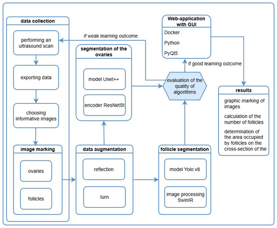
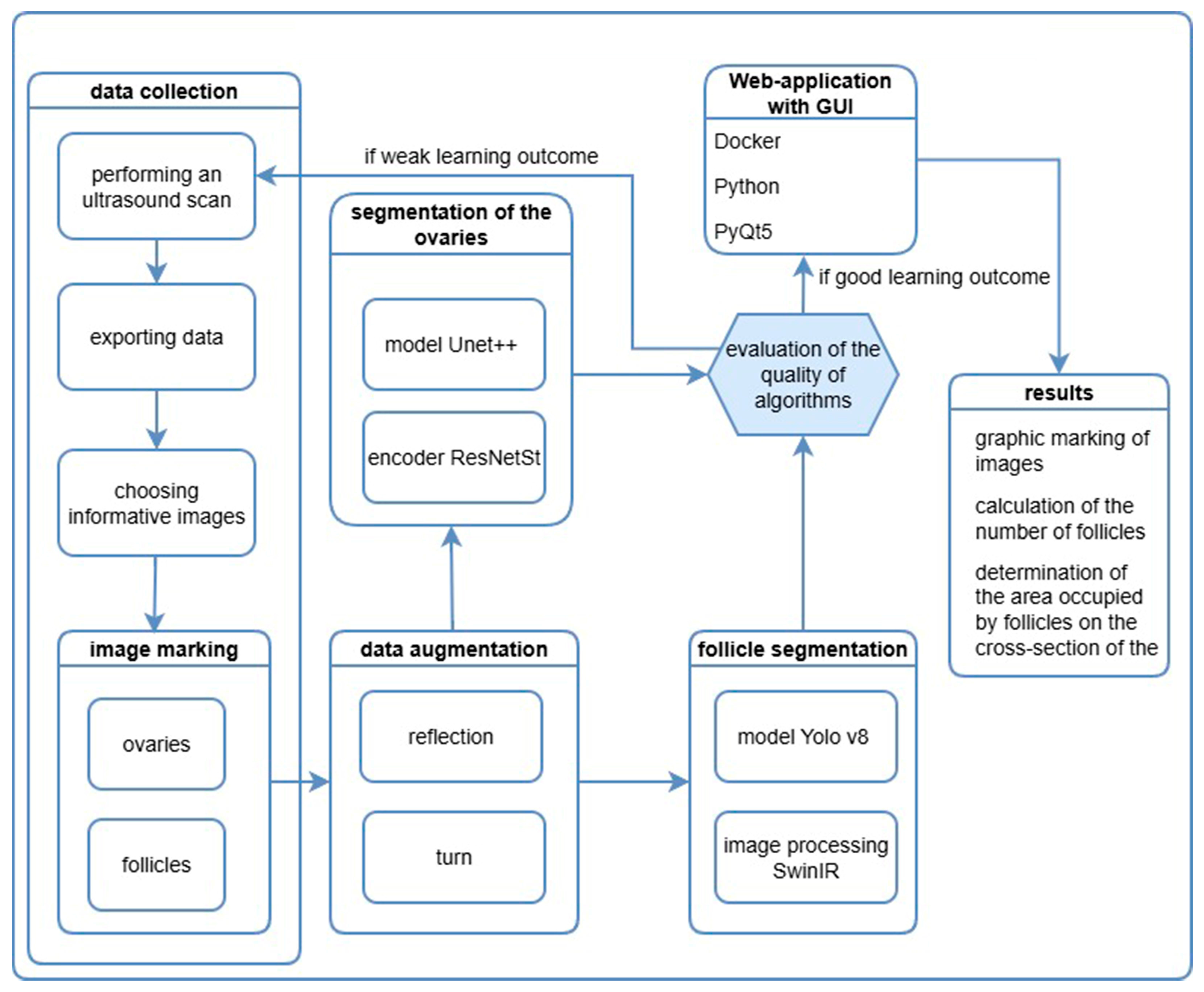
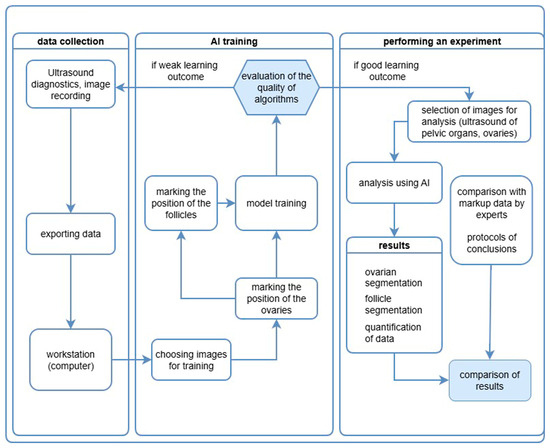
The diagram of the experiment organization model is shown in Figure 1.
Figure 1.
The scheme of the model of organization of the experiment analysis of ultrasound data for segmentation of ovaries and follicles.
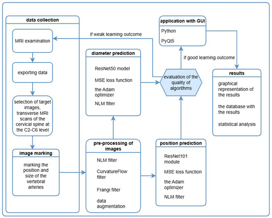
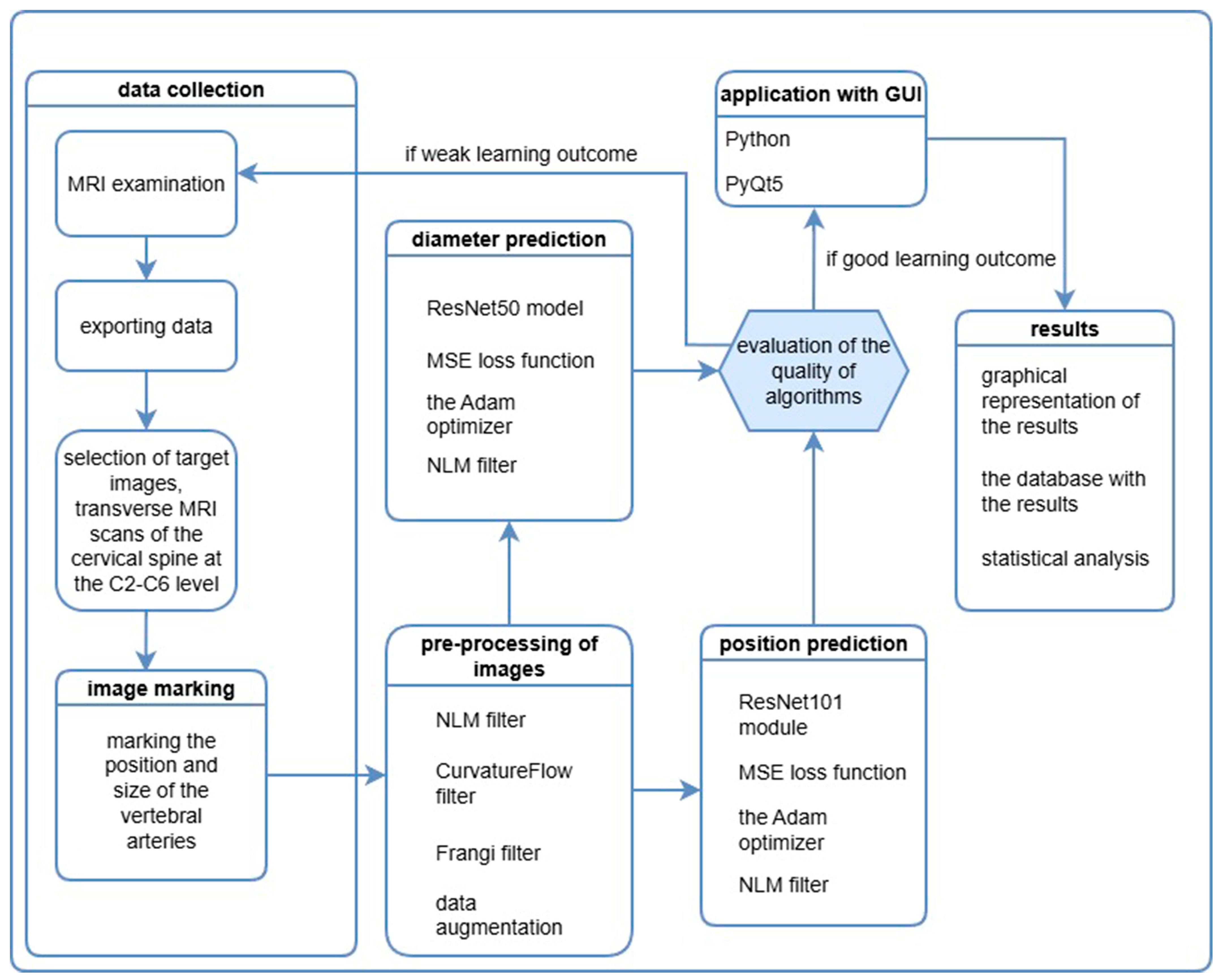
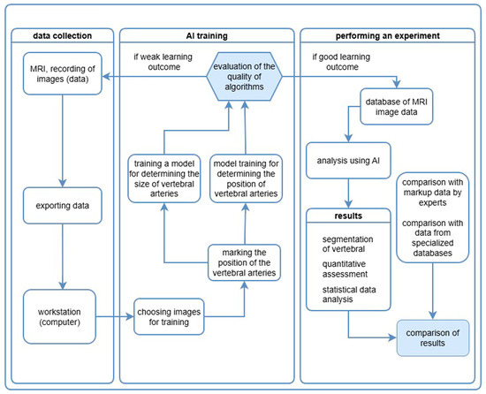
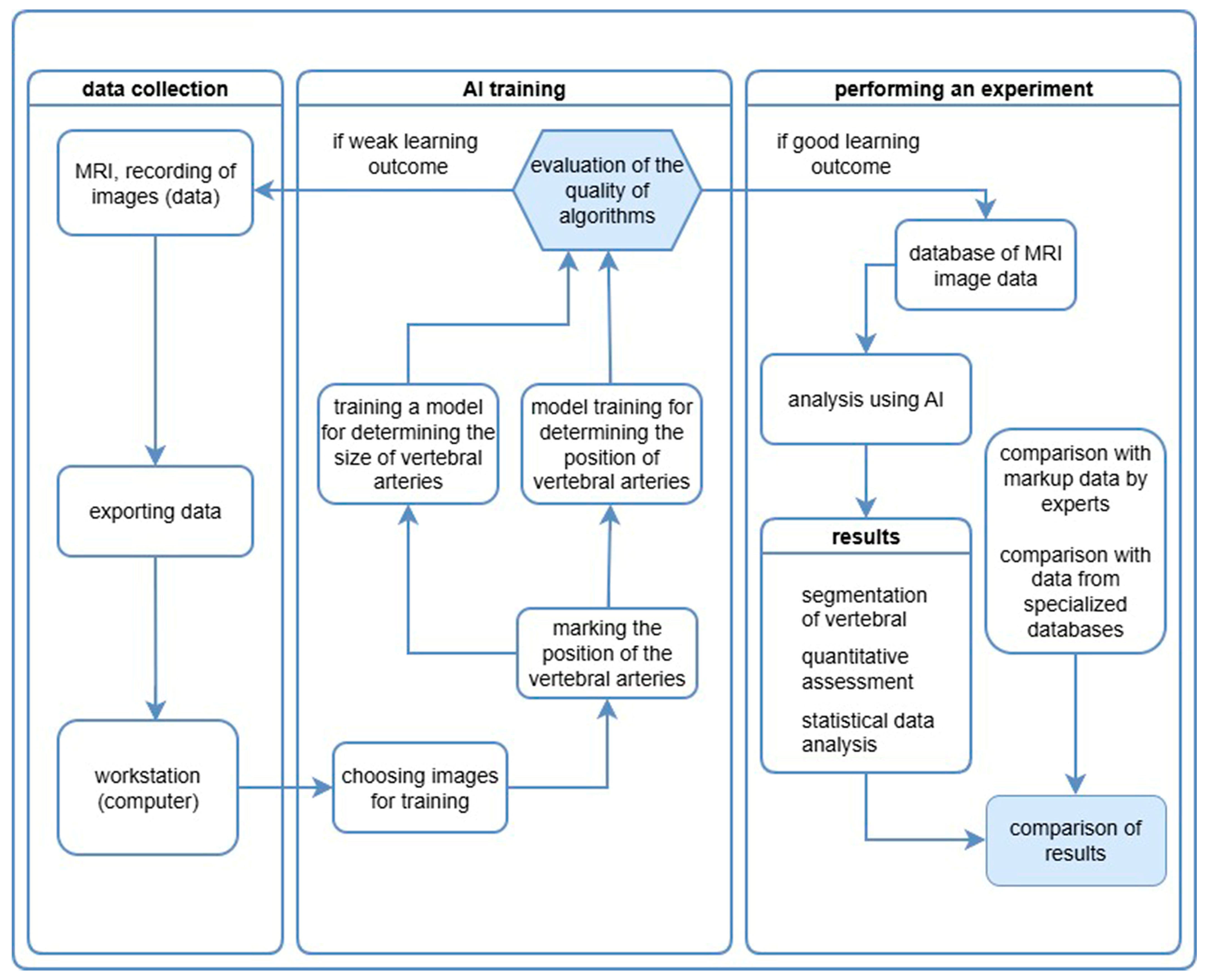
During the preparation of the training dataset, 1100 original images with marking of the position of the vertebral arteries were used to determine and measure the size of the vertebral arteries. The improvement of the analysis accuracy was achieved by consistent use of image processing algorithms and specialization of the vascular structure search algorithm. The size of the image validation database was at least 20% of the volume used for training. Then, 220 additional markup examples were used for MRI images for validation. The size of the images was 512 × 512. In total, 200 training epochs have been completed. The diagram of the experiment organization model is shown in Figure 2.
Figure 2.
The scheme of the model of organization of the experiment of the analysis of MRI data for segmentation of vertebral arteries.
Anonymous MRI results were exported as images for processing. A medical staff member performed manual annotation of the vertebral artery positions on transverse images of the cervical spine at the C2–C6 vertebral levels. During the training of the AI algorithm, a series of preliminary image optimization steps (pre-processing of images) was conducted.
The training of AI algorithms for segmentation of vertebral artery images and for determining their position was performed separately. This separation of stages was due to different accuracy requirements. The IoU parameter was given greater importance for determining the vertebral artery diameter. The algorithm for determining vessel position required higher Dice scores. As a result, the best practical results were obtained for artery segmentation using ResNet50, and for determining vessel position using ResNet101 with custom filter tuning.
The difficulty of identifying vertebral arteries by computer vision systems is due to the small size of the structures and the relatively low image resolution. Therefore, when model training performance was low, in addition to optimizing preprocessing, extra images were annotated and training stages were repeated until the required accuracy level was achieved.
The final stage was the development of a GUI for the convenient practical use of the software, with the output of reports containing the necessary AI results.
The separation of the segmentation and position-determination systems was deliberately designed to enable the creation of independent subsystems and to reduce the computational load when performing routine tasks. Determining vessel position allows for successful diagnosis of positional anomalies. The use of more sensitive segmentation algorithms is employed on demand to search for causes of possible hemodynamic changes in complex clinical cases. Through this approach, we were able to reduce the potential computational burden on equipment when solving similar practical tasks based on related computer image analysis algorithms.
The hardware used for testing the project was Intel 10400F CPU, 64 GB DDR4 Ram, 12 Gb RTX 3060 GPU, 1Tb SSD, Win10.
Statistical processing included an assessment of the detection efficiency of the evaluated structures, calculation of the average values of morphometric indicators and the average error (M ± m), the first and third quartiles of the distribution (Q1–Q3), standard deviation (σ), and coefficient of variation (CV, %). The reliability of the results obtained was assessed using distribution diagrams, and the student’s t-test was calculated to determine the significance of the differences between the compared indicators. IBMSPSS Statistics 20.0 was used for statistical calculations.
3. Results
Despite the fact that the repeated analysis of US imaging results was carried out retrospectively using static images, including low-quality ones, the errors in detection and analysis of anatomical structures did not exceed 6% on the left and 8% on the right. These results were obtained by comparing the data obtained by computer analysis with the results of clinical trials performed by a medical expert.
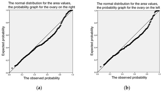
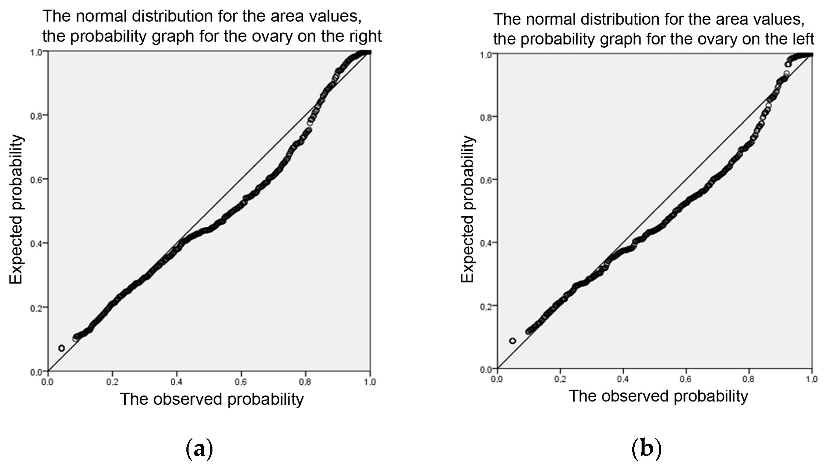
The retrospective analysis process did not require additional technical training for the operator, allowing processing of all submitted material in a relatively short time. As a result of the computer analysis, unique data were obtained on the ratio of the organ section (ovary) to the total area occupied by all follicles in the image. This indicator is extremely time-consuming when using traditional image analysis tools and therefore could not be used in scientific research of significant groups of participants and in clinical practice. The partially obtained observation results were published [11,12]. General information about the results of the analysis is presented in Table 1 and in Figure 3.
Table 1.
Results of computer analysis using AI of the area occupied by follicles on diagnostic images of ovaries obtained by US imaging.
Figure 3.
Distribution normality diagrams for the area occupied by follicles on the US image of the ovary (%). Probability calculation: (a) For the ovary on the right; (b) For the ovary on the left.
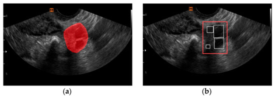
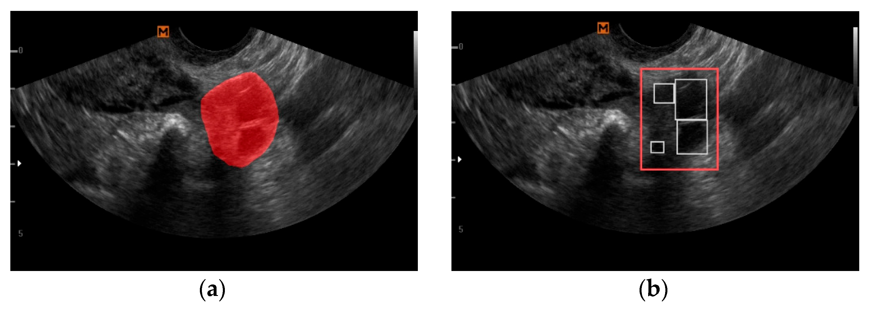
An example of the operation of the presented software solution is shown in Figure 4.
Figure 4.
Demonstration of the effectiveness of ovarian and follicle segmentation on a real diagnostic US image: (a) Ovarian detection (the red area); (b) Follicle segmentation (the red rectangle represents the position of the ovary; the white rectangles represent the position of the follicles).
The analysis of the data from 1030 magnetic resonance imaging studies made it possible to solve an equally difficult practical problem, while significantly reducing the time required for specialized analysis.
In only 0.19% of cases, it was not possible to obtain reliable image analysis results. Using AI tools, the diameter of the vertebral arteries was studied on transversely oriented tomograms of the cervical spine, which corresponded to the second segment of the artery according to anatomical classification. Depending on the individual characteristics and the nature of the changes identified during the examination, each study included from 5 to 14 images for analysis (a total of 8486 tomograms). For each series, the values of the largest, smallest and average diameters were calculated, taking into account the side position of the vessels. The observation shows the variability of the largest diameter of the arteries. In the daily work of specialists, this is the task that requires a significant increase in the amount of work; so, we will present a comparison with a similar set of measurements of the diameter of the vertebral arteries performed by US imaging.
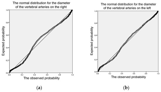
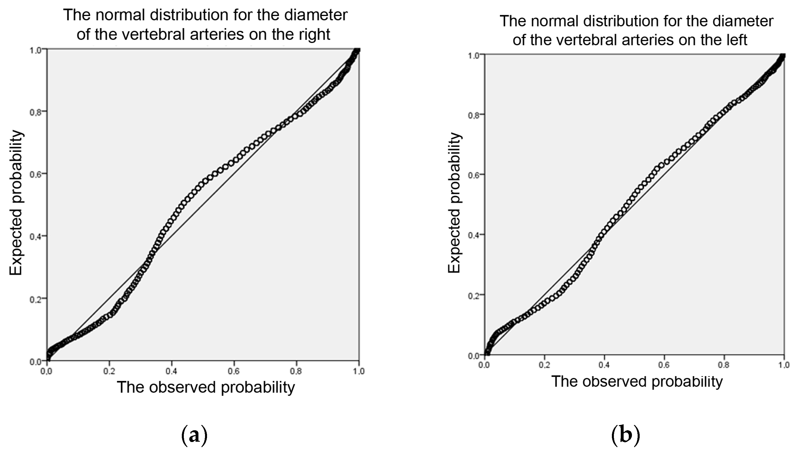
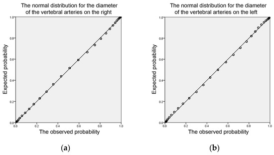
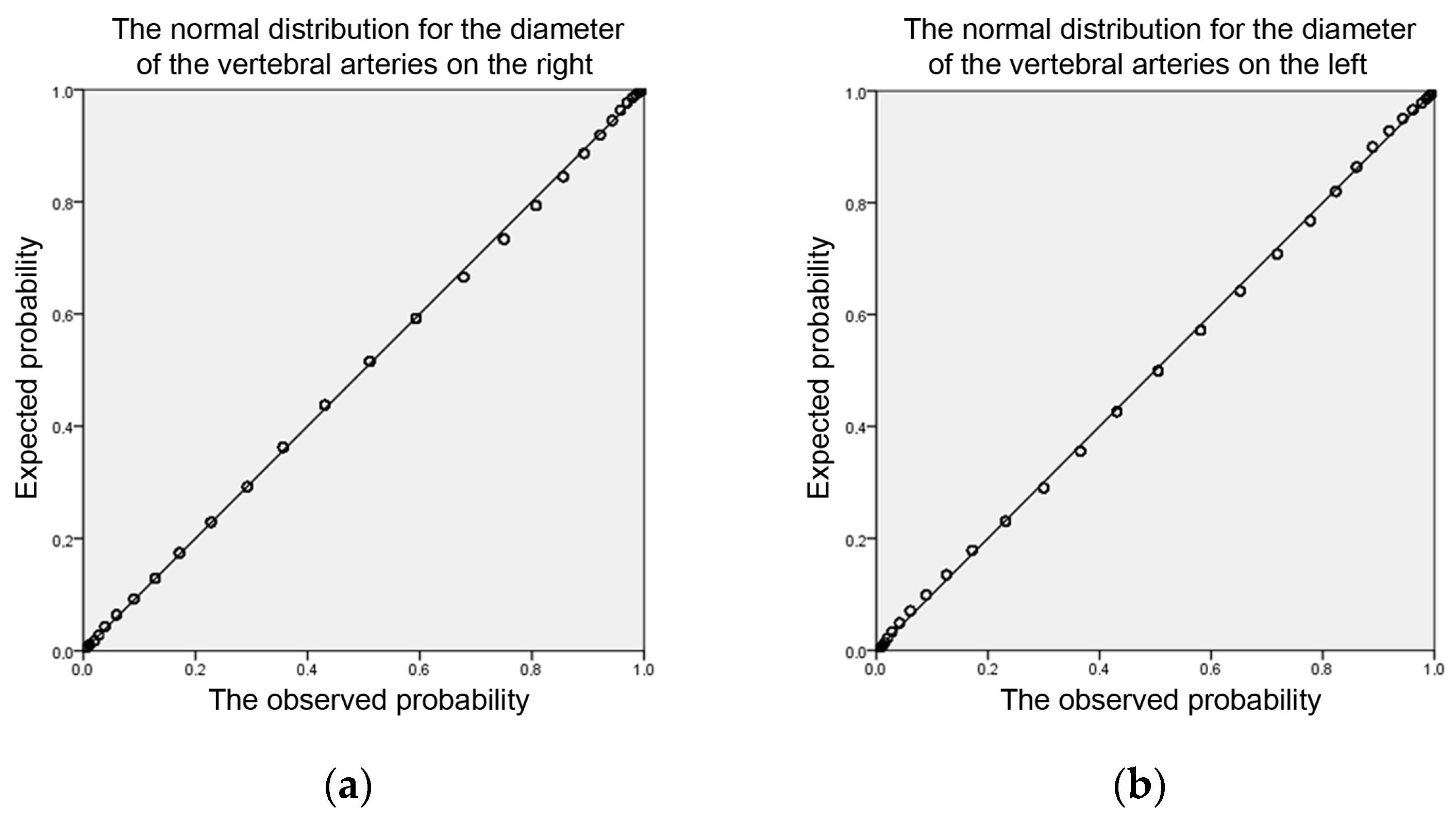
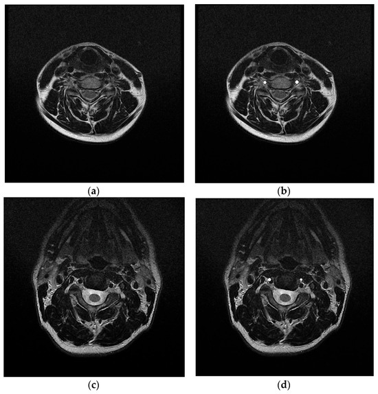
General information about the observation results is presented in Table 2; graphs of the distribution of measurement results are shown in diagrams (Figure 5 and Figure 6). A visual demonstration of the segmentation of the vertebral arteries on MRI scans is shown in Figure 7.
Table 2.
The results of the assessment of the diameter of the vertebral arteries at the level of the second segment by computer analysis of MRI and US imaging.
Figure 5.
Distribution normality diagrams for the diameter of vertebral arteries according to MRI data with AI analysis (mm): (a) Distribution of results for the vertebral artery on the right; (b) Distribution of results for the vertebral artery on the left.
Figure 6.
Distribution normality diagrams for the diameter of vertebral arteries in US imaging (data analysis without AI) (mm): (a) Distribution of results for the vertebral artery on the right; (b) Distribution of results for the vertebral artery on the left.
Figure 7.
Demonstration of the effectiveness of the determination of vertebral arteries on MR tomograms: (a) The original image; (b) The result of segmentation of vertebral arteries; (c) the original image in which a significant segmentation fault occurred; (d) an example of erroneous segmentation in an image.
The differences in the average values for the dimensional characteristics of the studied vessels were 0.11–0.12 mm, due to differences in imaging technologies. The diagrams of the normality of the distribution of measurement results show differences due to the density of values due to the fact that US imaging data in the initial version are presented with an accuracy of tenths, and computer analysis operated with values up to hundredths of a millimeter.
4. Discussion
The use of AI as an assistant in practical healthcare and scientific activities takes an increasingly confident position every year. The introduction of computer image analysis methods is most successful for computer and magnetic resonance imaging [23,24,25]. This is facilitated by relatively standardized research conditions and the rapid accumulation of the necessary dataset for training neural networks.
The process of collecting diagnostic information during US examinations is characterized by significant differences in comparison with the processing of digital radiographs and computer and magnetic resonance imaging data. The easiest way is to save images of organs in standard projections before performing routine measurements. With this method of data collection, images are the clearest, structures are easily identified, and they are easy to read. The disadvantage is training on an “idealized” dataset; as a result, the models will be less sensitive to interpretation when working with video, less suitable for analyzing images from other devices and evaluating images in poor visualization quality; also, they will be of a different level of training (compared to the established methodology) for the specialist performing diagnostics. The next option for collecting diagnostic data is to make a video capture from a working diagnostic device for a certain period of time (a work shift). In this case, a separate workstation with significant storage space for the results is required; difficulties in identifying participants in the video array are inevitable; and with a low level of streaming data, images will be preserved with significant loss of quality. The advantage is that such data collection does not affect the work of the employee performing the research. The third option is to record and save the data as a video loop. Thus, it is possible to prepare the visualization data for target organs in several planes and collect the data from a single patient using various equipment settings (different frequencies, contrast, diagnostic modes, etc.). The data obtained allows us to train a software algorithm for analyzing the results on the fly and makes it possible to reduce the number of study participants, while increasing the importance of the training sample. The disadvantage of this method is an increase in the duration of routine research in the process of collecting information, which requires focused work by the employee performing the survey. Post-processing of a video sequence requires its decomposition into separate frames. Assessing the possibilities of exporting images, it is necessary to take into account the importance of image compression tools for the loss of some information with a significant reduction in the volume of files received. The application of masks can be performed using specialized software (RoboFlow), as well as any available image editors. In our work, we used binary image masks to reduce the amount of training files. Initially, the area of the organ under study (ovary) was isolated and the “mask” of its image was preserved. Subsequently, in the next layer of the graphic editor, the follicles were marked within the boundaries of the selected area. The received data was saved as “*.png” files. The disadvantage of the method was the high time required to complete the training sample and the reduced accuracy of marking the edges of the selected areas (due to manual processing). Thus, our experience of using AI to interpret and analyze real single clinical images of organs vividly demonstrated the strengths and weaknesses of the technology: poor image quality in retrospective analysis can become an insurmountable obstacle; and the specialist, who performs the diagnosis from experience, is only able to perform a limited set of manipulations for standard observation.
The first stage used the YOLOv8 model [26,27,28]. It was used for training on a compiled dataset with the final map of 0.995 demonstrating high accuracy. Noise reduction in images was achieved by using median Gauss filters and a Lie filter. At the same time, the overall information content of the images was significantly reduced; so, instead of filters designed to smooth the image, neural network approaches, trained to improve image quality, were used. One of the most effective architectures turned out to be SwinIR. Using the basic YOLOv8 architecture, without prior preparation of the dataset, demonstrated learning outcomes with values of IoU 0.36, Dice 0.43. To increase the amount of data in the learning process, transformation methods with rotation and reflection of the source data were used. The YOLOv8 model training allowed us to achieve 0.6 mAP50 quality for ovarian detection and segmentation. A simplified scheme for organizing the training of the model is shown in Figure 8.
Figure 8.
The scheme for organizing the training of a model based on ultrasound examination data for segmentation of ovaries and follicles.
The segmentation problem was solved using U-net++ [29]. The main feature of U-net is the use of bandwidth connections. These connections allow passing layers in the encoder with transmission directly to certain levels of the decoder. This way, detailed information about the location of objects in the image is saved, which is especially useful for segmentation. ResNet34 was used as an encoder for the U-net model, which made it possible to achieve a quality of 0.5 IU. In general, various methods of pre-processing US images (highlighting an area using a detection model, magnification, and quality improvement) were developed and implemented as a system; original models of segmentation and detection of ovarian localization, and a set of tools for detecting follicles were developed.
The use of AI technology for segmentation of anatomical structures is currently being actively implemented in the daily work of radiologists. The example, presented in our observation, demonstrates the high efficiency of the technology comparable with other results of instrumental clinical diagnostics. Achieving such high performance became possible due to the formation of a pipeline for preprocessing diagnostic images both at the training stage and directly during image analysis [30,31,32]. This need is due to the complexity of interpreting the results, the wide variety of shapes of anatomical objects, and the wide range of color palettes (even for gray shades of the image). The scheme of organization of model training based on MRI examination data for segmentation of vertebral arteries is shown in Figure 9.
Figure 9.
A simplified scheme for organizing the training of a model based on MRI examination data for segmentation of vertebral arteries.
Preprocessing of MR tomograms was performed by sequential application of filters: Non-local means (NLM), Curvature Flow (CF), Gauss, and Frangi. The segmentation task was implemented using a modified ResNet convolutional architecture [33,34,35]. After combining each individual preprocessing method and the loss function, each instance was trained using an Adam optimizer at a learning rate of 1 × 10⁻³ in 200 epochs. It was then evaluated using precision, recall, Dice, and IoU. This made it possible to raise the values of the model’s learning index from IoU 0.035, Dice 0.068 to IoU 0.595, Dice 0.964.
The beginning of the use of AI methods in medicine was based on the search for characteristic image anomalies, primarily for the detection and differential diagnosis of benign and malignant neoplasms [16,36,37]. To do this, it was necessary to create datasets sufficient for training and based on visualization of certain manifestations of diseases. A certain compromise between the need for an impressive dataset was achieved by convolutional and convolutional neural networks implemented using U-Net technology [11,29].
The growth of computing power of computer equipment, expansion of technological capabilities, range of software solutions, and pre-trained models in the public domain currently allow us to reconsider the role of AI from an expert assistant for the targeted search for a certain pathology to an assistant in solving everyday tasks [38]. The development of advances in computer analysis of diagnostic images with a rich set of graphical data to reduce noise levels and accentuate the necessary indicators represent the beginning of a new era in the clinical diagnosis of pathology, which promises to increase accuracy and efficiency through the use of affordable digital tools with artificial intelligence. Such changes should contribute to a significant increase in the quality of diagnostics and an overall increase in the efficiency of medical service [39,40]. The introduction of AI technologies is necessary and inevitable for the transformation of educational and research activities in accordance with the level of improvement and dissemination of digital tools in every field of our lives [41,42,43]. Currently, the training of specialists in medicine requires the formation of professional skills in working with information systems and the development of experience in analyzing diagnostic images. The use of computer technologies, including methods of intelligent analysis and image generation, make it possible to develop and control the formation of professional skills in the process of training specialists.
5. Conclusions
The results presented demonstrate the high efficiency of AI technology to facilitate routine clinical tasks. An undeniable advantage is the integration of computer technologies into scalable systems that allow us to identify and demonstrate numerous patterns and raise the level of medical diagnostics to a new qualitative level.
A Unified AI Approach for medical images analysis is presented. Example cases of this approach for the detection of ovaries, follicles, and vertebral arteries are presented. It was shown that detection errors in the analysis of 550 US cases did not exceed 6–8% and were associated with technical difficulties due to image quality. When studying 1030 MRI studies, only 0.19% of cases failed to obtain reliable image analysis results. The differences in the average values for the dimensional characteristics of the studied vessels were 0.11–0.12 mm.
In the presented work, we described the experience of using various models to solve similar problems, which allows us to discuss the elements of universality in processing medical diagnostic images obtained from various sources based on empirical experience, comparing the results in the implementation of various systems, and in the future we will certainly continue to study this problem.
Author Contributions
Conceptualization, A.R. and A.N.; methodology, M.F. and A.M.; software, A.M. and A.R.; validation, M.F., O.K., A.N. and D.R.; formal analysis, V.G.; investigation, A.M.; resources, V.A.; data curation, A.M., V.G. and D.R.; writing—original draft preparation, M.F. and A.M.; writing—review and editing, A.R. and A.M.; visualization, A.M.; supervision, A.R. All authors have read and agreed to the published version of the manuscript.
Funding
This research received no external funding.
Institutional Review Board Statement
The authors confirm that they respect the rights of the people who participated in the study, including obtaining informed consent when it is necessary. The study was approved by the Ethics Committee of Orel State University named after I. S. Turgenev, Ministry of Science and Higher Education of Russia (Protocol № 25, 16 November 2022).
Informed Consent Statement
Informed consent was obtained from all subjects involved in the study. Written informed consent has been obtained from the patients to publish this paper.
Data Availability Statement
The raw data supporting the conclusions of this article will be made available by the authors on request.
Conflicts of Interest
The authors declare no conflicts of interest.
Abbreviations
The following abbreviations are used in this manuscript:
| AI | Artificial intelligence |
| MRI | Magnetic resonance imaging |
| US | Ultrasound |
References
- Chen, K.; Shao, A.; Burapacheep, J.; Li, Y. Conversational AI and equity through assessing GPT-3’s communication with diverse social groups on contentious topics. Sci. Rep. 2024, 14, 1561. [Google Scholar] [CrossRef] [Scilit] [PubMed]
- Chun, Y.; Hur, J.; Hwang, J. AI technology specialization and national competitiveness. PLoS ONE 2024, 19, e0301091. [Google Scholar] [CrossRef] [Scilit] [PubMed]
- Sosnin, E.I.; Vasil’ev, Y.L.; Solovyev, R.A.; Stempkovskiy, A.L.; Telpukhov, D.V.; Vasil’ev, A.A.; Amerikanov, A.A.; Romanov, A.Y. AlphaDent: A dataset for automated tooth pathology detection. Comput. Opt. 2025, 49, 1129–1137. [Google Scholar] [CrossRef] [Scilit]
- Zhang, X.E.; Liu, C.; Dai, J.; Yuan, Y.; Gao, C.; Feng, Y.; Wu, B.; Wei, P.; You, C.; Wang, X.; et al. Enabling technology and core theory of synthetic biology. Sci. China Life Sci. 2023, 66, 1742–1785. [Google Scholar] [CrossRef] [Scilit] [PubMed]
- Zhou, J.; Zhang, B.; Li, G.; Chen, X.; Li, H.; Xu, X.; Chen, S.; He, W.; Xu, C.; Liu, L.; et al. An AI Agent for Fully Automated Multi-Omic Analyses. Adv. Sci. 2024, 11, e2407094. [Google Scholar] [CrossRef] [Scilit] [PubMed]
- Gutiérrez-Cirlos, C.; Bermúdez-González, J.L.; Carrillo-Pérez, D.L.; Hidrogo-Montemayor, I.; Martínez-González, A.; Carrillo-Esper, R.; Sánchez-Mendiola, M. Medicine and the metaverse: Current applications and future. Gac. Med. Mex. 2023, 159, 280–286. [Google Scholar] [CrossRef] [Scilit] [PubMed]
- Tam, W.; Huynh, T.; Tang, A.; Luong, S.; Khatri, Y.; Zhou, W. Nursing education in the age of artificial intelligence powered Chatbots (AI-Chatbots): Are we ready yet? Nurse Educ. Today 2023, 129, 105917. [Google Scholar] [CrossRef] [Scilit] [PubMed]
- Sami, A.; Tanveer, F.; Sajwani, K.; Kiran, N.; Javed, M.A.; Ozsahin, D.U.; Muhammad, K.; Waheed, Y. Medical students’ attitudes toward AI in education: Perception, effectiveness, and its credibility. BMC Med. Educ. 2025, 25, 82. [Google Scholar] [CrossRef] [Scilit] [PubMed]
- Jackson, P.; Ponath Sukumaran, G.; Babu, C.; Tony, M.C.; Jack, D.S.; Reshma, V.R.; Davis, D.; Kurian, N.; John, A. Artificial intelligence in medical education—Perception among medical students. BMC Med. Educ. 2024, 24, 804. [Google Scholar] [CrossRef] [Scilit] [PubMed]
- Kents, A.S.; Hamad, Y.A.; Simonov, K.V. Approbation of the shiarlet transformation technique for visualization of pathological lung changes on CT images for diagnosis of COVID-19. Med. High Technol. 2021, 3, 5–13. [Google Scholar] [CrossRef] [Scilit]
- Moshkina, L.V.; Novikov, M.S.; Bochkarev, A.B.; Sheverdin, N.N.; Stupin, A.S. Analysis of the effectiveness of computer vision methods in assessing follicular reserve. Tver Med. J. 2024, 6, 84–88. [Google Scholar]
- Laputin, F.A.; Sidorov, I.V.; Moshkin, A.S. Determination of ovarian follicular reserve according to ultrasound examination based on machine learning methods. Digit. Diagn. 2024, 5, 40–42. [Google Scholar] [CrossRef] [Scilit]
- Kingma, D.P.; Ba, J.L. Adam: A Method for Stochastic Optimization. arXiv 2014, arXiv:1412.6980. [Google Scholar] [CrossRef] [Scilit]
- Kourounis, G.; Elmahmudi, A.A.; Thomson, B.; Hunter, J.; Ugail, H.; Wilson, C. Computer image analysis with artificial intelligence: A practical introduction to convolutional neural networks for medical professionals. Postgrad. Med. J. 2023, 99, 1287–1294. [Google Scholar] [CrossRef] [Scilit] [PubMed]
- Liang, J.; Cao, J.; Sun, G.; Zhang, K.; Van Gool, L.; Timofte, R. SwinIR: Image restoration using swin transforme. In Proceedings of the 2021 IEEE/CVF International Conference on Computer Vision Workshops (ICCVW), Montreal, BC, Canada, 11–17 October 2021; pp. 1833–1844. [Google Scholar] [CrossRef] [Scilit]
- Tran, K.A.; Kondrashova, O.; Bradley, A.; Williams, E.D.; Pearson, J.V.; Waddell, N. Deep learning in cancer diagnosis, prognosis and treatment selection. Genome Med. 2021, 13, 152. [Google Scholar] [CrossRef] [Scilit] [PubMed]
- Sung, J. Artificial intelligence in medicine: Ethical, social and legal perspectives. Ann. Acad. Med. Singap. 2023, 52, 695–699. [Google Scholar] [CrossRef] [Scilit] [PubMed]
- Weidener, L.; Fischer, M. Teaching AI Ethics in Medical Education: A Scoping Review of Current Literature and Practices. Perspect. Med. Educ. 2023, 12, 399–410. [Google Scholar] [CrossRef] [Scilit] [PubMed]
- Hofmann, B. The role of philosophy and ethics at the edges of medicine. Philos. Ethics Humanit. Med. 2021, 16, 14. [Google Scholar] [CrossRef] [Scilit] [PubMed]
- Shafi, S.; Parwani, A.V. Artificial intelligence in diagnostic pathology. Diagn. Pathol. 2023, 18, 109. [Google Scholar] [CrossRef] [Scilit] [PubMed]
- Thangaraj, P.M.; Benson, S.H.; Oikonomou, E.K.; Asselbergs, F.W.; Khera, R. Cardiovascular care with digital twin technology in the era of generative artificial intelligence. Eur. Heart J. 2024, 45, 4808–4821. [Google Scholar] [CrossRef] [Scilit] [PubMed]
- Huang, W.; Shu, N. AI-powered integration of multimodal imaging in precision medicine for neuropsychiatric disorders. Cell Rep. Med. 2025, 6, 102132. [Google Scholar] [CrossRef] [Scilit] [PubMed]
- Quanyang, W.; Yao, H.; Sicong, W.; Linlin, Q.; Zewei, Z.; Donghui, H.; Hongjia, L.; Shijun, Z. Artificial intelligence in lung cancer screening: Detection, classification, prediction, and prognosis. Cancer Med. 2024, 13, e7140. [Google Scholar] [CrossRef] [Scilit] [PubMed]
- de Jong, C.M.M.; Kroft, L.J.M.; van Mens, T.E.; Huisman, M.V.; Stöger, J.L.; Klok, F.A. Modern imaging of acute pulmonary embolism. Thromb. Res. 2024, 238, 105–116. [Google Scholar] [CrossRef] [Scilit] [PubMed]
- Dayarathna, S.; Islam, K.T.; Uribe, S.; Yang, G.; Hayat, M.; Chen, Z. Deep learning based synthesis of MRI, CT and PET: Review and analysis. Med. Image Anal. 2024, 92, 103046. [Google Scholar] [CrossRef] [Scilit] [PubMed]
- Ruiz, A.J.; Hernández Torres, S.I.; Snider, E.J. Development of Deep Learning Models for Real-Time Thoracic Ultrasound Image Interpretation. J. Imaging 2025, 11, 222. [Google Scholar] [CrossRef] [Scilit] [PubMed]
- Sahoo, P.K.; Gupta, P.; Lai, Y.C.; Chiang, S.F.; You, J.F.; Onthoni, D.D.; Chern, Y.J. Localization of Colorectal Cancer Lesions in Contrast-Computed Tomography Images via a Deep Learning Approach. Bioengineering 2023, 10, 972. [Google Scholar] [CrossRef] [Scilit] [PubMed]
- Cascella, M.; Shariff, M.N.; Lo Bianco, G.; Monaco, F.; Gargano, F.; Simonini, A.; Ponsiglione, A.M.; Piazza, O. Employing the Artificial Intelligence Object Detection Tool YOLOv8 for Real-Time Pain Detection: A Feasibility Study. J. Pain Res. 2024, 17, 3681–3696. [Google Scholar] [CrossRef] [Scilit] [PubMed]
- Zhou, Z.; Rahman Siddiquee, M.M.; Tajbakhsh, N.; Liang, J. Unet++: A nested u-net architecture for medical image segmentation. In Deep Learning in Medical Image Analysis and Multimodal Learning for Clinical Decision Support; Lecture Notes in Computer Science (Including Subseries Lecture Notes in Artificial Intelligence and Lecture Notes in Bioinformatics); Springer: Cham, Switzerland, 2018; Volume 11045, pp. 3–11. [Google Scholar] [CrossRef] [Scilit] [PubMed]
- Abdellatif, H.; Al Mushaiqri, M.; Albalushi, H.; Al-Zaabi, A.A.; Roychoudhury, S.; Das, S. Teaching, Learning and Assessing Anatomy with Artificial Intelligence: The Road to a Better Future. Int. J. Environ. Res. Public Health 2022, 19, 14209. [Google Scholar] [CrossRef] [Scilit] [PubMed]
- Jin, J.; Zhu, H.; Teng, Y.; Ai, Y.; Xie, C.; Jin, X. The Accuracy and Radiomics Feature Effects of Multiple U-net-Based Automatic Segmentation Models for Transvaginal Ultrasound Images of Cervical Cancer. J. Digit. Imaging 2022, 35, 983–992. [Google Scholar] [CrossRef] [Scilit] [PubMed]
- Lin, T.Y.; Goyal, P.; Girshick, R.; He, K.; Dollar, P. Focal Loss for Dense Object Detection. IEEE Trans. Pattern Anal. Mach. Intell. 2020, 42, 318–327. [Google Scholar] [CrossRef] [Scilit] [PubMed]
- Zhao, S.; Xiao, F.; Griffith, J.F.; Li, R.; Chen, W. Denoising of volumetric magnetic resonance imaging using multi-channel three-dimensional convolutional neural network with applications on fast spin echo acquisitions. Quant. Imaging Med. Surg. 2024, 14, 6517–6530. [Google Scholar] [CrossRef] [Scilit] [PubMed]
- Golub, I.S.; Thummala, A.; Morad, T.; Dhaliwal, J.; Elisarraras, F.; Karlsberg, R.P.; Cho, G.W. Artificial Intelligence in Nuclear Cardiac Imaging: Novel Advances, Emerging Techniques, and Recent Clinical Trials. J. Clin. Med. 2025, 14, 2095. [Google Scholar] [CrossRef] [Scilit] [PubMed]
- Tayyil Purayil, A.L.; Joseph, R.M.; Raj, A.; Kooriyattil, A.; Jabeen, N.; Beevi, S.F.; Lathief, N.; Latheif, F. Role of Artificial Intelligence in MRI-Based Rectal Cancer Staging: A Systematic Review. Cureus 2024, 16, e76185. [Google Scholar] [CrossRef] [Scilit] [PubMed]
- Lyutkin, D.A.; Romanov, A.Y.; Nasonov, N.D. Segmenting prostate cancer on TRUS images with a small dataset: A comprehensive methodology. In Proceedings of the 2023 International Russian Smart Industry Conference (SmartIndustryCon), Sochi, Russia, 27–31 March 2023; pp. 454–459. [Google Scholar] [CrossRef] [Scilit]
- Wang, X.; Zhao, J.; Marostica, E.; Yuan, W.; Jin, J.; Zhang, J.; Li, R.; Tang, H.; Wang, K.; Li, Y.; et al. A pathology foundation model for cancer diagnosis and prognosis prediction. Nature 2024, 634, 970–978. [Google Scholar] [CrossRef] [Scilit] [PubMed]
- MacEachern, S.J.; Forkert, N.D. Machine learning for precision medicine. Genome 2021, 64, 416–425. [Google Scholar] [CrossRef] [Scilit] [PubMed]
- Raguram, A.; Banskota, S.; Liu, D.R. Therapeutic in vivo delivery of gene editing agents. Cell 2022, 185, 2806–2827. [Google Scholar] [CrossRef] [Scilit] [PubMed]
- Knudsen, J.E.; Ghaffar, U.; Ma, R.; Hung, A.J. Clinical applications of artificial intelligence in robotic surgery. J. Robot. Surg. 2024, 18, 102. [Google Scholar] [CrossRef] [Scilit] [PubMed]
- Zhang, D.Y.; Venkat, A.; Khasawneh, H.; Sali, R.; Zhang, V.; Pei, Z. Implementation of Digital Pathology and Artificial Intelligence in Routine Pathology Practice. Lab. Investig. 2024, 104, 102111. [Google Scholar] [CrossRef] [Scilit] [PubMed]
- Li, Q.; Qin, Y. AI in medical education: Medical student perception, curriculum recommendations and design suggestions. BMC Med. Educ. 2023, 23, 852. [Google Scholar] [CrossRef] [Scilit] [PubMed]
- Xu, Y.; Jiang, Z.; Ting, D.S.W.; Kow, A.W.C.; Bello, F.; Car, J.; Tham, Y.C.; Wong, T.Y. Medical education and physician training in the era of artificial intelligence. Singap. Med. J. 2024, 65, 159–166. [Google Scholar] [CrossRef] [Scilit] [PubMed]

Disclaimer/Publisher’s Note: The statements, opinions and data contained in all publications are solely those of the individual author(s) and contributor(s) and not of MDPI and/or the editor(s). MDPI and/or the editor(s) disclaim responsibility for any injury to people or property resulting from any ideas, methods, instructions or products referred to in the content. |
© 2026 by the authors. Licensee MDPI, Basel, Switzerland. This article is an open access article distributed under the terms and conditions of the Creative Commons Attribution (CC BY) license.