


Abstract
Arthroscopic-assisted reduction is one recommended technique for pediatric distal tibial epiphyseal fractures. A 15-year-old male patient with a triplane distal tibial epiphyseal fracture (Tillaux type) was scheduled for arthroscopic reduction and pinning, which failed to provide adequate fracture reduction. Open surgery was subsequently performed, which revealed a capsular interposition in the fracture line attributable to an anterior capsular tear of the ankle. The possibility of capsular interposition should always be taken into consideration when problems such as those described in this case presentation are encountered in such patients. This is precisely why direct visualization either through arthroscopy or open arthrotomy is sometimes necessary.
Distal tibial epiphyseal fracture is common among other epiphyseal fractures in the pediatric population.[1] The main objective of epiphyseal fracture treatment is to avoid any damage to the physis. Invasive procedures are consequently avoided in the treatment of epiphyseal fractures, although anatomical restoration is necessary in order to permit the continued growth of the epiphyseal plate.[2] Closed reductions with fixation[3,4] or arthroscopic-assisted fixation[5–9] have previously been described as treatments for distal tibial epiphyseal fractures.
In epiphyseal fractures, the tissues surrounding the fracture line can be interposed because of separation of the physeal line[10–14]; this can cause an arrest of growth in the epiphyseal articulation.[15] We present a case of triplane distal tibial epiphyseal fracture (Tillaux type) that was scheduled for arthroscopic-assisted pinning but was subsequently treated by an open reduction and fixation because of the interposition of the anterior ankle capsule.
Case Report
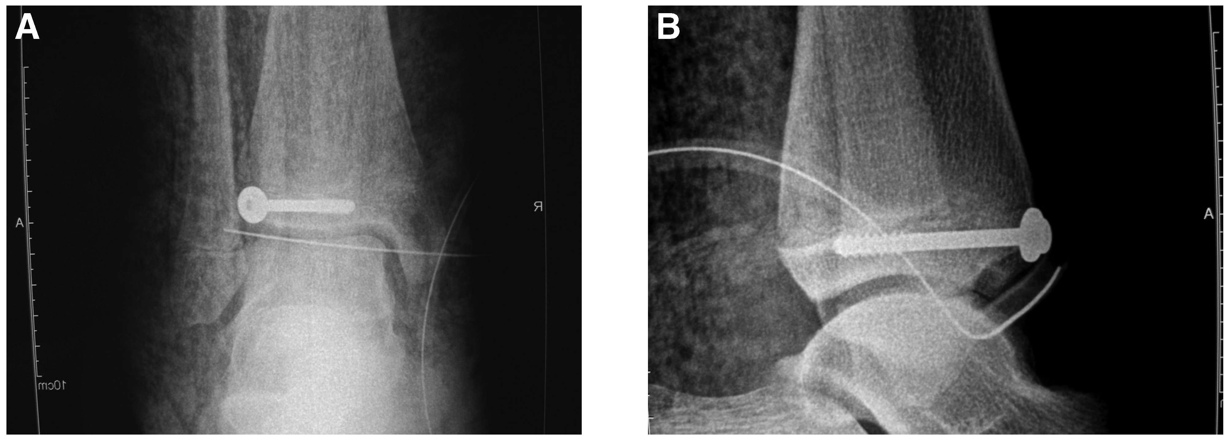
A 15-year-old male was admitted with pain and swelling in his left ankle. He described an ankle inversion sustained in a soccer tackle. A physical examination and radiographs revealed a triplane epiphyseal fracture (Tillaux type) in the distal tibia (Fig. 1A and B). An arthroscopic-assisted technique was planned prior to surgery. After the standard anteromedial entry to the ankle joint, however, it was noticed that the passage of the scope to the lateral compartment of the ankle was impeded. There was a mechanical blockade and a sudden swelling in the anterior compartment of the leg. In view of the extensive damage to the joint capsule, open surgery with an anterior longitudinal incision was performed. It was observed that the anterior capsule had been torn with the resulting tissue margins invading the fracture line (Fig. 2). The interposing capsule was removed and fixation was established with a cancellous screw (Fig. 3A and B).
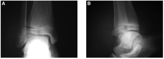
Figure 1.
Preoperative anteroposterior (A) and lateral (B) radiographs of the distal tibial epiphyseal fracture.
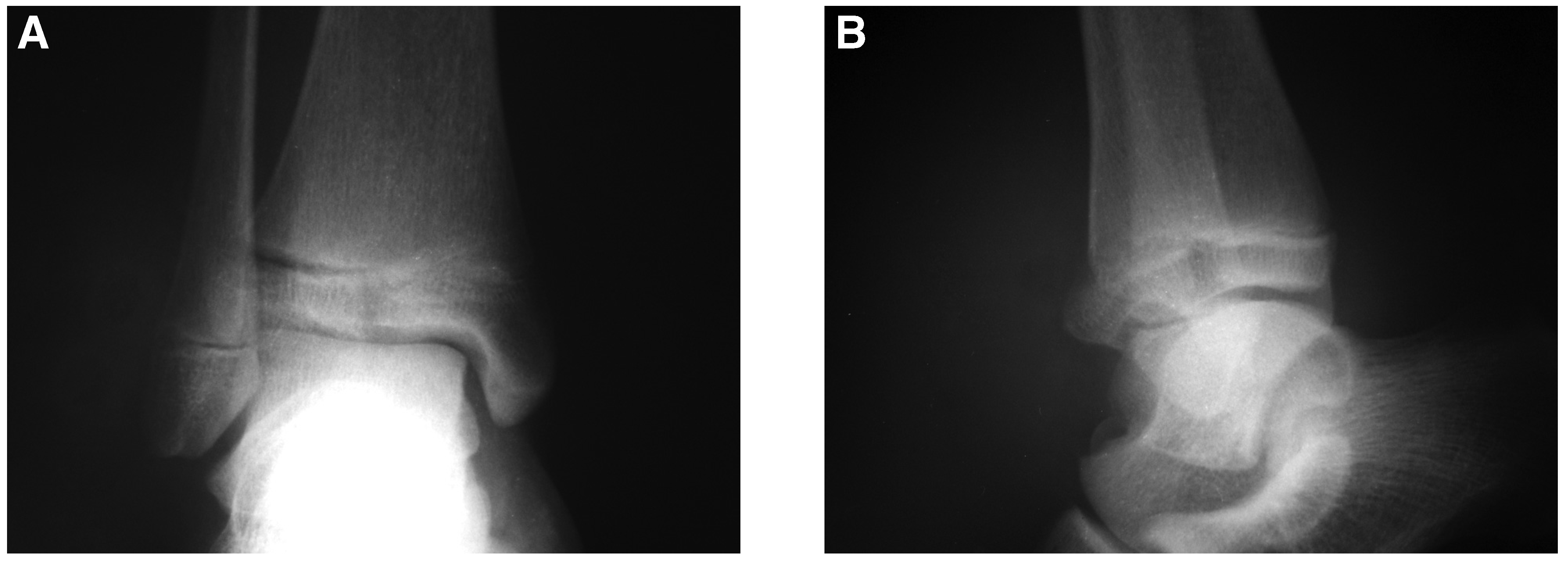
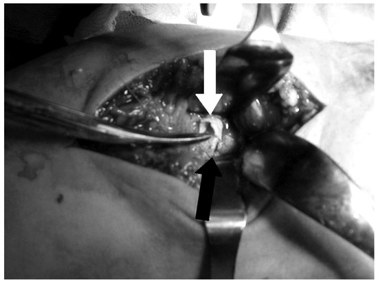
Figure 2.
A photograph showing the capsular interposition. The black arrow indicates the fracture line; the white arrow, the interposed capsule.
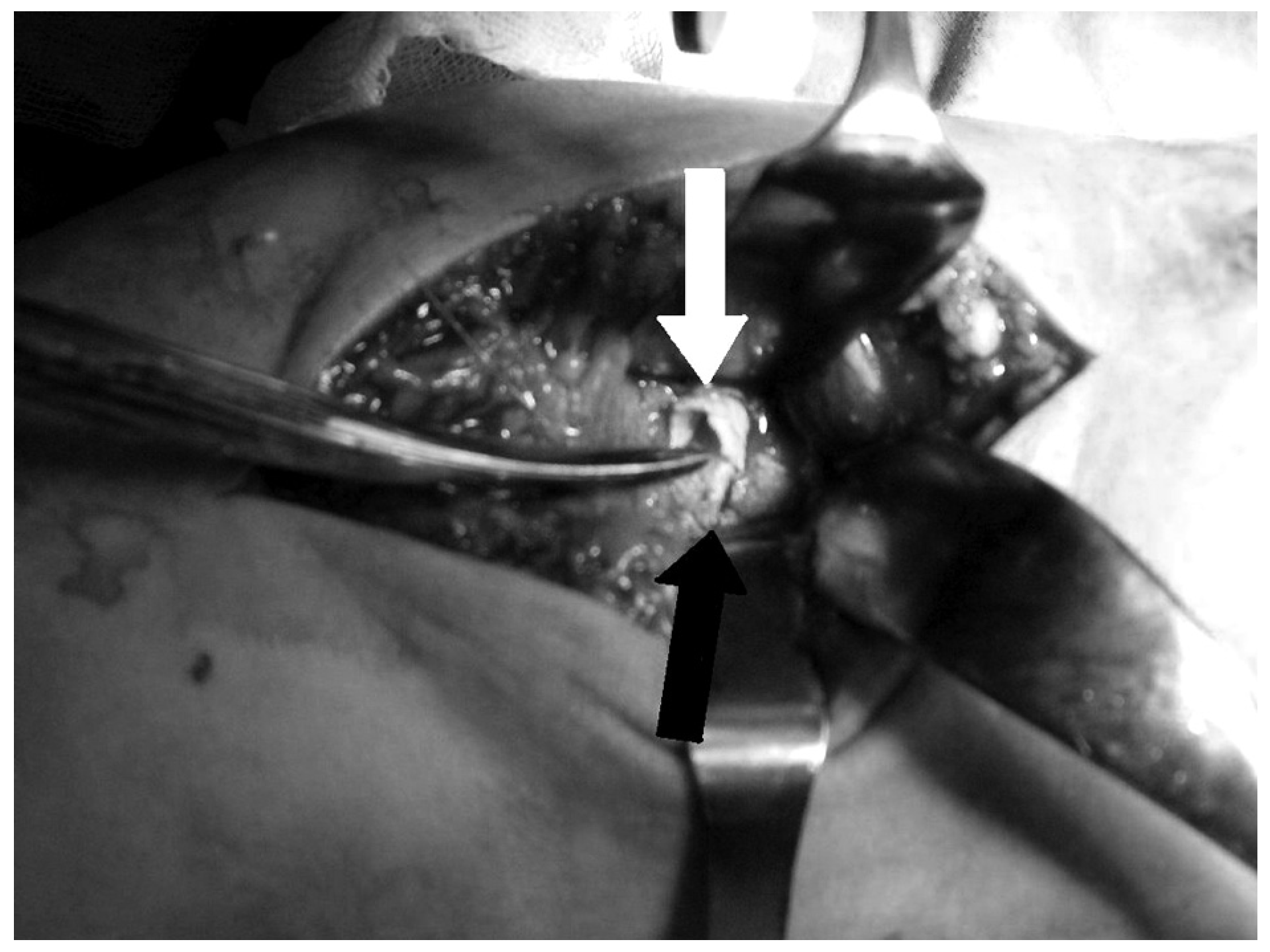
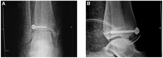
Figure 3.
Preoperative anteroposterior (A) and lateral (B) radiographs of the distal tibial epiphyseal fracture.
Discussion
It is not uncommon for soft tissue or capsular material to become interposed within intra-articular fractures.[10–14] This possibility should always be taken into consideration because some fracture types encourage surgeons to undertake closed reduction. In the presence of an interposition, however, a closed reduction maneuver may damage the physis.
Arthroscopic intervention may be used in the case of intra-articular epiphyseal fractures to confirm absence of soft-tissue interposition.[5–9] On the other hand, inadequate reduction has been reported, thus questioning the application of certain reduction techniques.[7–9] No case of capsular interposition to the fracture line was reported in these cases. Nevertheless, one should always consider the possibility of a capsular tear in the presence of an intra-articular mechanical blockade or extra-articular fluid leak encountered during ankle arthroscopy. If there is a problem with reduction, this may alert the surgeon to the need for removal of the capsular interposition, which may in turn necessitate open surgery.
We presented a case of Tillaux fracture with a capsular interposition to the fracture line, which was associated with an intra-articular mechanical blockade and extra-articular fluid leakage noticed during arthroscopic intervention.
Financial Disclosure
None reported.
Conflicts of Interest
None reported.
References
- Goldberg VM, Aadalen R: Distal tibial epiphyseal injuries: the role of athletics in fifty-three cases. Am J Sports Med6: 263, 1978.
- Herring MD: “General Principles of Managing Orthopaedic Injuries,” in Tachdjian’s Pediatric Orthopaedics, 4th Ed, Vol 3, ed by MD Herring, p 2355, Saunders, Philadelphia, 2008.
- Schlesinger I, Wedge JH: Percutaneous reduction and fixation of displaced juvenile Tillaux fractures: a new surgical technique. J Pediatr Orthop13: 389, 1993.
- Manderson EL, Ollivierre CO: Closed anatomic reduction of a juvenile tillaux fracture by dorsiflexion of the ankle. A case report. Clin Orthop Relat Res276: 262, 1992.
- Miller MD: Arthroscopically assisted reduction and fixation of an adult Tillaux fracture of the ankle. Arthroscopy13: 117, 1997.
- Leetun DT, Ireland ML: Arthroscopically assisted reduction and fixation of a juvenile Tillaux fracture. Arthroscopy18: 427, 2002.
- Panagopoulos A, van Niekerk L: Arthroscopic assisted reduction and fixation of a juvenile Tillaux fracture. Knee Surg Sports Traumatol Arthrosc15: 415, 2007.
- Thaunat M, Billot N, Bauer T, et al: Arthroscopic treatment of a juvenile tillaux fracture. Knee Surg Sports Traumatol Arthrosc15: 286, 2007.
- Jennings MM, Lagaay P, Schuberth JM: Arthroscopic assisted fixation of juvenile intra-articular epiphyseal ankle fractures. J Foot Ankle Surg46: 376, 2007.
- Visser JD, Rietberg M: Interposition of the tendon of the long head of biceps in fracture separation of the proximal humeral epiphysis. Neth J Surg32: 12, 1980.
- Grace DL: Irreducible fracture-separations of the distal tibial epiphysis. J Bone Joint Surg Br65: 160, 1983.
- Wood KB, Bradley JP, Ward WT: Pes anserinus interposition in a proximal tibial physeal fracture. A case report. Clin Orthop Relat Res264: 239, 1991.
- Faraj AA, Kumar MS, Ketzer B, et al: An irreducible Salter-Harris type IV distal ulna fracture. Injury31: 746, 2000.
- Beslikas TA, Dadoukis DJ, Nenopoulos SP, et al: A completely displaced Salter-Harris I fracture of the distal radial epiphysis with intact ulna: a case report and review of the literature. Acta Chir Belg105: 539, 2005.
- Phieffer LS, Meyer RA Jr, Gruber HE, et al: Effect of interposed periosteum in an animal physeal fracture model. Clin Orthop Relat Res376: 15, 2000.
© 2009 American Podiatric Medical Association