


Abstract
We report an unusual case of a variant of Lisfranc injury, plantar dislocation of the medial cuneiform with plantar fracture-dislocation of the intermediate cuneiform and dorsal fracture-dislocation of the lateral cuneiform, which has never been reported, to our knowledge. The entire pathologic abnormality was treated by open reduction and fixation with Kirschner wires, which were removed 8 weeks postoperatively because of pin-tract infection. Complex regional pain syndrome, which was a problem early in the recovery process, is now in remission, and at the 25-month follow-up examination, the patient was almost symptom free.
Plantar dislocation of the medial cuneiform in Lisfranc joint complex injury is a rare variant. Only one case of plantar medial subluxation of the medial cuneiform has been reported,[1] and no frank dislocation of the medial cuneiform medially, plantar to the navicular bone, has been described. Plantar fracture-dislocation of the intermediate cuneiform is also rare; Doshi et al[2] recently reported a dorsal type.
According to the classification of Myerson[3] for Lisfranc fracture-dislocations, the injury types are divided into three groups: type A, total incongruity; type B, partial incongruity; and type C, divergent. Type B is further subclassified as B1, in which only the medial ray is involved, and B2, in which one or more of the lesser metatarsals are affected. Type C is further subclassified as C1, in which the first metatarsal is displaced medially and partial incongruity of the lesser four metatarsals is seen, and C2, which shows the diversion pattern with total incongruity of the lesser metatarsals.
The aims of this study are to report a rare Lisfranc joint complex injury treated with open reduction and Kirschner wire fixation and to discuss the mechanisms, treatment options, and associated problems. In a search of the literature, we did not encounter any cases of partially divergent (Myerson type C1) Lisfranc fracture-dislocation with an associated injury pattern of plantar dislocation of the medial cuneiform, plantar fracture-dislocation of the intermediate cuneiform, and dorsal fracture-dislocation of the lateral cuneiform.
Case Report
A 30-year-old man was brought to the hospital emergency department with an injury to his left foot caused by a motor vehicle crash. His foot was swollen and diffusely tender. There were superficial abrasions and ecchymosis on the dorsolateral surface of the foot. Neurovascular examination demonstrated intact sensation and pulses. Radiographic examination revealed a Myerson type C1 Lisfranc joint fracture-dislocation associated with medial cuneiform plantar dislocation and lateral cuneiform dorsal dislocation on the nonweightbearing anteroposterior and lateral views (Fig. 1). Computed tomography demonstrated that the medial cuneiform was dislocated in a plantar direction under the navicular bone, and the intermediate cuneiform was comminuted and displaced. Some of the fragments of the intermediate cuneiform were at the posterosuperior part of the medial cuneiform. Moreover, the medial articular surface of the lateral cuneiform was fractured and dislocated dorsally (Fig. 2).
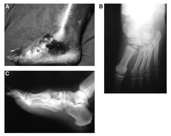
Figure 1.
Myerson type C1 Lisfranc joint fracture-dislocation associated with medial cuneiform plantar dislocation and lateral cuneiform dorsal dislocation. A, Clinical view of the left foot after trauma. B, Preoperative anteroposterior radiographic view. C, Preoperative lateral radiographic view.
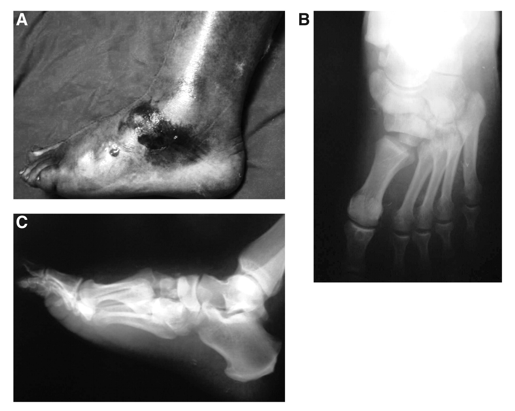
Figure 2.
Preoperative computed tomography. C indicates cuboid; IC, intermediate cuneiform; LC, lateral cuneiform; MC, medial cuneiform; 5MT, fifth metatarsal; and N, navicular.
Because the current literature advocates operative management,[4] we did not perform closed reduction to avoid the risk of further soft-tissue injury. We used the surgical approach, in which two dorsal longitudinal incisions, between the first and second metatarsal interspaces and over the fourth metatarsal bone were made. Through the first incision, the tibialis anterior tendon was found to be interposed between the medial cuneiform and navicular bones. After excision of the displaced small intermediate cuneiform fragments, the medial cuneiform was reduced and fixed by a Kirschner wire passing through the first metatarsal base to the navicular bone. Through the second incision, the third and fourth metatarsal bones were found to be dislocated dorsally, the medial articular surface of the lateral cuneiform was fractured and rotated dorsally, and the fracture of the intermediate cuneiform was detected. Anatomical reduction was performed to the lateral cuneiform and after excision of some of the small fragments blocking reduction, the remaining body of the intermediate cuneiform was then reduced. The first Kirschner wire was placed through the first metatarsal to the lateral cuneiform and the cuboid; the second wire went through the second metatarsal to the intermediate cuneiform and the navicular; the third wire went through the fifth metatarsal to the lateral cuneiform and the navicular. The final Kirschner wire was placed transversely through the first metatarsal to the third metatarsal (Fig. 3 A and B).
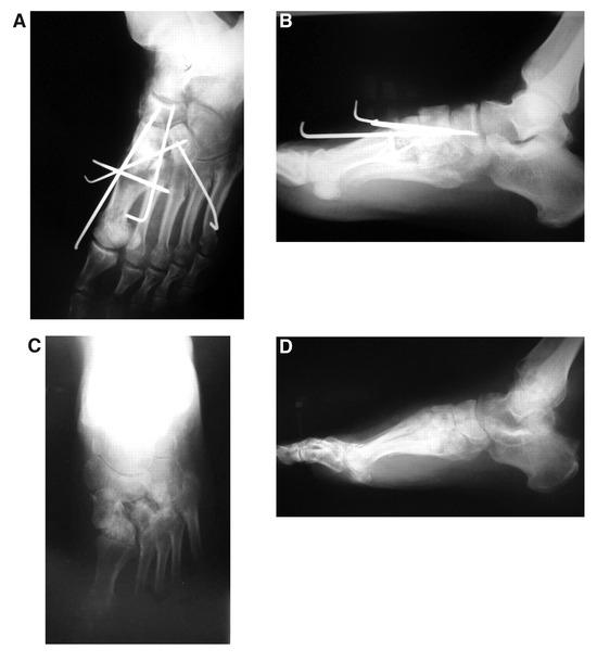
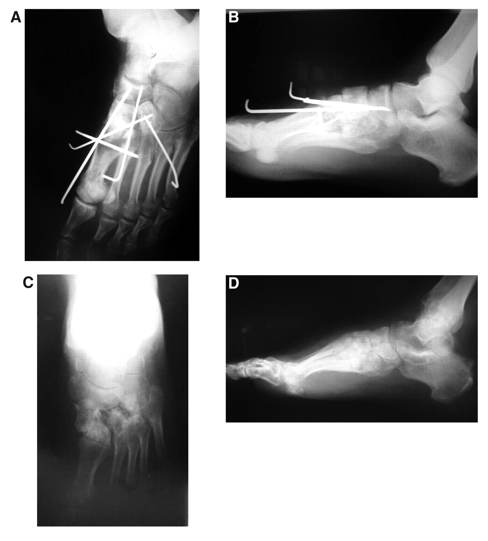
Figure 3.
A, Postoperative anteroposterior radiographic view. B, Postoperative lateral radiographic view. C, Anteroposterior radiographic view at postoperative month 25. D, Lateral radiographic view at postoperative month 25.
The leg was protected in a nonweightbearing cast-brace. All of the hardware, except the Kirschner wire through the first metatarsal to the medial cuneiform and the navicular, had to be removed 8 weeks postoperatively because of superficial pin-tract infection. Methicillin-sensitive Staphylococcus aureus was cultured from the pin tracts, and the patient was treated with the appropriate antibiotic drugs. Because complex regional pain syndrome type 1 was developing, calcitonin and calcium treatments were initiated, and the patient was allowed to begin partial weightbearing with a below-the-knee cast. Two weeks later, the remaining Kirschner wire was removed. The pin tracts all healed uneventfully. The patient then started walking in a shoe with an arch support in place.
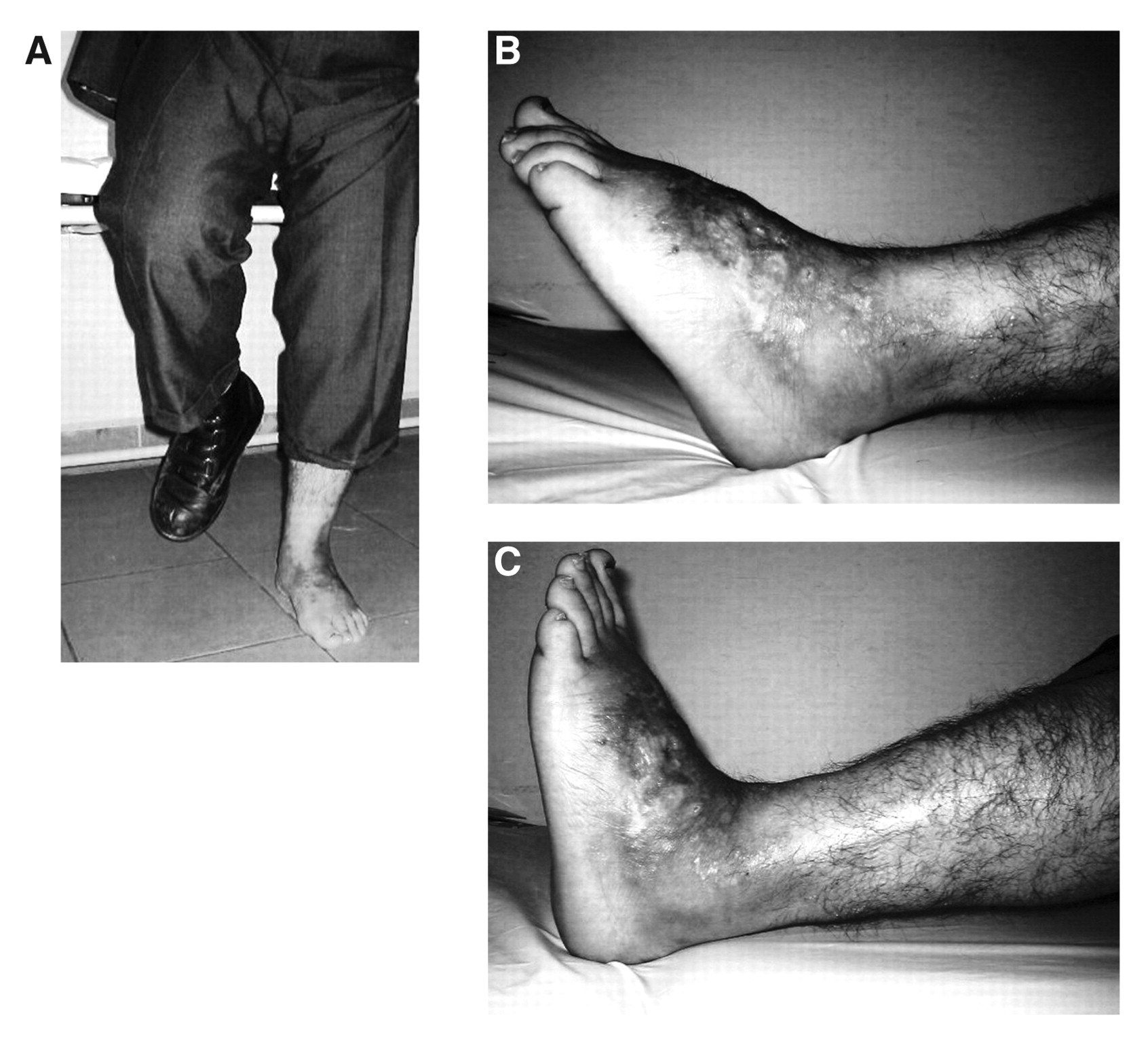
At the 25-month follow-up, radiographic images showed good anatomical alignment (Fig. 3 C and D). The American Orthopaedic Foot and Ankle Society midfoot score was 76. The medial longitudinal arch of the patient’s foot was restored, full range of motion of the ankle was achieved, and the only problem he has is minor plantar foot pain after vigorous activities (Fig. 4).
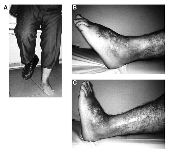
Figure 4.
Functional motion capacity of the ankle and foot at postoperative month 25. A, Medial longitudinal arch of the patient’s foot was restored. B, Full range of plantarflexion. C, Satisfactory dorsal flexion of the ankle was achieved.
Discussion
Tarsometatarsal fractures are sometimes associated with cuneiform, cuboid, and navicular fracture-dislocations, so Myerson[3] proposed to call them “Lisfranc joint complex” injuries. Vuori and Aro[5] reported the rate of midtarsal fracture-dislocations associated with Lisfranc joint injuries to be 39% and found that this association rate increases especially with high-energy trauma. Two mechanisms of injury were described for high-energy trauma: direct and indirect loading.[6] Direct loading is usually in the manner of a crush injury to the dorsal surface of the foot. Indirect loading is a more common mechanism. It is characterized by longitudinal loading of a plantarflexed foot, which causes hyperplantarflexion, disrupting first the dorsal ligaments and then the plantar. The presence of secondary forces, which is common in motor vehicle crashes, defines the final injury pattern. In the present case, the patient as the driver of the motor vehicle seems to be affected by the classic pedal injury, which is a dorsally directed force. There should also be secondary forces because the bones and the soft tissues were highly affected.
Cuneiform dislocations are reported to be few in number. Fujita et al[7] reported an isolated intermediate cuneiform dislocation. There were 13 intermediate cuneiform dislocations reported in the literature, and only three of them were plantar dislocations.[7] Levine et al[8] reported an isolated medial cuneiform plantar dislocation, and they encountered only one similar case in their literature review. Rareness of medial cuneiform plantar dislocation depends on the rigid anatomical structure. The medial cuneiform, articulating with the navicular, the intermediate cuneiform, and the first and second metatarsals, is located on a thick and extremely strong plantar ligament. It is reinforced by tibialis posterior, tibialis anterior, and peroneus longus tendon insertions and has strong ligamentous and capsular attachments to the navicular through naviculocuneiform ligaments.[8] However, the Lisfranc ligament, connecting the medial cuneiform to the second metatarsal basis, is the main stabilizer of the tarsometatarsal joint.[3] Therefore, because of such strong ligamentous structures, plantar and interosseous ligaments must be disrupted for plantar dislocation of the medial cuneiform.[7,8]
Stable anatomical reduction is important to obtain maximum success in the treatment of Lisfranc joint complex injuries.[9] Kuo et al[10] found an increasing risk of secondary osteoarthritis in tarsometatarsal fracture-dislocations that are not reduced anatomically. However, even after satisfactory reduction and fixation, forefoot stiffness, loss of metatarsal arch, intrinsic contracture, reflex sympathetic dystrophy, and causalgia (complex regional pain syndrome types 1 and 2) can be a problem.[9] For fixation, proponents of both Kirschner wire and screw fixation exist in the literature.[3] The advantages of Kirschner wire fixation include its ease of insertion and removal; disadvantages are risk of infection, pin migration, and loss of reduction. The advantage of screw fixation is improved fixation, and the main disadvantages are removal and difficult surgery. In this case, wires were used because of communition of the cuneiform and to decrease operating time. The pin-tract infection became a problem. Because of the inability to achieve rigid fixation, complex regional pain syndrome, which was seen in this case, may be triggered by the relatively early removal of the wires or by the original injury causing traction injury to the superficial and deep peroneal nerves. Adelaar[9] warned that with injuries to the dorsal surface, the superficial peroneal nerve, which can be injured easily, can be a source of causalgia (complex regional pain syndrome type 2).
In the present case, we performed open reduction and internal fixation and tried to maintain the normal foot anatomy as much as possible. Recently, Ly and Coetzee[11] reported superior short- and medium-term outcomes with primary arthrodesis than with open reduction and internal fixation; however, the series consisted of patients who had pure ligamentous Lisfranc joint injury, and it is generally accepted that this subgroup of patients has worse outcomes with open reduction and internal fixation.[10] Mulier et al[12] advocated open reduction and internal fixation or partial arthrodesis as a treatment of choice in severe tarsometatarsal fracture-dislocations while keeping primary complete arthrodesis as a salvage procedure. In the present patient, an active young man, we tried to achieve anatomical reduction, and because follow-up was relatively short, we did not see any osteoarthritis. If in longer-term follow-up osteoarthritis is seen, arthrodesis may be needed for the affected joints.
We have encountered the handicaps of using Kirschner wires (pin-tract infection leading to early removal of the wires and complex regional pain syndrome type 1) because of the inability to proceed with aggressive and early physiotherapy. In a biomechanical study, Lee et al[13] advised on the use of screws after finding better stability on cadaveric feet. We think that anatomical reduction is the mainstay of treatment in young patients, with the fixation methods being of secondary importance. The patient experienced the benefits of performing an anatomical reduction, because in long-term follow-up he is almost symptom free, with the complex regional pain syndrome in full remission.
We did not encounter in the literature search any reports on Myerson type C1 fracture-dislocation associated with medial cuneiform plantar fracture-dislocation, plantar fracture-dislocation of the intermediate cuneiform, and dorsal dislocation of the lateral cuneiform. An operative approach to maintain anatomical congruity was preferred and had been successful, despite there having been some problems encountered with the use of Kirschner wires.
Acknowledgment
Jeffrey C. Christensen, DPM, for his critical review of the manuscript.
Financial Disclosure
None reported.
Conflict of Interest
None reported.
References
- Lynch JR Sr, Cooperstein LA, DiGioia AM: Plantar medial subluxation of the medial cuneiform: case report of an uncommon variant of the Lisfranc injury.Foot Ankle Int16:299,1995.
- Doshi D, Prabhu P, Bhattacharjee A: Dorsal dislocation of the intermediate cuneiform with fracture of the Lisfranc joint: a case report. J Foot Ankle Surg47:60,2008.
- Myerson M: The diagnosis and treatment of injuries to the Lisfranc joint complex.Orthop Clin North Am20:655,1989.
- Buzzard BM, Briggs PJ: Surgical management of acute tarsometatarsal fracture dislocation in the adult.Clin Orthop Relat Res353:125,1998.
- Vuori JP, Aro HT: Lisfranc joint injuries: trauma mechanisms and associated injuries.J Trauma35:40,1993.
- Early JS: “Fractures and Dislocations of the Midfoot and Forefoot,” in Rockwood & Green’s Fractures in Adults, Vol 2, ed by RW Bucholz, JD Heckman, CM Court-Brown, p 2337, Lippincott Williams & Wilkins, Philadelphia,2006.
- Fujita M, Yamamoto H, Kariyama K, et al: Isolated plantar dislocation of the middle cuneiform: a case report.J Orthop Sci8:875,2003.
- Levine BP, Stoppacher R, Kristiansen TK: Plantar lateral dislocation of the medial cuneiform: a case report.Foot Ankle Int19:118,1998.
- Adelaar RS: Complications of forefoot and midfoot fractures.Clin Orthop Relat Res391:26,2001.
- Kuo RS, Tejwani NC, DiGiovanni CW, et al: Outcome after open reduction and internal fixation of Lisfranc joint injuries.J Bone Joint Surg Am82:1609,2000.
- Ly TV, Coetzee JC: Treatment of primarily ligamentous Lisfranc joint injuries: primary arthrodesis compared with open reduction and internal fixation: a prospective, randomized study.J Bone Joint Surg Am88:514,2006.
- Mulier T, Reynders P, Dereymaeker G, et al: Severe Lisfrancs injuries: primary arthrodesis or ORIF?. Foot Ankle Int23:902,2002.
- Lee CA, Birkedal JP, Dickerson EA, et al: Stabilization of Lisfranc joint injuries: a biomechanical study.Foot Ankle Int25:365,2004.
© 2009 American Podiatric Medical Association