


Abstract
Background: Research addressing the effect of running shoe type on the low- or high-arched foot during gait is limited. We sought 1) to analyze mean plantar pressure and mean contact area differences between low- and high-arched feet across three test conditions, 2) to determine which regions of the foot (rearfoot, midfoot, and forefoot) contributed to potential differences in mean plantar pressure and mean contact area, and 3) to determine the association between the static arch height index and the dynamic modified arch index.
Methods: Plantar pressure distributions for 75 participants (40 low arched and 35 high arched) were analyzed across three conditions (nonshod, motion control running shoes, and cushioning running shoes) during treadmill walking.
Results: In the motion control and cushioning shoe conditions, mean plantar contact area increased in the midfoot (28% for low arched and 68% for high arched), whereas mean plantar pressure decreased by approximately 30% relative to the nonshod condition. There was moderate to good negative correlation between the arch height index and the modified arch index.
Conclusions: Cushioning and motion control running shoes tend to increase midfoot mean plantar contact area while decreasing mean plantar pressure across the low- or high-arched foot. (J Am Podiatr Med Assoc 99(4): 330–338, 2009)
The opinions or assertions contained herein are the private views of the authors and are not to be construed as official or as reflecting the views of the Departments of the Army or Defense. AAFES was not involved in the study design, data collection, analysis, interpretation of the data, or writing of the manuscript.
An estimated 30 million Americans run to develop or maintain aerobic fitness; approximately one-half to two-thirds of these individuals will sustain at least one injury that will limit running for 1 week or longer. [1] In the military population, nearly half of all injuries have been reported to be associated with physical fitness training; approximately 30% of these injuries have been linked to running. [2] Researchers [2] have found that soldiers with running-related training injuries miss more than seven times the number of military training days as do those who sustain injuries unrelated to running.
Although there are conflicting reports in the literature, researchers [3,4] have suggested an association between arch type and risk of overuse-related training injuries. Kaufman et al [1] reported that individuals with either a low- or a high-arched foot were twice as likely to sustain an overuse-related injury as were those with normal arches. Several authors [5]– [7] have reported that individuals with low-arched feet are at greater risk for stress fractures, whereas others [4,8–10] have concluded that individuals with high-arched feet are at greater risk. Williams et al [11] found that both low- or high-arched feet were associated with specific injuries. Individuals with high-arched feet were more likely to experience ankle injuries, bony-related injuries, and injuries associated with the lateral aspects of the lower extremity. Individuals with low-arched feet were more likely to report knee injuries, soft-tissue–related injuries, and injuries associated with the medial aspects of the lower extremity.
Several static arch indices have been developed and described in the literature to classify arch type. [12–15] The arch height index used by Williams and McClay [16] incorporated foot length to normalize arch height values. This tool has been found to be a reliable index that can help identify subgroups of individuals at risk for specific injuries based on arch height. [11]
Although lower-extremity overuse injuries are multifactorial, running shoes and orthotic devices are the only equipment-related control measures used to help prevent or minimize these injuries. In an observational study, Knapik et al [12] noted a 50% reduction in lower-extremity injuries and low-back pain after implementation of a running shoe selection program designed to match an individual’s foot type to a specific running shoe recommendation. Although the confounding variables in this study prevent establishment of a cause-effect relationship, further research into the runner-footwear interaction is warranted. Recently, Butler et al [17] demonstrated that motion control shoes control rearfoot motion better than cushioning shoes, whereas cushioning shoes are more effective at attenuating shock. Although disagreement exists among researchers, [18–20] these findings help support previous biomechanical studies [21–24] suggesting that the midsole properties of motion control shoes may help limit or control pronation, while the midsole properties of cushioning shoes may help decrease the magnitude of lower-extremity shock.
Direct in-shoe pressure measurements may allow researchers to further explore the relationship between arch type and running shoe design. Although technology exists to measure plantar mean contact areas and mean pressures during stance or gait, this technology has primarily been applied to assess the diabetic foot. [25–27] Using pressure measurements, Chu et al [28] found that the modified arch index had a moderate negative correlation with arch height. However, limited data exist that address the effect of running shoe type on modified arch index pressure distributions.
Given the potential that arch type may affect running-related overuse injury risk and the limited data that address the effect of running shoe type on dynamic foot motion, the primary purpose of this study was to analyze differences in plantar pressure and mean contact area values between low- and high-arched individuals across three conditions: nonshod, motion control running shoes, and cushioning running shoes. A secondary purpose was to determine which regions of the foot (rearfoot, midfoot, and forefoot) contributed to potential differences in mean plantar pressure and mean contact area. A tertiary purpose was to determine whether an association exists between the static arch height index and the dynamic modified arch index.
Methods
Participants
As part of a larger epidemiologic study, the foot type of 543 individuals was assessed. Individuals with an arch height index greater or less than 1.5 SD from the group mean and whose foot size fell within the range of shoe sizes available to the researchers (women’s shoe sizes: 6–10; men’s shoe sizes: 7–14) were invited to participate in this study. The researchers used the strict inclusion criterion of an arch height index of ±1.5 SD from the group mean to minimize the potential for including normal-arched individuals with no more than slight arch height deviations. Williams et al [11] previously used this criterion to limit their focus to patients “whose capacity for compensation may be more limited in scope.”(p343) Participants were excluded if they demonstrated an analgesic gait, could not bear weight independently through both lower extremities, had any digital deformities, or were currently being treated for any lower-extremity condition. This study was approved by the Brooke Army Medical Center institutional review board. All of the participants signed an informed consent form approved by the same institutional review board.
Experimental Procedures
The arch height index was used to identify individuals with a high- or low-arched foot. The arch height index was calculated by measuring with a caliper device the dorsum height at 50% of total foot length and dividing that value by the truncated foot length measured from the heel to the first metatarsal head. [11,16] The arch height index was measured with the patient at 50% weightbearing (the patient stood with each foot on a separate digital scale until his or her weight was evenly distributed). During a pilot study, we found arch height index to have an intraclass correlation coefficient [3,1] of 0.96 (95% confidence interval, 0.93–0.98). Cutoff values to define low- and high-arched individuals were calculated on the basis of the mean arch height index for all 543 individuals, subtracting 1.5 SD to identify the low-arched group (<0.306) and adding 1.5 SD (>0.386) to identify the high-arched group. These values were similar to previous reports that used the arch height index to categorize individuals based on arch height. [16] If a participant met the arch height index inclusion criterion bilaterally, the foot with the extreme value was analyzed in this study.
Mean plantar contact area and mean plantar pressure measurements were recorded with capacitance-based pressure-sensing insoles (Pedar; Novel Electronics Inc, Munich, Germany). Each pressure-sensing insole was 2.5 mm thick, with a sampling rate of 50 Hz. Before each participant’s arrival for data collection, the insoles were calibrated across a pressure range of 0 to 60 N/cm2 at intervals of 5 N/cm2. Hsiao et al [29] reported that this range of calibration pressures allowed for measurements that were as accurate as those taken from insoles calibrated across a larger range of pressures. The insoles were secured to the participants’ feet with antiembolism stockings (CAP; Carolon Co, Rural Hall, North Carolina), which remained in place for all of the shod conditions. This method of securing the insoles is based on a similar technique that used a nylon sock and tape to secure the insoles. [30]
Mean plantar contact area and mean plantar pressure measurements were recorded while the participant walked on a calibrated treadmill (Biodex Rehabilitation Treadmill; Biodex Medical Systems, Shirley, New York) at 3.0 mph across three different test conditions (nonshod, motion control shoe, and cushioning shoe). Plantar contact area and pressure measurements were assessed during the stance phase of gait. The researchers elected to measure area and pressure based on the findings of Butler et al [17] that these variables change in different manners in response to changes in footwear type.
The shoes were manufactured by New Balance Athletic Shoe Inc (Boston, Massachusetts) and were classified by the manufacturer as cushioning (model 880) or motion control (model 1122). The motion control shoe had a dual-density midsole with medial and lateral rearfoot posting, whereas the cushioning shoe had a single-density midsole and lacked rearfoot posting. After the participant was placed in one of the three test conditions (motion control shoe, cushion shoe or insoles, and stockings for nonshod), the insoles were zeroed by unweighting the lower extremity one side at a time. For familiarization, the participants then walked on the treadmill at 3.0 mph for 2 min in the selected condition. After familiarization, plantar pressure data were recorded for 30 sec during treadmill walking. This procedure was completed for each test condition. The order of testing for the three conditions was counterbalanced.
Data Analysis
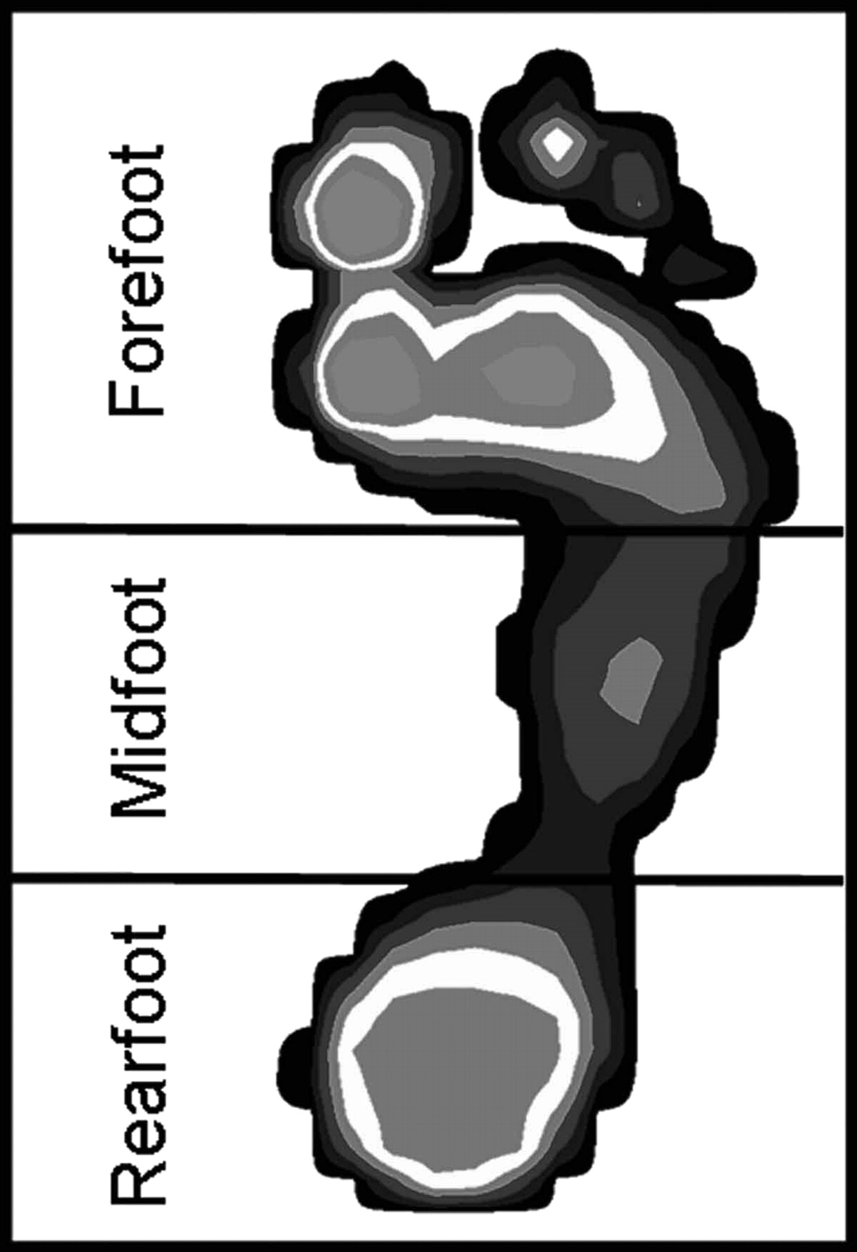
Mean plantar contact area and mean plantar pressure measurements were collected across a ten-step interval. A ten-step interval was selected based on the findings of Kernozek et al, [31] who reported a reliability coefficient of 0.90 across all foot regions in as few as nine steps while using Pedar insoles. For analysis of mean plantar contact area and mean plantar pressure, the foot was divided into three regions on the basis of the work of Cavanagh et al. [32,33] These divisions were used to calculate the two dependent measures: modified arch index and modified pressure index. The modified arch index was calculated by dividing the mean plantar contact area of the midfoot by that of the entire plantar foot. [34] In contrast to the determination by Chu et al [34] of the modified arch index while participants stood on a force plate, modified arch index determination in this study occurred as previously described through in-shoe pressure sensor measures during walking. The modified pressure index was calculated by dividing the mean contact pressure of the midfoot by that of the entire foot (Fig. 1).
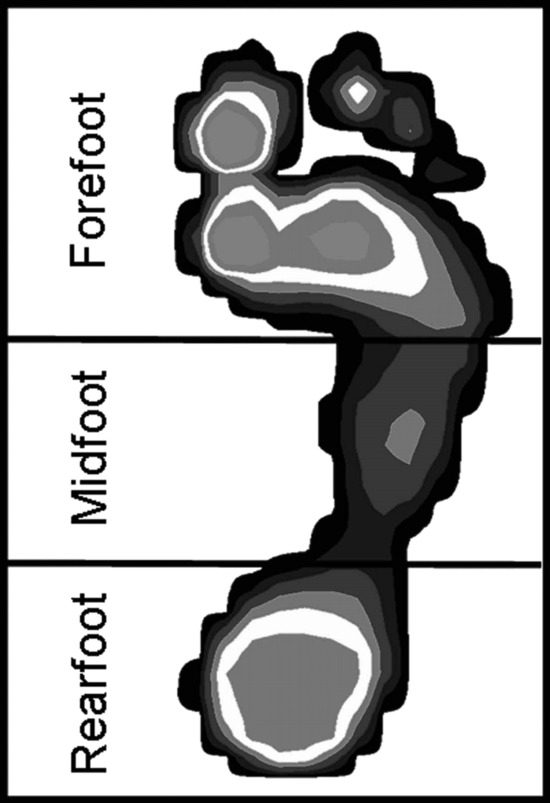
Figure 1
.
Three anatomical mask regions for evaluation of the modified arch and pressure indices: rearfoot (0%–27%), midfoot (27%–55%), and forefoot (55%–100%).
A 2 × 3 mixed-model repeated-measures analysis of variance (α = .05) with a Greenhouse-Geyser correction was performed for each dependent measure. Independent variables were arch type (low and high) and shod condition (nonshod, cushioning shoe, and motion control shoe). Post hoc paired t tests with Bonferroni correction were performed for the high-and low-arched groups to identify significant differences in modified arch index or modified pressure index across the shod conditions.
A secondary analysis of individual foot regions (rearfoot, midfoot, and forefoot) was performed to determine which foot regions contributed to the variations in modified arch index and modified pressure index found on the initial analysis. Separate 2 × 3 mixed-model analyses of variance for mean plantar contact area and mean plantar pressure were used to delineate significant differences among arch type and the three test conditions. Post hoc paired t tests with Bonferroni correction were performed for rearfoot, midfoot, and forefoot mean contact areas and mean pressures to identify significant differences across the test conditions.
Correlation coefficients were derived to analyze the relationship between the static arch height index and the two dynamic measures (modified arch index and modified pressure index) for all three test conditions. The criteria for evaluation of correlation coefficients were in accordance with those cited by Portney and Watkins. [35] Correlation coefficient values less than 0.25 were categorized as having little or no relationship. Correlation coefficient values of 0.25 to 0.50 were categorized as being fair correlations; values greater than 0.50 to 0.75 were categorized as being moderate to good correlations. Finally, correlation coefficient values greater than 0.75 were categorized as being good to excellent. Statistical analysis was performed with Microsoft Excel spreadsheet software (Microsoft Corp, Redmond, Washington) and SPSS 11.5 statistical software (SPSS Inc, Chicago, Illinois). Novel Scientific software (Novel Electronics Inc) was used for management and analysis of mean plantar contact areas and mean plantar pressure distributions.
Results
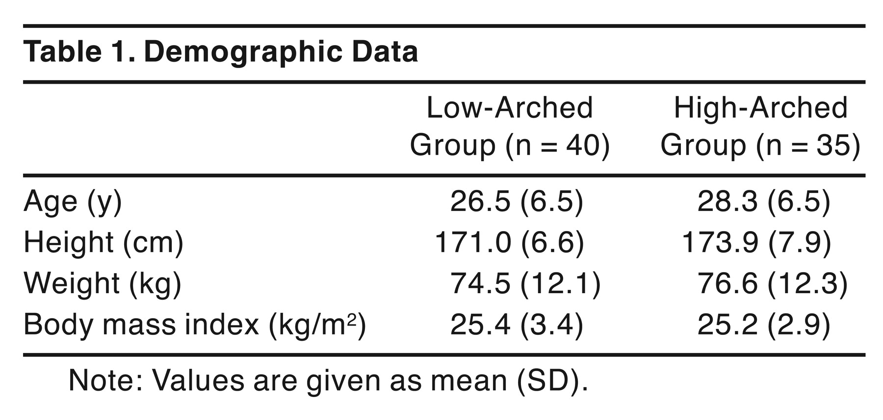
Of 543 individuals screened, 73 were identified as having a low arch and 53 as having a high arch. Seventy-eight participants (43 low arched and 35 high arched) volunteered for data collection in this study. Because of errors in data files, three participants were excluded from data analysis. Final data analysis was performed on 75 participants (54 male and 21 female), with a mean (SD) age of 27.4 (6.5) years. Forty of these participants had a low arch and 35 had a high arch. Demographic data are presented in Table 1.

Table 1
.
Demographic Data.
Effect of Arch Height and the Shod Condition on the Modified Arch Index
A significant interaction effect was found for modified arch index between the shod condition and arch height (P = .017). In addition, there were significant main effects for shod condition (P < .001) and arch height (P < .001). For participants with a low-arched foot, there was a significant difference in modified arch index among all pairwise comparisons for the shod condition (P < .001) (Table 2), demonstrating a decrease in modified arch index values from the cushioning to the motion control to the nonshod conditions. For participants with a high arch, modified arch index values were increased in the cushioning and motion control conditions compared with the nonshod condition (P < .001). No significant difference in modified arch index was found between the cushioning and motion control conditions for high-arched participants (P = .737).
Mean plantar contact area for low-arched participants was significantly different among the shod conditions in the midfoot (P < .001) and forefoot (P < .001) (Table 2 and Fig. 2). For the rearfoot, the mean plantar contact area was significantly different between the nonshod and cushioning shoe conditions (P < .001) and between the nonshod and motion control shoe conditions (P < .001). Mean plantar contact area in the rearfoot was not significantly different between the cushioning and motion control shoe conditions (P = .052). Differences in midfoot mean plantar contact areas in participants with low-arched feet were greatest when comparing the nonshod and the cushioning shoe conditions (33% increase) and the nonshod and the motion control shoe conditions (24% increase). There was a 7% increase in midfoot contact area from the motion control to the cushioning shoe condition in low-arched feet.
Mean plantar contact area in high-arched feet was significantly different in the rearfoot, midfoot, and forefoot for the nonshod compared with the cushioning (P < .001) and motion control (P < .001) conditions (Table 2 and Fig. 2). The greatest differences were found in the midfoot, with increases in mean contact area of 69% from the nonshod to the cushioning condition and of 68% from the nonshod to the motion control shoe condition. There was no difference in mean contact area between the cushioning and motion control shoe conditions for the rearfoot (P = .362), midfoot (P = .848), or forefoot (P = .582). The increase in midfoot contact area from the motion control to the cushioning shoe condition was 0.4%.
Table 2
.
MAI and Mean Contact Area Values for the Forefoot, Midfoot, and Rearfoot Regions by Arch Type and Test Condition.
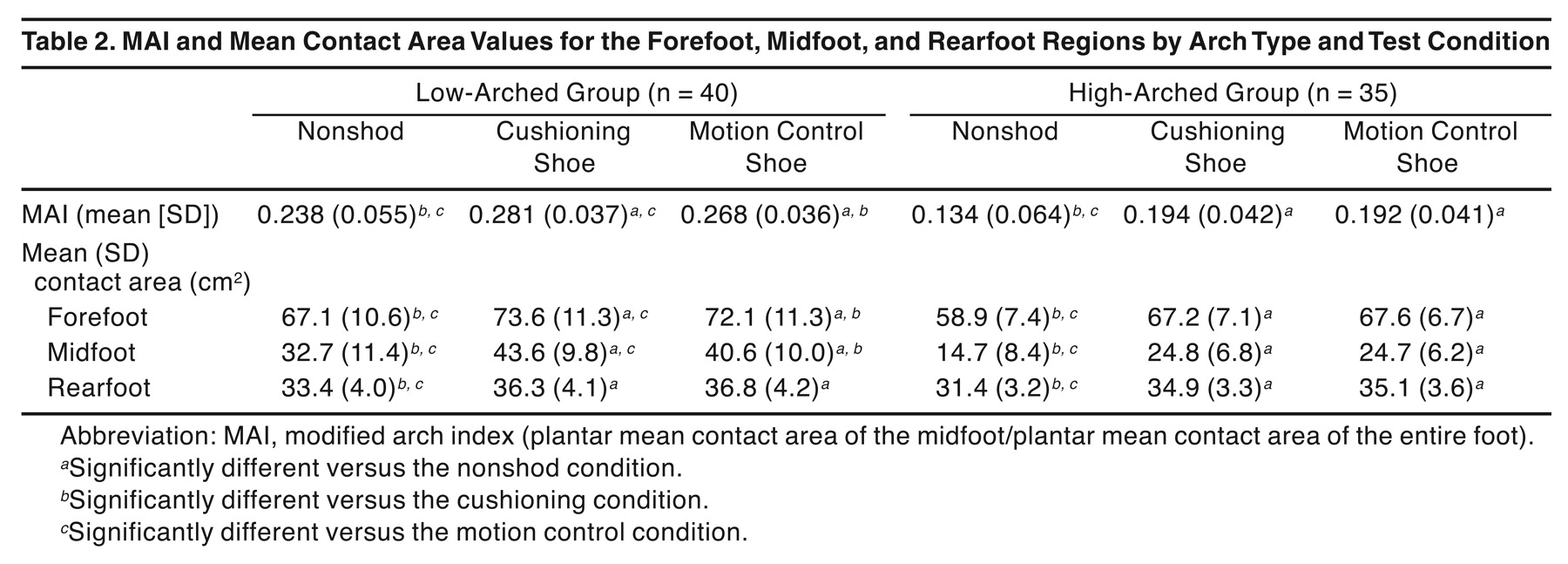
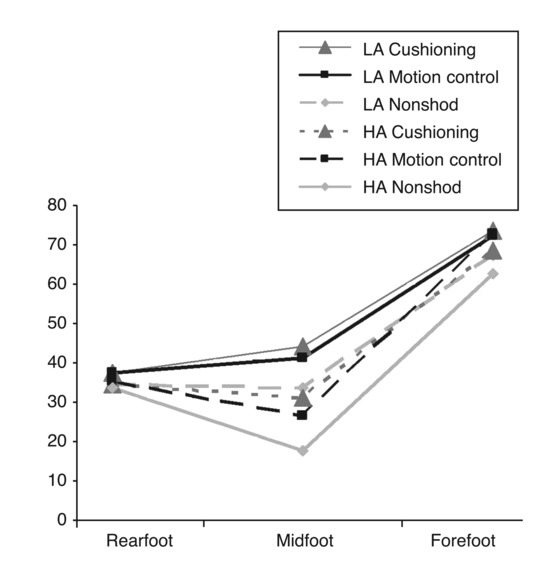
Figure 2
.
Mean contact area of the three foot regions for individuals with low-arched (LA) and high-arched (HA) feet during each of the three test conditions: cushioning, motion control, and nonshod. Compared with the nonshod condition, the mean midfoot contact area increased in those with a low-arched foot by 33% in cushioning shoes and by 24% in motion control shoes; in those with a high-arched foot, the mean contact area increased 69% in cushioning shoes and 68% in motion control shoes.
Effect of Arch Height and the Shod Condition on the Modified Pressure Index
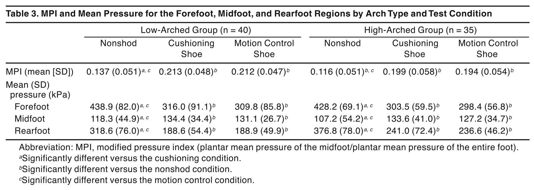
There was no significant interaction effect between the modified pressure index and arch height (P = .752). There also was no significant main effect for arch height (P = .110). A significant main effect existed for the shod condition (P < .001) (Table 3). For low- and high-arched feet, a significant difference existed in the modified pressure index between the cushioning and nonshod conditions (P < .001) and between the motion control and nonshod conditions (P < .001). No significant difference existed between the cushioning and motion control shoe conditions (P > .3).
Overall, mean plantar pressure decreased by approximately 40% in the forefoot and rearfoot for the cushioning and motion control shoe conditions compared with the nonshod condition. Mean plantar pressure increased by approximately 15% to 20% in the midfoot from the nonshod to either shod condition (Fig. 3). Regardless of arch type, mean plantar pressure was significantly different in the forefoot and rearfoot between the nonshod and cushioning conditions (P < .001) (Table 3) and between the nonshod and motion control conditions (P < .001). Mean plantar pressure was significantly different in the midfoot between the nonshod and motion control shoe conditions (P < .002) for individuals with low- or high-arched feet. Mean plantar pressure was also significantly different between the nonshod and cushioning shoe conditions (P < .001) for those with high-arched feet. However, for individuals with low-arched feet, mean plantar pressure was not significantly different between the nonshod and cushioning shoe conditions (P = .018).
Table 3
.
MPI and Mean Pressure for the Forefoot, Midfoot, and Rearfoot Regions by Arch Type and Test Condition.
Relationship Between the Static and Dynamic Measures
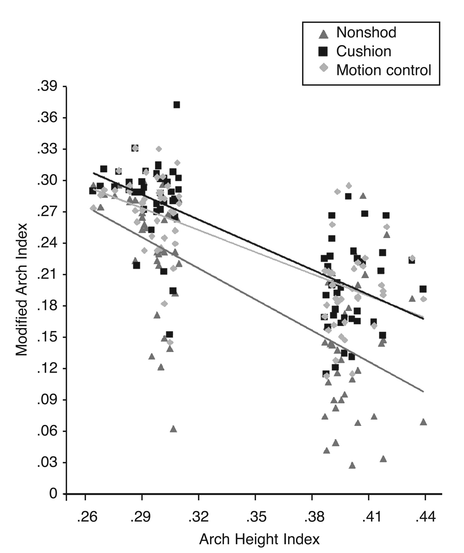
There was a moderate to good negative correlation between the modified arch index and the arch height index for the cushioning (r = −0.72), motion control (r = −0.70), and nonshod (r = −0.68) conditions (Fig. 4). There was little or no correlation between the modified pressure index and the arch height index for the cushioning (r = −0.15), motion control (r = −0.18), and nonshod (r = −0.23) conditions.
Discussion
The primary focus of this study was to address the effect of different running shoe types on individuals with a low- or high-arched foot while walking. Modified arch index values, which measure the relative contribution of the mean contact area of the midfoot as related to the total contact area of the foot, significantly increased in all of the participants regardless of foot type or running shoe type compared with the nonshod condition. Specifically, modified arch index values increased from 12.6% to 18.6% for low-arched individuals and from 43.3% to 44.7% in high-arched individuals when tested in either of the running shoes compared with the nonshod condition.
Regardless of foot type, mean contact area across both shod conditions increased in all three regions of the foot when compared with the nonshod conditions. Changes in the midfoot contact area accounted for most of the differences observed in modified arch index values. The midfoot contact area increased by 24.1% to 33.3% in low-arched individuals and by 68.1% to 69.1% in high-arched individuals compared with the nonshod condition.
In addition, there was a 4.9% decrease in the modified arch index for individuals with a low-arched foot when wearing a motion control shoe compared with a cushioning shoe. When cushioning and motion control shoes were compared, no differences were found in modified arch index values for individuals with a high-arched foot. Although motion was not measured in this study, a decrease in the modified arch index or midfoot contact area may indicate lesser medial midfoot pressure and, thus, a lesser degree of pronation during gait. Should this be true when walking and running, the decrease in the modified arch index in the low-arched group when wearing motion control shoes provides support for recommending a motion control shoe when attempting to limit pronation in runners with low-arched feet.
Although not universally true, individuals with low-arched feet have been characterized to pronate to a greater degree than those with higher-arched feet. [36] Therefore, efforts to control overuse injuries in low-arched runners have traditionally focused on limiting pronation with motion control running shoes. Although the 4.9% decrease in the modified arch index for low-arched individuals was statistically significant, the actual difference in modified arch index values was small. However, the relatively large size of individual pressure sensors in these insoles may have limited the Pedar system’s sensitivity in terms of measuring contact surface area differences between the conditions. Further research is required to better address the relationship between modified arch index and specific running shoe type in the low-arched foot.
As contact area increased, mean plantar pressure across the foot decreased by approximately one-third from the nonshod to both shod conditions regardless of foot type. In addition, motion control and cushioning shoe wear resulted in a redistribution of mean plantar pressures across the foot. The greatest redistribution occurred from rearfoot to midfoot, with a 6% decrease in rearfoot pressure and a 7% increase in midfoot pressure from the nonshod to either shod condition (Fig. 3).
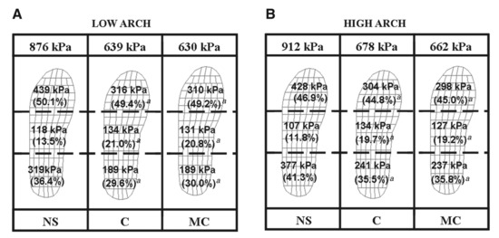
Figure 3
.
Mean pressure over the total foot, forefoot, midfoot, and rearfoot for individuals with a low-arched (A) or high-arched (B) foot during each of the three test conditions: nonshod (NS), cushioning (C), and motion control (MC). Values represent mean (percentage of total foot pressure). Pressure decreased from the NS to the C and MC conditions for overall, forefoot, and rearfoot pressures. In the midfoot region, mean pressure increased while patients wore either C or MC shoes. aP < .05 compared with the NS condition.
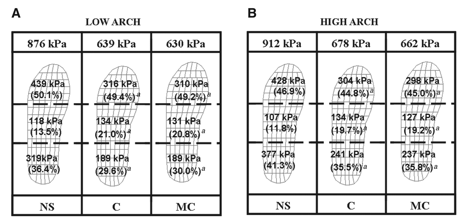
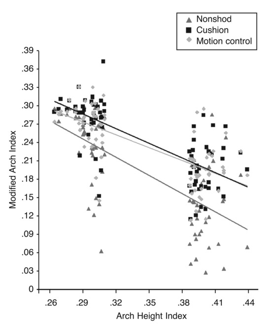
Figure 4.
Comparison of the modified arch index and the arch height index across all 75 participants, demonstrating a moderate to good negative correlation for the cushioning (r = −0.72), motion control (r = −0.70), and nonshod (r = −0.68) conditions.
The combined increase in midfoot mean contact area, decrease in mean plantar pressure, and mean plantar pressure redistribution from rearfoot to midfoot that occurred with footwear has potential clinical relevance when considering that the vertical ground reaction force experienced during running may be as great as 2.5 times body weight. [37] This may be particularly important to the high-arched individual given the decreased shock attenuation capability and subsequent increase in forces typically thought to be transmitted through the high-arched foot. [38]
As part of a larger study, we identified individuals with extremes of arch height as defined by the static arch height index measure. In this study, for all shod conditions, individuals with the smallest arch height index demonstrated the greatest modified arch index, whereas individuals with the largest arch height index had the smallest modified arch index while walking across all shod conditions. Although the effect of normal arch heights (arch height index within 1.5 SD of the group mean) on the relationship between the modified arch index and the arch height index is unknown, a relationship between extremes of arch height was evident. This relationship indicates that individuals with a low-arched foot have a greater midfoot contact area than do high-arched individuals while walking (Fig. 4).
The moderate to good negative correlation between arch height index and modified arch index across all conditions also suggests that the static arch height index measure may be clinically applicable when addressing arch height under dynamic conditions such as walking or running. Chu et al [28] reported a similar correlation between static and modified arch indices (r = −0.71). However, they determined the static arch index via pedobarographs and digital imaging in individuals with a normal-arched foot in a nonshod condition. More recently, McPoil and Cornwall [39] found an association between static (longitudinal arch angle) and dynamic (digital images acquired from videotape) measures. The arch height index may provide the clinician with an objective, time-efficient method of assessing arch height under static and dynamic conditions.
Although running shoe recommendations based on arch type are typically intended to affect foot motion while running, we assessed the relative effects of shoe type on plantar pressure distributions while walking. Joint kinetic patterns during walking demonstrate minimal variability among individuals in ground reaction force distribution, whereas the distribution of ground reaction forces during running varies with individual running styles. [40] We, therefore, attempted to identify the effect of shoe type on plantar pressure distribution and magnitude under a condition in which there is less variability in ground reaction force distribution. Future studies that address dynamic measures at walking or running speeds greater than 3.0 mph may provide greater information about plantar pressure distributions and magnitudes during running. This could enable researchers to better assess the relative effects of shoe type on foot motion while running.
A potential limitation of this study is that the modified arch index is an anterior to posterior plantar foot measure. Although a decrease in the modified arch index may indicate a reduction in pronation, a lateral to medial pressure distribution measure of the foot could be a better potential indicator of pronation. Therefore, future researchers should focus on developing and testing lateral to medial measures to determine how shoe type affects foot motion during running.
In conclusion, individuals with low-arched feet in this study demonstrated a significant decrease in the modified arch index when wearing motion control shoes than when wearing cushioning shoes. Should a decrease in the modified arch index be associated with decreased pronation, more sensitive measures of contact surface area differences could better support the clinician’s recommendation of a motion control shoe when attempting to limit pronation in low-arched individuals. The combined increase in midfoot mean plantar contact area, decrease in mean plantar pressure, and mean plantar pressure redistribution from rearfoot to midfoot that occurred with shoe wear has potential clinical relevance in terms of decreased force transmission and enhanced shock attenuation capability through the lower extremities. Finally, the static arch height index measure has a moderate to good association with the modified arch index, which measures plantar pressure distribution under dynamic conditions. As a result, the arch height index provides the clinician with a valid means of assessing arch height under static and dynamic conditions.
Acknowledgments
John Childs, Rachelle Thomas, Candi Darst, Brian Gray, and Travis Robbins for their time and contributions to this study.
Financial Disclosure:
None reported.
Conflict of Interest:
None reported.
References
- Kaufman KR, Brodine SK, Shaffer RA, et al: The effect of foot structure and range of motion on musculoskeletal overuse injuries. Am J Sports Med 27: 585, 1999.
- Smith TA, Cashman TM: The incidence of injury in light infantry soldiers. Mil Med 167: 104, 2002.
- Jones BH, Knapik JJ: Physical training and exercise-related injuries: surveillance, research and injury prevention in military populations. Sports Med 27: 111, 1999.
- Burns J, Keenan AM, Redmond A: Foot type and overuse injury in triathletes. JAPMA 95: 235, 2005.
- Bennett JE, Reinking MF, Pluemer B, et al: Factors contributing to the development of medial tibial stress syndrome in high school runners. J Orthop Sports Phys Ther 31: 504, 2001.
- Montgomery LC, Nelson FR, Norton JP, et al: Orthopedic history and examination in the etiology of overuse injuries. Med Sci Sports 21: 237, 1989.
- Matheson GO, Clement DB, McKenzie DC, et al: Stress fractures in athletes: a study of 320 cases. Am J Sports Med 15: 46, 1987.
- Gellman R, Burns S: Walking aches and running pains: injuries of the foot and ankle. Prim Care 23: 263, 1996.
- Cowan DN, Jones BH, Robinson JR: Foot morphologic characteristics and risk of exercise-related injury. Arch Fam Med 2: 773, 1993.
- Giladi M, Milgrom C, Stein M: The low arch, a protective factor in stress fractures: a prospective study of 295 military recruits. Orthop Rev 14: 709, 1985.
- Williams DS, McClay IS, Hamill J: Arch structure and injury patterns in runners. Clin Biomech 16: 341, 2001.
- Knapik J, Feltwell D, Canham-Chervak M, et al: Evaluation of Injury Rates During Implementation of the Fort Drum Running Shoe Injury Prevention Program, US Army Center for Health Promotion and Preventive Medicine, Aberdeen Proving Grounds, MD. ,2001.
- Keenan AM, Redmond AC, Horton M, et al: The Foot Posture Index: Rasch analysis of a novel, foot-specific outcome measure. Arch Phys Med Rehabil 88: 88, 2007.
- Redmond AC, Crosbie J, Ouvrier RA: Development and validation of a novel rating system for scoring standing foot posture: the Foot Posture Index. Clin Biomech 21: 89, 2006.
- Clarke HH: An objective method of measuring the height of the longitudinal arch in foot examinations. Res Q Exerc Sport 4: 99, 1933.
- Williams DS, McClay IS: Measurements used to characterize the foot and the medial longitudinal arch: reliability and validity. Phys Ther 80: 864, 2000.
- Butler RJ, Davis IS, Hamill J: Interaction of arch type and footwear on running mechanics. Am J Sports Med 34: 1998, 2006.
- Reinschmidt C, Nigg BM: Current issues in the design of running and court shoes. Sportverletz Sportschaden 14: 71, 2000.
- Clarke TE, Frederick EC, Hamill CL: The effects of shoe design parameters on rearfoot control in running. Med Sci Sports Exerc 15: 376, 1983.
- Stacoff A, Reinschmidt C, Nigg BM, et al: Effects of shoe sole construction on skeletal motion during running. Med Sci Sports 33: 311, 2001.
- Heil B: Running shoe design and selection related to lower limb biomechanics. Physiotherapy 78: 406, 1992.
- DeWit B, DeClerq D, Lenoir M: The effect of varying midsole hardness on impact forces and foot motion during foot contact in running. J Appl Biomech 11: 395, 1995.
- Hamill J, Bates BT, Holt KG: Timing of lower extremity joint actions during treadmill running. Med Sci Sports 24: 807, 1992.
- Perry SD, Lafortune MA: Influences of inversion/ eversion of the foot upon impact loading during locomotion. Clin Biomech 10: 253, 1995.
- Viswanathan V, Sivagami M, Seena R, et al: Increased forefoot to rearfoot plantar pressure ratio in South Indian patients with diabetic foot ulceration. Diabet Med 21: 396, 2004.
- Praet SF, Louwerens JW: The influence of shoe design on plantar pressures in neuropathic feet. Diabetes Care 26: 441, 2003.
- Perry JE, Ulbrecht JS, Derr JA, et al: The use of running shoes to reduce plantar pressures in patients who have diabetes. J Bone Joint Surg Am 77: 1819, 1995.
- Chu WC, Lee SH, Chu W, et al: The use of arch index to characterize arch height: a digital image processing approach. IEEE Trans Biomed Eng 42: 1088, 1995.
- Hsiao H, Guan J, Weatherly M: Accuracy and precision of two in-shoe pressure measurement systems. Ergonomics 45: 537, 2002.
- Burnfield JM, Few CD, Mohamed OS, et al: The influence of walking speed and footwear on plantar pressures in older adults. Clin Biomech 19: 78, 2004.
- Kernozek TW, LaMott EE, Dancisak MJ: Reliability of an in-shoe pressure measurement system during treadmill walking. Foot Ankle Int 17: 204, 1996.
- Cavanagh PR, Rodgers MM: The arch index: a useful measure from footprints. J Biomech 20: 547, 1987.
- Cavanagh PR, Rodgers MM, Iiboshi A: Pressure distribution under symptom-free feet during barefoot standing. Foot Ankle 7: 262, 1987.
- Chu WC, Lee SH, Chu W, et al: The use of arch index to characterize arch height: a digital image processing approach. IEEE Trans Biomed Eng 42: 1088, 1995.
- Portney LG, Watkins MP: Foundations of Clinical Research: Applications to Practice, p 439, Appleton & Lange, Norwalk, CT. ,1993.
- Boozer MH, Finch A, Waite LR: Investigation of the relationship between arch height and maximum pronation angle during running. Biomed Sci Instrum 38: 203, 2002.
- Mann RA, Hagy J: Biomechanics of walking, running, and sprinting. Am J Sports Med 8: 345, 1980.
- Williams DS, Davis IM, Scholz JP, et al: High-arched runners exhibit increased leg stiffness compared to low-arched runners. Gait Posture 19: 263, 2004.
- McPoil TG, Cornwall MW: Use of the longitudinal arch angle to predict dynamic foot posture in walking. JAPMA 95: 114, 2005.
- Ounpuu S: The biomechanics of walking and running. Clin Sports Med 13: 843, 1994.
© 2009 American Podiatric Medical Association