


Abstract
Background: Verrucae are caused by infection of epidermal keratinocytes by human papilloma virus (HPV). Although there are currently more than 100 known types of HPV, certain lesions are consistently caused by infection with one or a few types. Recent studies have identified the presence of unusual HPV types in anogenital and cervical condylomata (warts) of patients infected with human immunodeficiency virus (HIV). Although cutaneous verrucae are typically caused by HPV-1, HPV-2, and HPV-4, infection with HIV may predispose an individual to infection with an unusual HPV type. Methods: We report the detection of a rare HPV type in a clinically aggressive plantar verruca from an HIV-positive patient. The viral DNA from this specimen was analyzed to identify the predominant HPV type. To complete this analysis, HPV DNA was extracted from the formalin-fixed specimen, followed by polymerase chain reaction with consensus HPV primers and digestion with a specific group of restriction endonucleases. The fragments were separated on an agarose gel, and the restriction fragment length polymorphism pattern was compared with known patterns for identification of the specific HPV type. Results: Identification of HPV-69, an HPV type previously reported to be rare and associated with dysplastic lesions, was confirmed by HPV DNA dot-blot hybridization with specific DNA probes for each known HPV type. Conclusions: Plantar verrucae in HIV-positive patients may be associated with unusual HPV types and should be analyzed and treated aggressively given the potential for a more distinct clinical manifestation. Additional lesional analysis studies are needed. (J Am Podiatr Med Assoc 99(1): 8–12, 2009)
Plantar verrucae, commonly known as plantar warts, are the most common benign soft-tissue lesions found in the foot and ankle. [1] These lesions are typically accompanied by significant symptoms because of their location on a heavily innervated and weight-bearing surface. In addition, owing to their viral etiology, plantar verrucae also typically present a significant challenge to the health-care provider.
The etiology of all cutaneous verrucae is human papilloma virus (HPV), a double-stranded DNA virus enclosed in a nonenveloped protein capsid. [2-4] More than 100 different types of HPV exist, and they can be differentiated by DNA sequence, protein production, host membrane proteins, and, for some, host antibody profile. [5-10] Infection by HPV types 1, 2, and 4 has been associated with the development of plantar and other cutaneous warts. [2-4,6,11] The life cycle of HPV includes exposure of the basal keratinocyte layer of skin to HPV, entrance of HPV into the basal cells, production of early viral proteins, production of late viral proteins in the outer layers of the epidermis, and shedding of the new viral particles from the cells of the stratum corneum. The early viral proteins coordinate the replication of the viral genome and act to transform the basal cell into a hyperplastic cell type as it differentiates and passes through the stratum spinosum and stratum granulosum cell layers. [2-4] The late viral proteins coordinate the packaging of the viral genome and constitute the viral capsid. [2-4] The uninhibited proliferation of the transformed keratinocytes contributes to the epidermal thickness, folding, and hyperkeratosis seen clinically and histologically in these lesions. Also, the hyperplastic response results in compression of the papillary dermis, which is responsible for the clinical signs of punctate or pinpoint bleeding, lateral compression tenderness, and absence of the epidermal skin lines used to make a clinical diagnosis of cutaneous verruca. [11] Definitive diagnosis is made histologically after excision at the epidermal-dermal junction.
Many different modalities have been used to treat these lesions. [11] Conservative therapies include superficial debridement, topical keratolysis with and without occlusion, and hydrotherapy. More invasive modalities include cryotherapy, electrical and thermal desiccations, and bleomycin injections. Invasive treatments include surgical curettage and full-thickness excision.
The pathologic process of plantar verrucae in human immunodeficiency virus (HIV)–positive patients is complicated by the presence of HIV. The effect of a positive HIV status on the clinical presentation and treatment response of plantar verrucae is well reported. [12,13] The number and size of lesions have been found to be greater in HIV-positive patients compared with HIV-negative patients. [5,7-10,12-17] In addition, lesions in HIV-positive patients have been shown to be less responsive to the curettage modality. [12]
There are many possible reasons for the adverse effect of HIV on the clinical presentation and treatment response of plantar verrucae, including an immunocompromised state affecting cell-mediated immunity and the possibility of an interaction between HIV proteins and HPV regulation sequences. [18] Also, the presence of HIV could predispose patients to cutaneous infection with an unusual HPV type responsible for a more aggressive keratinocyte. This is a phenomenon previously reported in anogenital warts in HIV-positive patients (eg, the discovery of HPV-16, 18 instead of HPV-6, 11 in anogenital lesions). [19-21]
Infection of HIV-positive patients with an unusual, more aggressive HPV type is the focus of our study. We report the finding of an unusual HPV type (HPV-69) in a clinically aggressive plantar verruca from an HIV-positive patient.
Materials and Methods
A 57-year-old man with a long history of HIV infection and a CD4 count less than 50 cells/mm3 presented to the California College of Podiatric Medicine, currently the California School of Podiatric Medicine at Samuel Merritt College, for evaluation and treatment of a painful verrucous skin lesion located at the plantar aspect of the first metatarsal head of the left foot. The lesion was reported to be recalcitrant to multiple previous attempts of conservative treatments. On physical examination, the lesion was observed to be circular and measured 18 mm in diameter. The lesion was tender to plantar and side-to-side compression, was noted to lack epidermal skin lines, and displayed punctate or pinpoint bleeding with superficial debridement. A diagnosis of clinically aggressive plantar verruca was made, and the lesion was treated with curettage. The curettage was performed at the epidermal-dermal junction, and the lesion was preserved in 10% buffered formalin. The lesion was sent to the Western Pathology Medical Group (Dublin, California) for histologic confirmation of the diagnosis of cutaneous verruca.
DNA Extraction from the Plantar Wart Biopsy Sample
For the purpose of HPV typing, the biopsy tissue was removed from the 10% buffered formalin and was soaked in phosphate-buffered saline for 3 days to remove the formalin. The tissue was added to a homogenization buffer (500mM Tris hydrochloride, 20mM EDTA, and 10mM sodium chloride; pH 8.5–9.0) and was homogenized at 15,000 rpm using a Polytron Homogenizer (Brinkmann Instruments Inc, Westbury, New York). The solution was then treated with sodium dodecyl sulfate and proteinase K at final concentrations of 1% and 500 μg/mL, respectively, and was incubated for 14 hours at 48°C. The previous step was repeated but with a final sodium dodecyl sulfate concentration of 2%, a final proteinase K concentration of 1 mg/mL, and only 3 hours of incubation at 48°C. The mixture was then centrifuged to separate out the denatured proteins and cell membrane debris.
The supernatant was then subjected to three phenol extractions with aqueous phase preservation. Buffered phenol was added to achieve a 1:1 mixture, and the solution was mixed by inversion. The mixture was centrifuged at 14,000 rpm for 2 min. The upper aqueous layer and any white precipitate at the interface were collected, and the lower organic phase was discarded. A similar extraction protocol was then performed twice on the aqueous phase using a 24:1 mixture of chloroform:isoamyl alcohol. To the final aqueous phase, 0.1 volumes of 3.3M ammonium acetate, 2.5 volumes of 100% ethanol, and 4 μL of glycogen were added. The solution was then stored at −20°C overnight to stimulate the precipitation of human and HPV DNA.
The mixture was thawed and then was subjected to centrifugation at 14,000 rpm for 1 hour. The supernatant was removed, and the DNA precipitate was washed twice with 70% ethanol. The ethanol was evaporated off, and the precipitate was resuspended in 100 μL of TE suspension buffer (10mM Tris hydrochloride and 1mM EDTA).
Purification/Amplification of HPV DNA
Polymerase chain reaction (PCR) was used to obtain DNA for analysis. The PCR primers used included MY09 and MY11, which are consensus primers that amplify the L1 regulatory region of HPV DNA. [22] The 100-μL amplification reactions were performed containing 10mM Tris (pH 8.3), 50mM potassium chloride, 4.0mM magnesium chloride, 200μM each dNTP, 500nM each primer, and 2.5 U of AmpliTaq (Perkin-Elmer, Foster City, California). To each reaction tube, 5 μL of crude DNA preparation was added. The PCR was performed using an Uno II Thermocycler (Biometra, Tampa, Florida). The PCR conditions were as follows: 95°C for 3 min hot start; 30 cycles at 95°C for 30 sec, 55°C for 30 sec, and 72°C for 1 min; an additional 0.5 μL of AmpliTaq was added to each tube; 40 cycles at 95°C for 30 sec, 55°C for 30 sec, and 72°C for 2 min; terminal extension at 72°C for 10 min. The HPV-16 DNA was used as a positive control along with no DNA as a negative control. The PCR products were then separated using a 1.5% agarose test gel to confirm the presence of amplified HPV DNA.
Restriction Endonuclease Digestion of PCR Products
Amplified HPV L1 DNA sequences were subjected to restriction endonuclease digestion for the purpose of building a restriction fragment length polymorphism fragment collection that could be compared with the restriction fragment length polymorphism fragment collections of known HPV types. The reaction tubes comprised 84 μL of PCR water, 56 μL of PCR product, 16.8 μL of 10X REact 3 buffer, and 1.4 μL of restriction enzyme. The restriction endonucleases used included Bam H1, Dde I, Hae III, Hinf I, Pst I, Rsa I, and Sau 3AI. The tubes were incubated in a water bath at 37°C for 3 hours. The fragments were run on a 4% NuSieve/agarose (3:1) gel. Photographs of the restriction fragment length polymorphism patterns were taken, and, with the assistance of DNASIS software (MedProbe, Oslo, Norway), the fragment sizes (in base pair) were compared with those of the fragments generated by using the same restriction enzymes on known HPV types.
Dot-Blot Hybridization
Confirmation of HPV type was performed by subjecting the PCR products from the sample lesion to dot-blot hybridization using HPV L1 DNA probes specific for each known HPV type.
Results
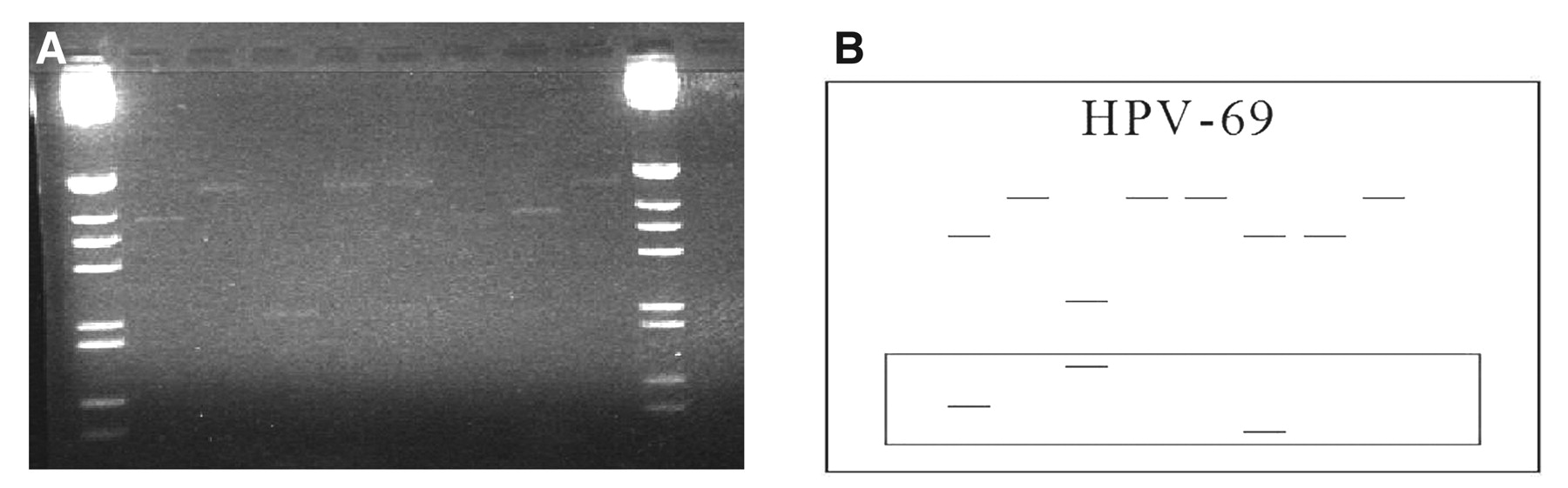
Histologic analysis revealed keratinocyte hyperplasia and hyperkeratosis consistent with the diagnosis of benign cutaneous verrucae (Fig. 1). The lesional HPV DNA fragmentation pattern produced by the restriction enzymes is shown in Figure 2A. The known fragmentation pattern for HPV-69 using the same enzymes is shown along with fragment sizes (in base pair) in Figure 2B. Visual analysis and the matching of fragment sizes for each known HPV type led to the identification of HPV-69 as the unknown HPV type from the sample lesion. This finding was confirmed by a positive binding reaction between the L1 sequence of the lesional HPV DNA and a probe specific for the L1 sequence of HPV-69.
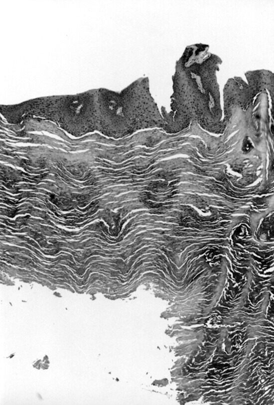
Figure 1.
Histologic analysis revealed keratinocyte hyperplasia and hyperkeratosis consistent with the diagnosis of benign cutaneous verrucae.
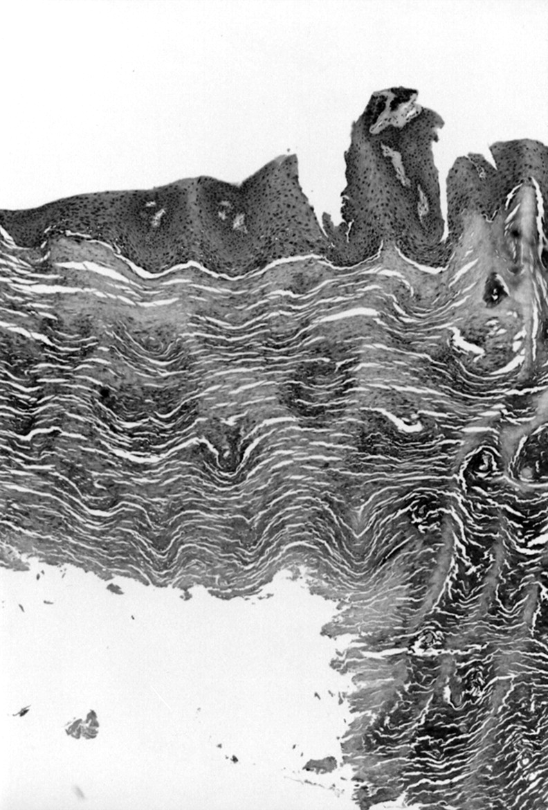
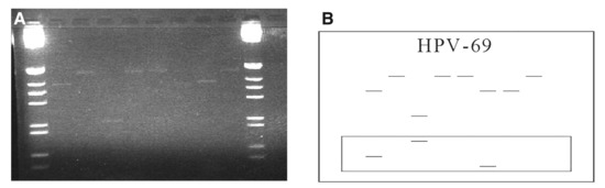
Figure 2.
A, Agarose gel containing the restriction fragments of the human papilloma virus (HPV) DNA from the sample lesion. B, Schematic diagram of the restriction fragment pattern for HPV-69. In both parts, the lanes are as follows from left to right: Bam H1, Dde I, Hae III, Hinf I, Pst I, Rsa I, Sau 3AI, and control (no enzyme). The uncut HPV L1 sequence comprises 455 base pair.
Discussion
Cutaneous verrucae are typically caused by keratinocyte infection with HPV types 1, 2, and 4. However, based on recent findings of atypical HPV infection in anogenital condylomata of HIV-positive patients, it must be considered possible that cutaneous verrucae are also associated with unusual HPV types in HIV-positive patients. The finding herein of HPV-69 in a plantar verruca from an HIV-positive patient, which is the first reported finding of HPV-69 in a plantar or other cutaneous verruca, confirms the likelihood of this possibility.
Given that some of the unusual HPV types display a greater tendency toward premalignant or malignant keratinocyte transformation (eg, the finding of HPV-69 in a dysplastic lesion of the tongue reported by Bernard et al [23]), the finding reported in this article also suggests the need for a more aggressive standard of care for plantar and other cutaneous verrucae in HIV-positive patients. Other studies that identify the HPV type associated with cutaneous verrucae from HIV-positive patients and those that provide histologic data to confirm the malignant nature of these unusual HPV types are necessary to support this suggested change in the standard of care of these lesions. The HIV-positive patients may have a more aggressive clinical pattern of plantar verrucae, and there is a need for more assertive treatment. Twenty-six years after the first documented case of acquired immunodeficiency syndrome, this deadly disease has transformed from a death sentence into a chronic illness with the help of highly active antiretroviral therapy. With the average life expectancy extended to 24.2 years, [24] clinical manifestations such as plantar warts have become of greater clinical importance in patient management. Previous studies have shown that plantar verrucae in HIV-positive patients tend to be greater in number and size and more resistant to traditional therapeutic interventions. Discovery of the unusual HPV-69 in this patient could be a possible reason for this more aggressive clinical presentation. The implications of this study include a more aggressive and attentive mode of treatment for HIV-positive individuals with plantar warts.
Acknowledgments
Dr. Carolyn Harvey and members of her staff at the California School of Podiatric Medicine for the collection of clinical lesions from patients; Dr. James Fitzpatrick and the Western Pathology Medical Group for histologic analysis of the sample lesion; Colleen Durkin of the California School of Podiatric Medicine for coordinating the histologic analysis of the sample lesion; and Daniel Felmlee of the Department of Laboratory Medicine at the University of California, San Francisco, for confirming the HPV typing result.
Financial Disclosure
This work was supported in part by a grant from the Center for AIDS Research.
Conflict of Interest
None reported.
References
- Kirby EJ, Shereff MJ, Lewis MM: Soft-tissue tumors and tumor-like lesions of the foot: an analysis of eighty-three cases. .J Bone Joint Surg Am71::621. ,1989.
- Fields B, Knipe D: Fields Virology, 2nd Ed, Raven Press, New York. ,1990.
- Brooks G, Butel J, Ornston L: Jawetz, Melnick, & Adelberg’s Medical Microbiology, 19th Ed, Appleton & Lange, Norfolk, CT. ,1991.
- Bunney M, Benton C, Cubie H: Viral Warts: Biology & Treatment, 2nd Ed, Oxford Medical Publications, Oxford. ,1992.
- Meberg R, Kenyon E, Bierman R, et al: Characterization of plantar verrucae among individuals with human immunodeficiency virus. .JAPMA88::442. ,1998.
- Viac J, Chomel J, Chardonnet Y, et al: Incidence of antibodies to human papillomavirus type 1 in patients with cutaneous and mucosal papillomas. .J Med Virol32::18. ,1990.
- Barbosa P: Plantar verrucae and HIV infection. .Clin Podiatr Med Surg15::317. ,1998.
- Soltani S, Kenyon E, Barbosa P: Chronic and aggressive plantar verrucae in a patient with HIV. .JAPMA86::555. ,1996.
- Kenyon E, Loveland L, Kilpatrick R, et al: Epidemiology of plantar verrucae in HIV-infected individuals. .J Acquir Immune Defic Syndr Hum Retrovirol17::94. ,1998.
- Barbosa P, Kneass Z: Molecular biology of HIV. .Clin Podiatr Med Surg15::189. ,1998.
- Jenkin W: “Management of Viral Infections of the Skin and Nail,” in Surgery Lecture Manual, p 14, California College of Podiatric Medicine, San Francisco. ,1994.
- Whitaker JM, Gaggero GL, Loveland L, et al: Plantar verrucae in patients with human immunodeficiency virus: clinical presentation and treatment response. .JAPMA91::79. ,2001.
- Adler J: Pedal infectious disease associated with acquired immunodeficiency syndrome. .Clin Podiatr Med Surg9::837. ,1992.
- Memar O, Cirelli R, Lee P, et al: Cutaneous manifestations of HIV-1 infection. .JAPMA85::362. ,1995.
- Burns S: Podiatric manifestations of AIDS. .JAPMA80::15. ,1990.
- Dover J, Johnson R: Cutaneous manifestations of human immunodeficiency virus infection. .Arch Dermatol27::1383. ,1991.
- Kang S, Fitzpatrick T: Debilitating verruca vulgaris in a patient infected with the human immunodeficiency virus. .Arch Dermatol130::294. ,1994.
- Vernon SD, Hart CE, Reeves WC, et al: The HIV-1 tat protein enhances E2-dependent human papillomavirus 16 transcription. .Virus Res27::133. ,1993.
- Cuesta KH, Palazzo JP, Mittal KR: Detection of human papillomavirus in verrucous carcinoma from HIV-seropositive patients. .J Cutan Pathol25::165. ,1998.
- Sun XW, Kuhn L, Ellerbrock TV, et al: Human papillomavirus infection in women infected with the human immunodeficiency virus. .N Engl J Med337::1343. ,1997.
- Tan W, Felber BK, Zolotukhin AS, et al: Efficient expression of the human papillomavirus type 16 L1 protein in epithelial cells by using Rev and the Rev-responsive element of human immunodeficiency virus or the cis-acting transactivation element of simian retrovirus type 1. .J Virol69::5607. ,1995.
- Ting Y, Manos MM: “Detection and Typing of Genital Human Papillomaviruses,” in PCR Protocols and Applications, p 356, Academic Press, San Diego. ,1990.
- Bernard HU, Chan SY, Manos MM, et al: Identification and assessment of known and novel human papillomaviruses by PCR amplification, restriction fragment length polymorphisms, nucleotide sequence, and phylogenetic algorithms. .J Infect Dis170::1077. ,1994.
- Schackman BR, Gebo KA, Walensky RP, et al: The lifetime cost of current human immunodeficiency virus care in the United States. .Med Care44::990. ,2006.
© 2009 American Podiatric Medical Association