


Abstract
Bilateral stress fractures of the fibula are very rare. We present an unusual case report of a 54-year-old osteoporotic woman with bilateral stress fractures involving the distal fibula. After conservative treatment, she made a good recovery with full motion and was free of pain.
Stress fractures are common overuse injuries that include fatigue and insufficiency fractures. Athletes, soldiers, and osteoporotic patients are some of the individuals at high risk for the development of this injury. Although stress fractures of the fibula are well recognized, bilateral distal fibular fractures are very rare. To our knowledge, no cases with bilateral distal fibular stress fractures attributable to osteoporosis have been reported in the English literature.
Case Report
A 54-year-old woman was admitted to Izmir Ataturk Training and Research Hospital (Izmir, Turkey) with complaints of pain and swelling at the lateral aspect of both ankles. She presented with osteoporosis and osteoarthritis diagnosed by a rheumatologist 2 months earlier. Her dual energy x-ray absorptiometry (DEXA) scan showed a spinal T score of –2.6 and a femoral T score of –1.3. She was prescribed alendronate 70 mg/ week, and was instructed to begin weightbearing exercises immediately. Thereafter, she started regular walking at high tempo.
After 1 month of weightbearing exercises, left ankle pain developed, followed immediately by right ankle pain. There was no history of trauma. Initially, the pain increased with physical activity, but it also began to occur at rest and during the night. Two weeks after the onset of pain she noticed a gradual swelling around the ankles. She was a nonsmoker and had been menopausal for 3 years. Laboratory investigations and biochemistry profile were within normal limits.
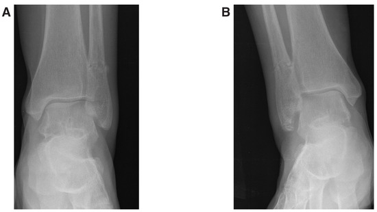
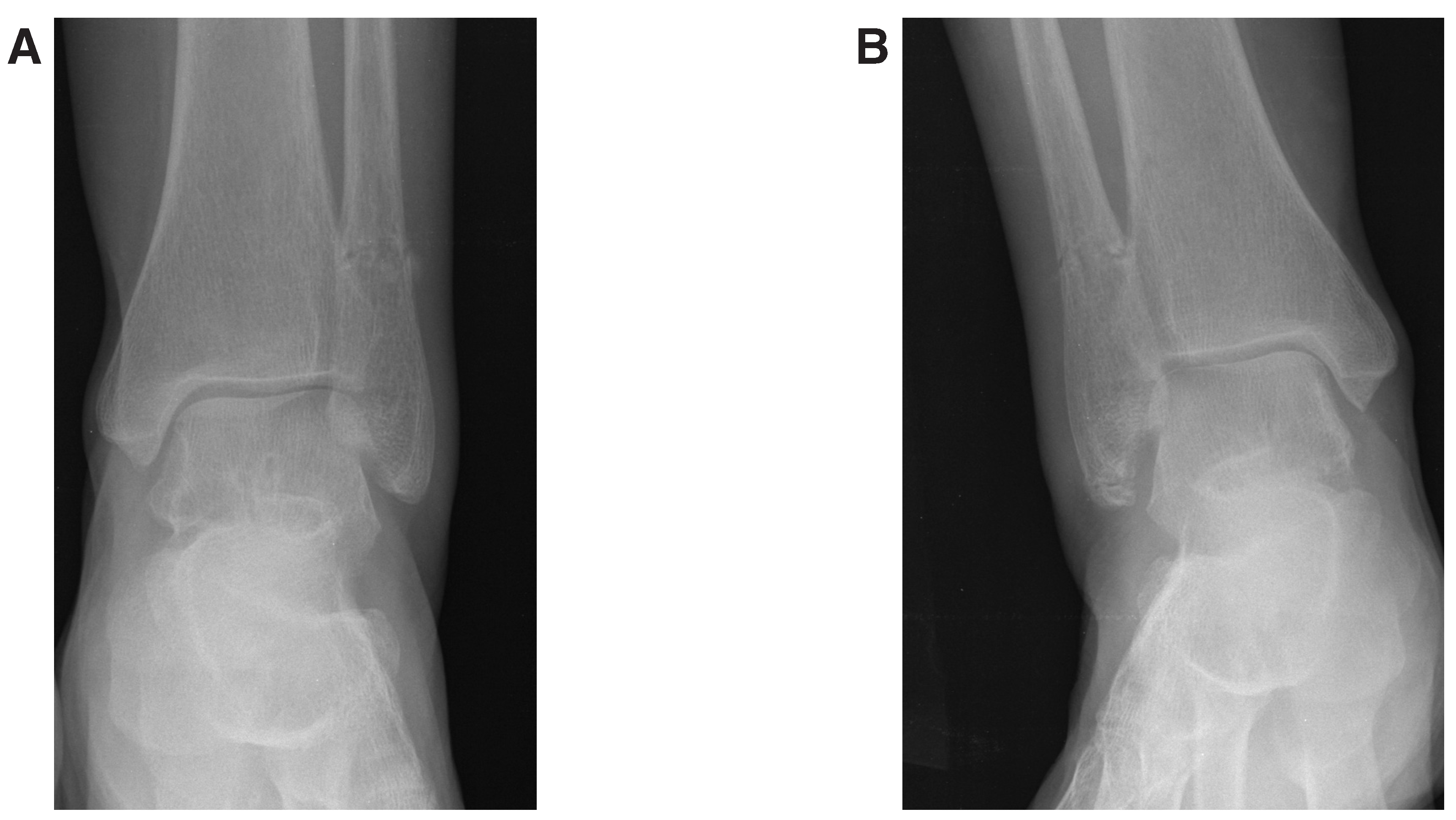
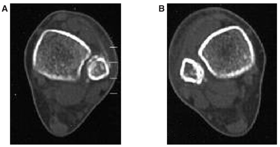
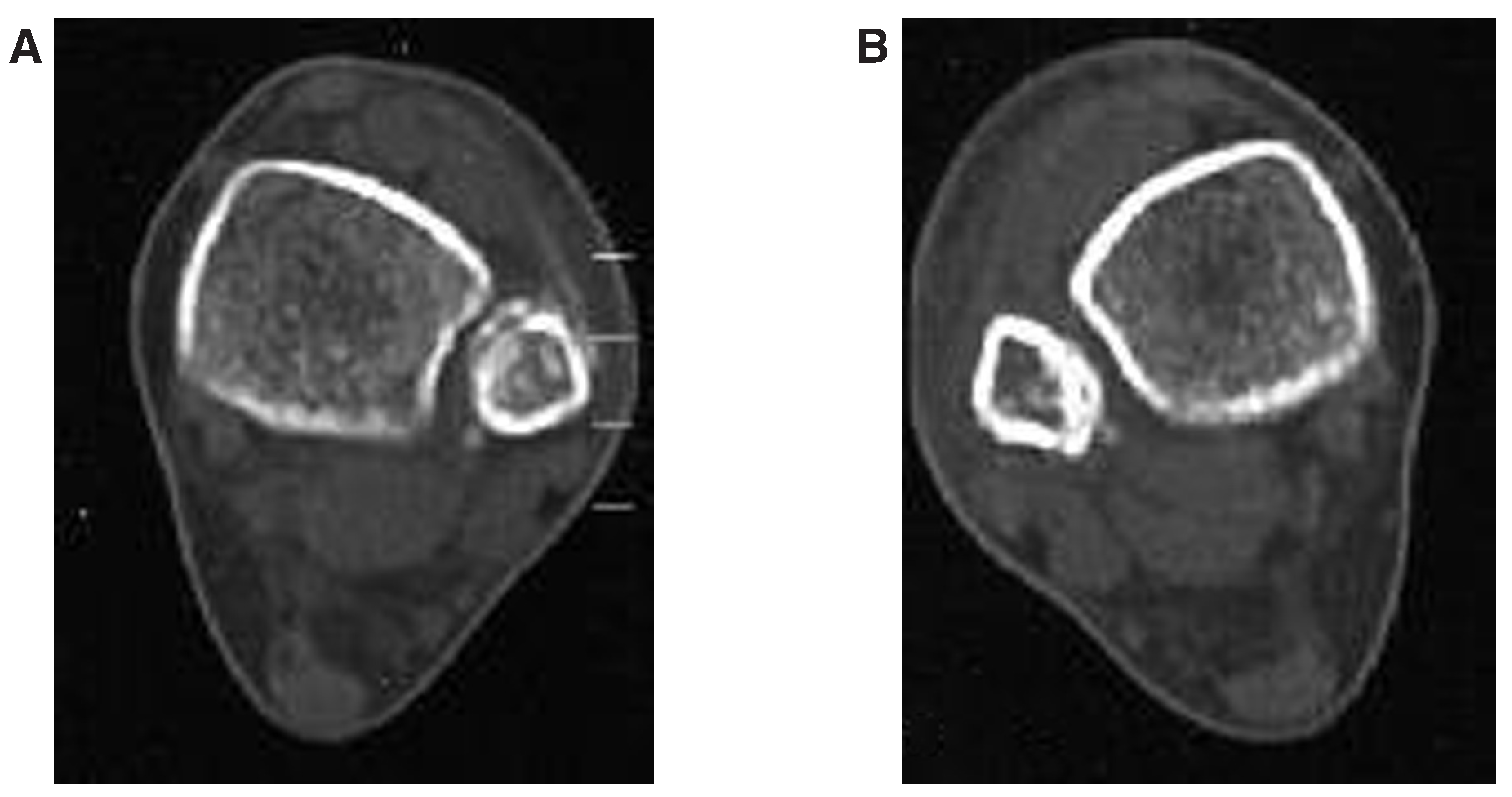
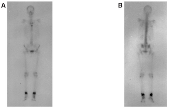
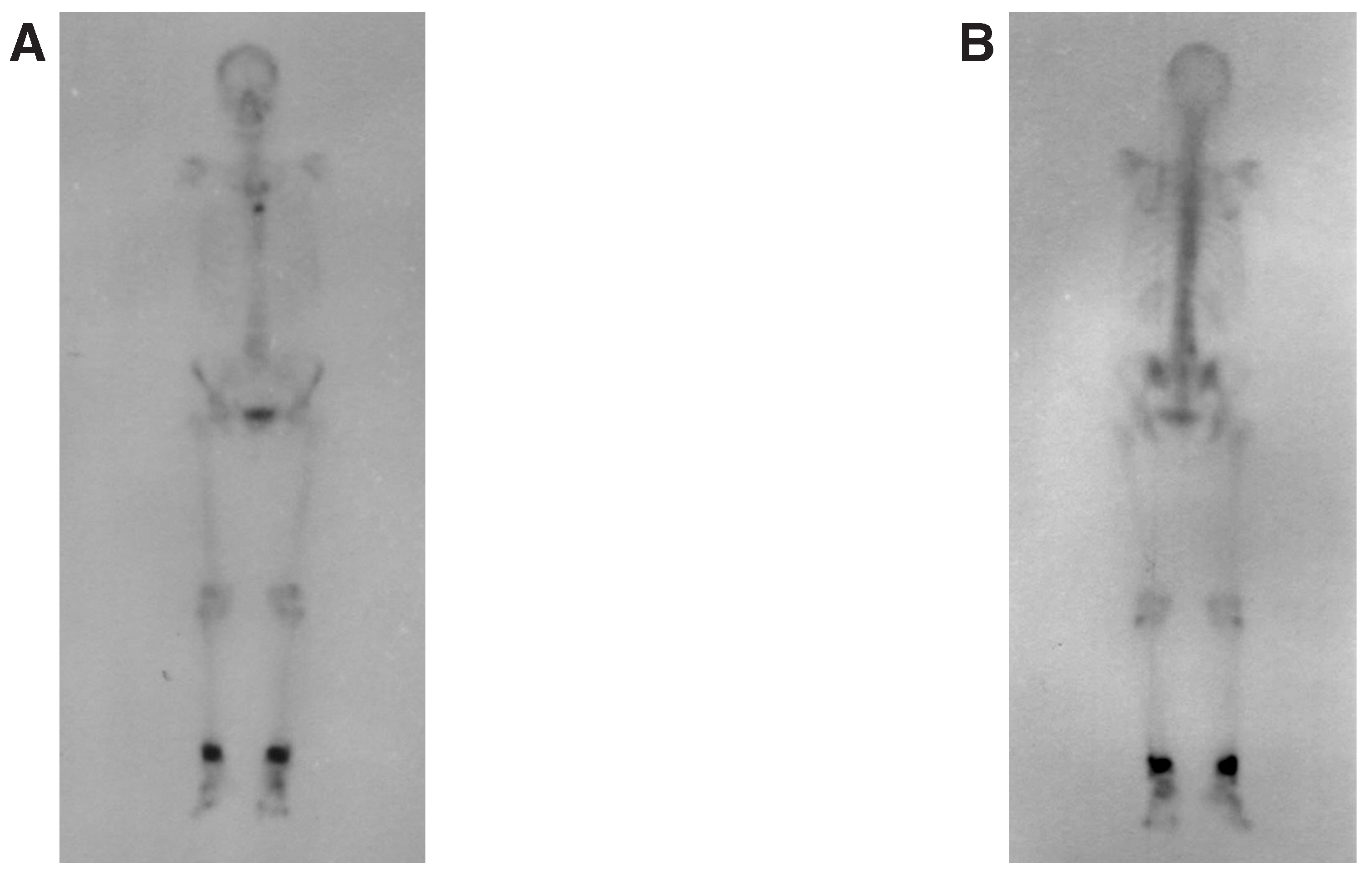
Examination revealed swelling over both ankles and tenderness over the distal fibula bilaterally. She had a mild limitation of ankle motion with no neurovascular deficit. Radiographs of both ankles revealed almost symmetrical stress fractures of the distal fibula (Figure 1). The diagnosis was also confirmed with computed tomography (CT) and a technetium-99m bone scan (Figure 2 and Figure 3).
Figure 1.
Left (A) and right (B) ankle anteroposterior radiographs revealing bilateral stress fractures of both fibulae.
Figure 2.
Computed tomography scan of left (A) and right (B) ankles demonstrating a patchy bone formation at the posterolateral aspect of both fibulae at the inferior tibiofibular junction.
Figure 3.
Anterior (A) and posterior (B) whole-body technetium-99m images taken 3 weeks after the fracture, revealing increased osteoblastic activity at the supramalleolar region of both ankles.
The patient was treated conservatively with rest and cast immobilization for 4 weeks. Thereafter, she became free of pain with full range of motion. Weightbearing was restricted until control radiographs demonstrated progress toward union at 6 weeks. A rehabilitation program was then implemented, and she was able to walk normally at 8 weeks follow-up.
Discussion
Bone strength depends on many factors such as elasticity and stiffness, which are related to bone mineral density and bone composition and structure. Because of diminished mineral density, an osteoporotic bone is unable to resist normal mechanical loading. The remodeling capacity of bone, which enables bone to repair microdamages caused by cyclic loading, also diminishes with age.[1,2] Stress fractures commonly occur in athletes and military recruits. Osteoporotic women are also at high risk for the development of stress fractures, especially during the early years of menopause in the phase of rapid bone resorption. At the beginning of menopause, women are often advised to begin weightbearing exercises to reduce the loss of bone mass. These exercises may predispose to the development of stress insufficiency fractures.[1,2]
The antiresorptive agents—bisphosphonates—are supposed to diminish bone turnover in conditions in which bone turnover is higher than normal. Milgrom et al [3] revealed that short-term prophylactic treatment with risedronate does not lower the stress fracture risk among military recruits. In their study, they speculated that bisphosphonates may suppress bone remodeling in excess. This effect may provide an alternative basis for stress fracture formation. Goh et al [4] and Demiralp et al [5] suggest that prolonged suppression of bone remodeling with alendronate therapy may result in a new form of stress fracture of the femur. Inhibition of the osteoclastic activity of bone may change the structure of bone and some of its mechanical properties. Mashiba et al [6] also revealed in an animal study that suppression of bone turnover by bisphosphonates increases the accumulation of microdamage leading to bone fragility. Our patient received alendronate treatment for only 2 months. We think that 2 months is not enough time to see the benefits of the treatment in terms of preventing stress fractures, or to say that the fractures were related to prolonged alendronate treatment.
Wang et al [7] demonstrated that the fibula carries between 10% and 30% of the static axial load and suggests that fibular loading varies during normal activities. The increase in proportional loading of the fibula with increase in total limb loading would lead to high fibular loads during rapid walking, resulting in insufficiency fractures. He stated that the interosseous membrane modified the load distribution between the fibula and the tibia, with the distal portion of the fibula carrying a higher proportion of the axial load than the proximal. Wang et al [7] also demonstrated that under-loading causes distal migration of the fibula, which produces tension in the interosseous membrane. This tension may result in a bending movement of the fibula, which can lead to stress fractures at the distal fibula.
Because the fibula plays a minor role in the axial load of the lower extremity, stress fractures are more unusual in the fibula than in the tibia.[7,8] In a study of 320 athletes, the most commonly injured bones were the tibia (49.1%) and fibula (6.6%).[9] There have been several previous reports of stress fractures of the fibula,[10,11,12] but very few of them have been bilateral and distal.[13] In 1944, Hamilton and Finklestein [10] reported the first bilateral distal stress fracture in a soldier. In 1948, Burrows [11] subclassified stress fractures of the distal fibula into two groups. The first group involved young male athletes with fractures 5 to 6 cm proximal to the distal tip of the lateral malleolus. The second group involved middle-aged females with fractures 3 to 4 cm proximal to the distal tip of the lateral malleolus. Burrows stated that fractures in young male athletes tend to occur through the cortical region of the fibula, whereas the lower fractures in middle-aged women occur through the more osteopenic cancellous bone. In 1956, Devas and Sweetnam [12] described the mechanism of stress fractures of the distal fibula. In a study of 49 athletes with distal stress fractures of the fibula, only one was bilateral. Devas and Sweetnam discussed the “to and fro” movement of the fibula during running on the toes. According to their study, the recurrent and rhythmic contraction of the long toe flexors transmit, through their origins from the fibulae, the extra stress that is produced from fracture at the distal fibula. The greatest stress occurs near the inferior tibiofibular junction, which is the most frequent site of distal fibular stress fracture (72%). Other reports predominantly describe the fracture pattern in athletes. More distal fractures in older patients are uncommon with few reports. To our knowledge there is no other report of bilateral distal fibular stress fractures because of osteoporosis. On some occasions when surrounding soft tissues became swollen with limitation of joint motion, the condition may be easily misdiagnosed as arthritis or tendinitis.
This report shows that ankle pain and swelling should raise the possibility of stress fractures in osteoporotic patients even though the symptoms are bilateral. The knowledge and awareness of stress fractures, as well as their related risk factors, would improve the diagnosis of this frequently missed injury.
Funding
None reported.
Conflicts of Interest
None reported.
References
- PERIS, P. Stress fractures in rheumatologic practice: clinical significance and localizations. Rheumatol Int 2002, 22, 77. [Google Scholar] [CrossRef] [Scilit] [PubMed]
- SCHAFFLER, M.B.; CHOI, K.; MILGROM, C. Aging and bone matrix microdamage accumulation in human compact bone. Bone 1995, 17, 521. [Google Scholar] [CrossRef] [Scilit] [PubMed]
- MILGROM, C.; FINESTONE, A.; NOVACK, V.; et al. The effect of prophylactic treatment with risedronate on stress fracture incidence among infantry recruits. Bone 2004, 35, 428. [Google Scholar] [CrossRef] [Scilit] [PubMed]
- GOH, S.K.; YANG, K.Y.; KOH, J.S.; et al. Subtrochanteric insufficiency fractures in patients on alendronate therapy. J Bone Joint Sugr Br 2007, 89, 349. [Google Scholar] [CrossRef] [Scilit] [PubMed]
- DEMIRALP, B.; ILGAN, S.; KARACALIOGLU, A.O.; et al. Bilateral femoral insufficiency fractures treated with inflatable intramedullary nails: a case report. Arch Orthop Trauma Surg 2007, 12, 597. [Google Scholar] [CrossRef] [Scilit] [PubMed]
- MASHIBA, T.; HIRANO, T.; TURNER, C.H. Suppressed bone turnover by bisphosphonates increases microdamage accumulation and reduces some biomechanical properties in dog rib. J Bone Miner Res 2000, 15, 613. [Google Scholar] [CrossRef] [Scilit] [PubMed]
- WANG, Q.; WHITTLE, M.; CUNNINGHAM, J.; et al. Fibula and its ligaments in load transmission and ankle joint stability. Clin Orthop Relat Res 1996, 330, 260. [Google Scholar] [CrossRef] [Scilit] [PubMed]
- LAMBERT, L.K. The weight-bearing function of the fibula: a strain gauge study. J Bone Joint Surg Am 1971, 53, 507. [Google Scholar] [CrossRef] [Scilit] [PubMed]
- MATHESON, G.O.; CLEMENT, D.B.; MCKENZIE, D.C.; et al. Stress fractures in athletes: a study of 320 cases. Am J Sports Med 1987, 15, 46. [Google Scholar] [CrossRef] [Scilit] [PubMed]
- HAMILTON, A.S.; FINKLESTEIN, E.H. March fracture: report of a case involving both fibulae. J Bone Joint Surg Am 1944, 26, 146. [Google Scholar]
- BURROWS, C.H. Fatigue fractures of fibulae. J Bone Joint Surg Br 1948, 30, 266. [Google Scholar] [CrossRef] [Scilit]
- DEVAS, M.B.; SWEETNAM, R. Stress fractures of the fibulae. J Bone Joint Surg Br 1956, 38, 819. [Google Scholar]
- MILLER, M.D.; MARKS, F.H. Bilateral stress fractures of the distal fibulae in a 35-year-old woman. Foot Ankle Int 1995, 15, 450. [Google Scholar] [CrossRef] [Scilit] [PubMed]
© 2009 American Podiatric Medical Association