


Abstract
Background: We evaluated patients with unilateral clubfoot deformity who were treated by complete subtalar release according to Simons’ criteria and assessed the correlation between clinical and radiographic results. Methods: Eleven patients underwent a complete subtalar release through a Cincinnati incision. Evaluation included a questionnaire and clinical and radiographic examination. Results: Mean follow-up was 12 years 8 months. The radiographic measurement differences in the diagnostic angles between normal feet and clubfeet were not significant. Shortening of the talus and the navicular bone was significant. The talar dome was flattened in seven patients and was flattened, sclerotic, and irregular in one. Flattening of the talar head was detected in eight patients, irregularity in one, and deformity and sclerosis in one. Six patients had deformity in the talonavicular joint. The navicular bone was wedge shaped in nine patients and subluxated dorsally in seven. The talar head was congruent with the navicular bone semilunar in normal feet; this relation was not detected in patients treated for clubfoot. Conclusion: Radiographic changes, such as flattening of the talar, a wedge-shaped navicular bone, dorsal navicular migration, irregularity, and lack of congruence of the talonavicular joint, can be encountered postoperatively in clinically and cosmetically healthy patients. These changes may be caused by the nature of the disease, correcting manipulations or casting, or surgical techniques. Although complete subtalar release is an effective procedure for satisfactory clinical results, maintenance of anatomical configuration, but not normal anatomical development of tarsal bones, can be achieved with this method. (J Am Podiatr Med Assoc 98(6): 451–456, 2008)
Congenital clubfoot is the most common orthopedic deformity with a multifactorial etiology.[1] Most orthopedists agree that the initial treatment of clubfoot should be nonoperative and should be started as soon as possible after birth.[1] However, several treatment methods, from simple to extensive surgical procedures, are recommended after unsuccessful conservative treatment methods or for later-diagnosed cases.[2]–[7] There is controversy regarding the criteria used to evaluate the results of clubfoot treatment. According to some orthopedic surgeons, radiographic configuration and anatomical development of the tarsal bones should be considered, as should clinical outcomes, whereas others believe that a functional result is the most important factor.[1],[3],[6],[8]–[14]
The purpose of this study was to evaluate patients with unilateral clubfoot deformity treated by extensive surgical soft-tissue release and to examine the correlation between clinical and radiographic results.
Methods
Eleven patients (five girls and six boys) with unilateral clubfoot deformity were included in the study. None of the patients had any other congenital or neuromuscular anomaly. The first cast was applied as soon as possible after birth, and a new cast was applied every 5 to 7 days for 3 to 5 months. In all of the patients, the Cincinnati incision and complete subtalar release were performed by a single surgeon (H.A.) as described by McKay[5] and Simons.[7] Age at surgery was 4 to 6 months. The cast and Kirschner wires (talonavicular and talocalcaneal) were removed 8 weeks after surgery, followed by the use of a St. Germain orthosis for 6 months. The parents performed stretching exercises on the child postoperatively. All of the patients were followed up every 6 months for 2 years and then once a year postoperatively. They were evaluated clinically and radiographically. All of the clubfeet were improved consistently according to Simons’ criteria.[15]
Evaluation included a questionnaire (completed by patients and their parents) and clinical and radiographic examinations. The questionnaire consisted of six questions: 1) Do you have any pain? 2) Do you experience any difficulty during walking? 3) Can you climb stairs without difficulty? 4) Do you take part in sporting activities? 5) Did you receive any treatment other than ours? and 6) What is your opinion about the results of the operation? Only the responses to the last question were taken into account; the other questions served as aids for formulating the response to the last question.
The clinical examination included cosmetic appearance (Fig. 1), forefoot and hindfoot alignment, ankle and subtalar joint motion, manual muscle power testing (on a 0–5 scale), foot size (length), and circumference of the supramalleolar (circumference passing just over the most prominent parts of the medial and lateral malleoli) and forefoot (circumference passing over the most prominent parts of the first and fifth metatarsal heads) regions. Passive range of motion of the foot was assessed with a handheld goniometer (Fig. 2). Findings were compared with those of the contralateral normal foot.
Standard weightbearing anteroposterior and lateral radiographs of both feet were obtained. The following measurements were assessed on radiographs: talocalcaneal angle on the anteroposterior and lateral views, talar–first metatarsal angle on the anteroposterior view, Böhler’s angle, calcaneal pitch angle, talocalcaneal index, length of the navicular bone on the anteroposterior and lateral views, length of the talar bone on the lateral view, talonavicular joint space, congruence distance of the talonavicular joint on the anteroposterior view, and dorsal navicular migration (subluxation) on the lateral view. In addition, the sphericity of the head and dome of the talar bone and the relationship between the talus and the navicular, tibiotalar, talocalcaneal, talonavicular, and calcaneocuboid joints were evaluated by means of Moose circles on the anteroposterior and lateral views. The presence of flat-top talus, navicular wedging, and degenerative changes in joints was also assessed. Normal feet and clubfeet were compared on radiographs.
Wilcoxon signed rank and paired-sample t tests were used to determine overall differences between normal and surgical feet. Statistics were calculated with the SPSS 9.0 standard statistical version with Epi Info U 6.0 program (SPSS Inc, Chicago, Illinois). A P < .05 was considered statistically significant.
Results
Eleven patients with unilateral clubfoot were reviewed. Mean follow-up was 12 years 8 months (range, 10 years 3 months to 15 years 4 months). Results of the surgical treatment were satisfactory according to Simons’ criteria. All of the patients were able to participate in unlimited sporting activities, and they could walk any distance with no discomfort or pain in the foot. All of the patients and their parents were satisfied with the end result of the treatment. None of the patients had any additional surgical treatment.
The average foot size difference between clubfeet and normal feet was 9.5 mm (range, 0–20 mm), and the average supramalleolar circumference difference was 8.5 mm (range, 0–20 mm). Forefoot circumference was similar in normal feet and clubfeet (average, 18.5 cm; range, 16.5–22.0 cm) Average adduction deformity of the forefoot was 14.5° (range, 10°–20°) in clubfeet and 16.8° (range, 14°–20°) in normal feet.
Dorsiflexion of the ankle averaged 17° (range, 0°–30°) for clubfeet and 20° (range, 10°–40°) for normal feet. Plantarflexion averaged 27° (range, 10°–40°) for clubfeet and 30° (range, 20°–40°) for normal feet. In three of 11 patients, the evaluation of muscle power revealed the tibialis posterior and peroneal muscles to be 3/5. All of the muscles were normal in the remaining patients.
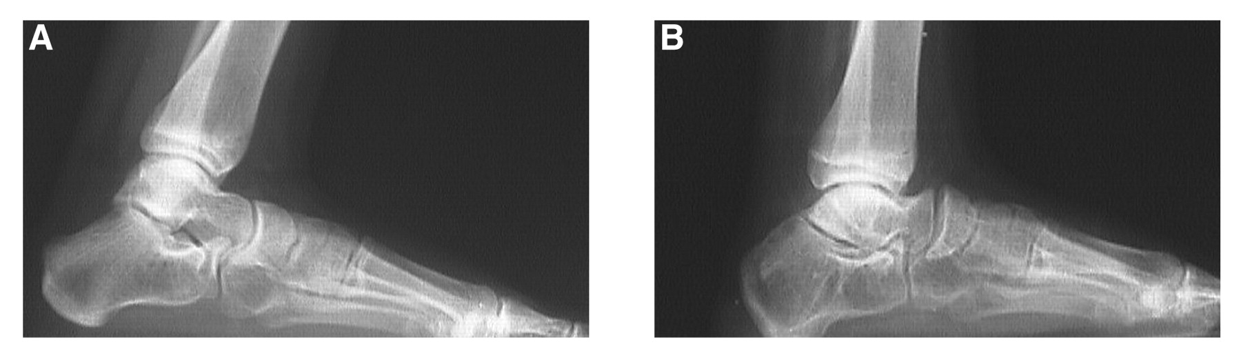
Radiographic angular measurements (anteroposterior and lateral talocalcaneal, talocalcaneal index, talar–first metatarsal, Böhler’s, and calcaneal pitch) were significantly similar between normal feet and clubfeet (P > .05) (Table 1). Whereas the length of the navicular bone ranged from 9 to 15 mm (mean, 12 mm) in normal feet, it measured 7 to 13 mm (mean, 10.5 mm) in clubfeet on anteroposterior views. It measured 8 to 15 mm (mean, 11.5 mm) and 6 to 13 mm (mean, 9.5 mm), respectively, on lateral views (Fig. 3A). The mean navicular bone length of clubfeet was found to be significantly smaller than that of normal feet (P < .001) (Table 2). The length of the talus ranged from 41 to 52 mm (mean, 48 mm) in normal feet and from 37 to 50 mm (mean, 43 mm) in clubfeet. Shortening of the talus was significant (P < .001) (Table 2).
Congruency of the tibiotalar joint relationship was not physiologic in two patients. The talar dome was flattened minimally in seven patients and was flattened, sclerotic, and irregular in one patient. Flattening of the talar head was detected in eight patients, irregularity in one, and deformity and sclerosis in one (Fig. 3B). Six patients had a deformity in the talonavicular joint relationship (P < .05). The articular distance of the talonavicular joint in the operated foot was similar to that in normal feet, and its average was 2.5 mm (range, 1–5 mm). Two patients had developmental retardation in the navicular bone. The navicular bone was wedge shaped, and the inferior aspect was hypoplastic and protruded dorsally approximately 3 mm (range, 2–5 mm) in seven patients (Table 2). The relation of the calcaneocuboid joint was normal in all of the patients. Cavus deformity in the calcaneal bone and increased calcaneal pitch angle (40°) due to 5 mm of dorsal subluxation of the navicular bone was found in one patient. Talonavicular congruency distance revealed that talar head coverage by the navicular bone was not detected in surgical feet versus normal feet (range, 1–6 mm; P < .001) (Table 2).
Discussion
Congenital clubfoot is one of the more common congenital deformities of the foot.[1] The foot is plantarflexed at the ankle and subtalar joints, the hindfoot is inverted, and the midfoot and forefoot are adducted, inverted, and in equinus position.[1] The aim of treatment is to provide a mobile foot with normal weightbearing and function.[1],[11]
Measuring the range of motion of the ankle is important in the assessment of corrected clubfeet.[9],[16] Bensahel et al[10] accepted a range of motion of 45° to achieve the best score result. Simons[15] indicated that ankle range of motion of at least 25° must be present for a satisfactory result. McKay[17] defined a range of motion less than 35° as unsatisfactory. In the present study, we used Simons’ criteria for assessment of patients clinically. In the present patients, mean ankle range of motion was 45° (range, 25°–60°). We agree that the best indication of a satisfactory result is that the patient and parents are pleased with the function and the cosmetic appearance of the foot after clubfoot surgery. When the foot is painless, the range of motion of the ankle is satisfactory, and the ankle does not hinder daily and sporting activities, the results of the clubfoot surgery are acceptable and successful.
Aronson and Puskarich[8] considered radiographs to be much more important than a clinical evaluation because of their reliability. During treatment, radiographs are obtained to decide whether concentric reduction has been achieved and is being maintained.[1] In this study, ultimate control radiographs revealed angles between the reference ranges (Table 1). However, Ponseti et al[6] reported decreased talocalcaneal angles in clubfeet compared with normal feet. Haas-beek and Wright[18] and Macnicol et al[19] stated that the total ranges of ankle motion and the talocalcaneal index were correlated with the eventual surgical outcome.
In this study, compared with normal feet, significant shortening of the talar and navicular bones was detected in treated patients. Ponseti et al[6] used a different surgical technique on their patients, although they detected wedge-shaped navicular bones, similar to the present patients. DePuy and Drennan[12] described patients who were treated with posteromedial release before 6 months of age and demonstrated better clinical results and less flattening and shortening of the talar head and navicular bone radiographically. Despite the use of three different surgical techniques, the same radiographic changes occurred in the navicular and talar bones. Therefore, the wedge shape of the navicular bone may be due to stretching exercises, manipulative casting, or the nature of the disease.
Avascular necrosis of the talus is a serious potential complication of clubfoot surgery, and the necrosis may involve the entire talus and result in progressive fragmentation and collapse.[20] Aplington and Riddle[21] reported 14.3% avascular necrosis of the talus after medial and lateral clubfeet surgery. In the literature, it has been reported that typical avascular necrosis of the talus is not common, but flattening of the dome is reported often.[17],[20] Decreased convexity of the talar dome has been reported in up to 100% of patients after the treatment of congenital clubfeet.[3],[6],[14],[22],[23] The incidence of talar flattening was inconsistent in different series. Cooper and Dietz[2] found 68% talar flattening in their patients after surgery; Hutchins et al,[4] 74%; Ponseti et al,[6] 56%; and Cummings et al,[20] 27%. Flattening of the talar dome does not indicate avascular necrosis.[20] The etiology of talar flattening is uncertain, but it may be an artifact of positioning, related to a pressure necrosis with manipulative casting, or from vascular disruption during surgery.[3],[9],[18] We detected flattening of the talar dome in 72% of the patients, but we never encountered typical avascular necrosis of the talus. Hjelmstedt and Sahlstedt[13] stated that if pronounced flattening of the trochlea of the talus occurred, normal mobility could not be seen in their arthrographic studies. However, Bach et al[9] reported that the degree of talar flattening correlated significantly with dynamic range of ankle motion but not with static mobility. We did not encounter this result in the present patient group.
In the present patients, flattening at the talar head and navicular bone was often seen radiographically. Bello et al[24] reported that flattening of the talar head, irregularity, and narrowing of the talonavicular joint occurred more frequently in the two-incision group (posteromedial and lateral incisions) but did not occur in the complete subtalar release group. However, a flat-top talus and a short talar neck were seen equally in the posteromedial, posteromedial and lateral, and complete subtalar release groups.[24] Ponseti et al[6] stated that flattening of the navicular bone was observed in 40% of patients on anteroposterior views only, in 6% on lateral views only, and in 21% on anteroposterior and lateral views. DePuy and Drennan[12] reported that flattening of the talar head and navicular bone increased depending on age at treatment, ranging from 7% to 35%.
A wedged-shaped navicular bone and dorsal migration of the bone were detected frequently in the present study. Ponseti et al[6] reported that the navicular bone was wedge shaped on 53% of the anteroposterior views only, on 9% of the lateral views only, and on 34% of anteroposterior and lateral radiographic views. Bello et al[24] stated that a wedge-shaped navicular bone and avascular necrosis of the navicular were noted in 37% of the two-incision group (posteromedial and lateral incisions) and in 6% of the posteromedial group but not in the complete subtalar release group. However, dorsal subluxation of the navicular bone occurred in 40% of each group.[24] Kuo and Jansen[25] found a 7% incidence of dorsal subluxation of the navicular bone after clubfoot release and required additional surgery to address residual cavovarus deformity. In the present series, dorsal migration of the navicular bone was encountered in 63% of the patients, with one patient showing radiographic changes of cavovarus deformity (calcaneal pitch angle, 40°). This patient had not suffered clinically. Dorsal subluxation of the navicular bone is a result of insufficient release of the tibionavicular ligament and the dorsolateral talonavicular joint capsule.[1]
The critical degree of migration and the timing of surgery have not been determined. It is proposed that the foot has to be operated on when the cavovarus deformity exists clinically.[1] The radiographic appearance of talonavicular congruence is different between normal and operated feet. These differences regarding the talonavicular joint may be due to anatomical variations (P < .001). The English-language literature did not reveal such data. Flattening of the talar head or the navicular bone or irregularity of the talonavicular joint may cause these differences.
In conclusion, radiographic changes, such as flattening (especially at the talar head and dome), a wedge-shaped navicular bone, dorsal navicular migration, irregularity, and lack of congruence of the talonavicular joint, can be encountered postoperatively despite clinically and cosmetically normal results. The lateral and anteroposterior talocalcaneal angles, the talocalcaneal index, and the talus–first metatarsal angles were found to be normal in the present patients. Radiographic changes may be caused by the nature of the disease, correcting manipulations or casting, and limited or extensive surgical techniques. Single or multiple factors may play a role.
This study revealed that poor radiographic results may accompany good functional results. This fact needs be supported by larger series with longer follow-up. In our opinion, radiographic findings may be important for the treatment and follow-up of clubfoot, but the satisfaction of the parents and patients who are free of complaints and have sufficient ankle range of motion are more important.
Complete subtalar release is an effective surgical procedure for satisfactory clinical results in the treatment of clubfoot. The maintenance of anatomical configuration but not the normal anatomical development of tarsal bones can be achieved by using this method.

Table 1.
Standard Radiographic Angles in the 11 Study Patients.
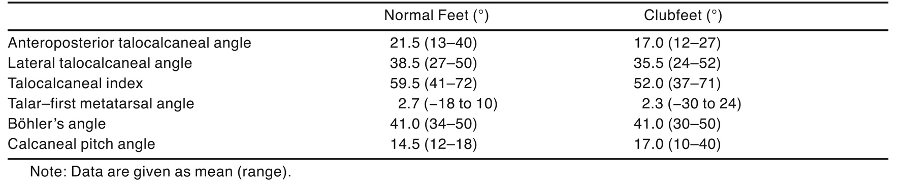
Table 2.
Radiographic Measurement Results in the 11 Study Patients.
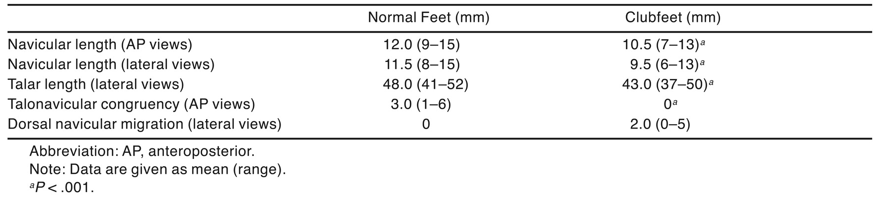
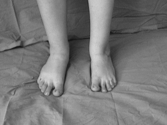
Figure 1.
Clinical appearance of normal (right) and operated (left) feet.
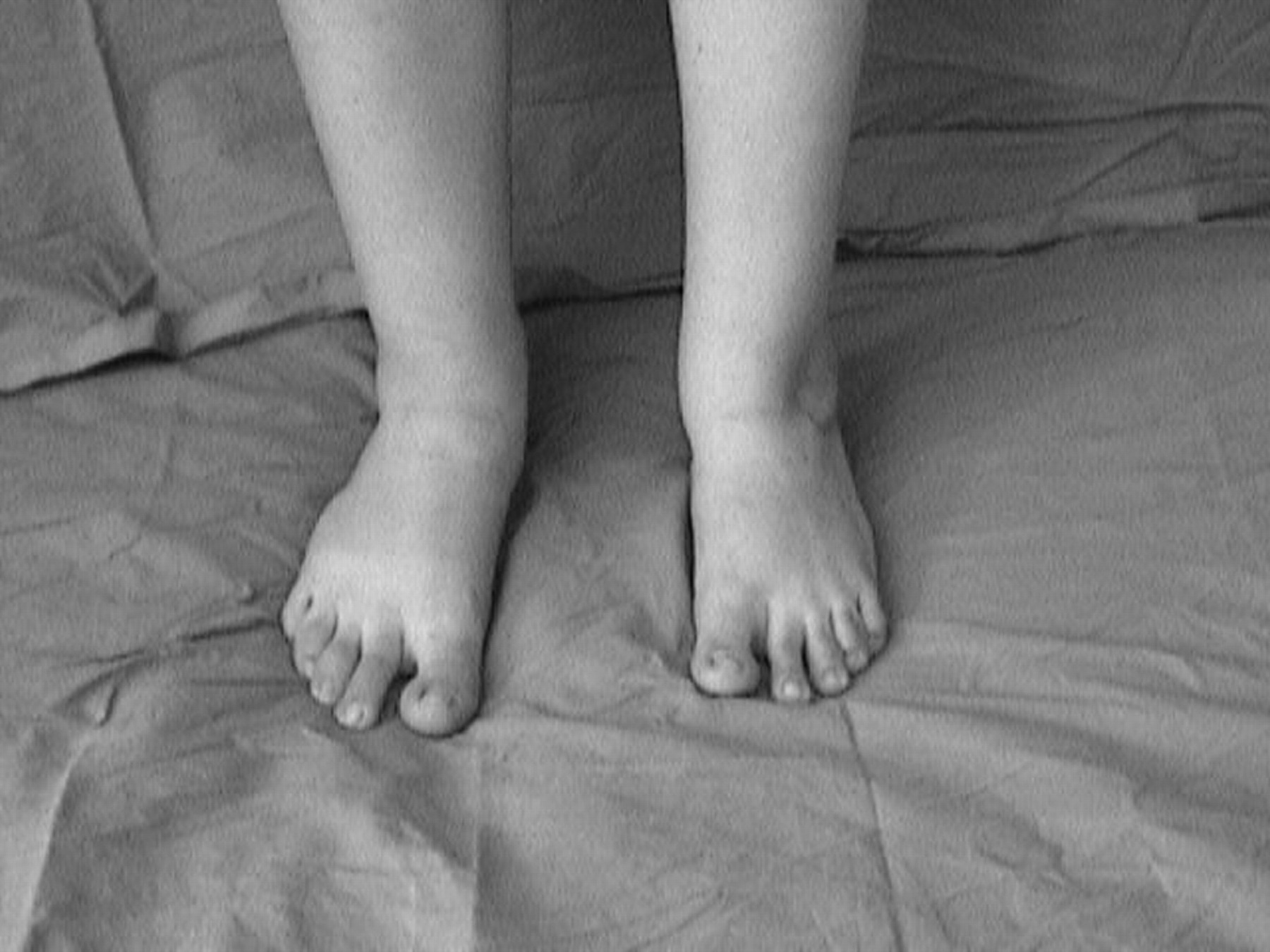
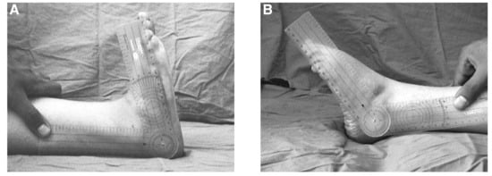
Figure 2.
Dorsiflexion (A) and plantarflexion (B) range of motion of the ankle of the operated foot.
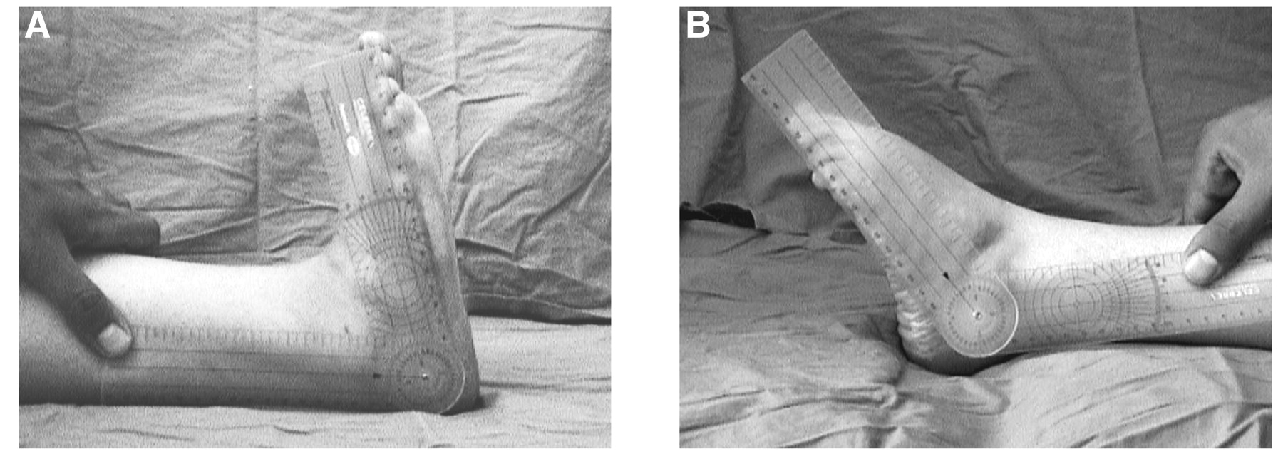
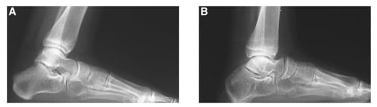
Figure 3.
A, Radiographic appearance of the normal foot. B, Wedge-shaped navicular bone and flattening of the talar head of the operated foot.
Acknowledgments
Nusret Köse, MD, Osmangazi University Hospital, Eskisehir, Turkey, for his contribution to the study.
Financial Disclosure: None reported.
Conflict of Interest: None reported.
References
- Herring JA (ed): Tachdjian’s Pediatric Orthopaedics: Disorders of the Foot, p 942, WB Saunders Co, Philadelphia. ,2002. .
- Cooper DM, Dietz FR: Treatment of idiopathic clubfoot: a thirty-year follow up note. .J Bone Joint Surg Am77::1477. ,1995. .
- Dunn HK, Samuelson KM: Flat top talus: a long-term report of twenty club feet. .J Bone Joint Surg Am56::57. ,1974. .
- Hutchins PM, Foster BK, Paterson DC, et al: Long-term results of early surgical release in club feet. .J Bone Joint Surg Br67::791. ,1985. .
- McKay DW: New concept and approach to clubfoot treatment: section II. Correction of the clubfoot. .J Pediatr Orthop3::10. ,1983. .
- Ponseti IV, El-Khoury GY, Ippolito E, et al: A radiographic study of skeletal deformities in treated clubfeet. .Clin Orthop160::30. ,1981. .
- Simons GW: Complete subtalar release in club feet: part I. A preliminary report. .J Bone Joint Surg Am67::1044. ,1985. .
- Aronson J, Puskarich CL: Deformity and disability from treated clubfoot. .J Pediatr Orthop10::109. ,1990. .
- Bach CM, Wachter R, Stockl B, et al: Significance of talar distortion for ankle mobility in idiopathic clubfoot. .Clin Orthop398::196. ,2002. .
- Bensahel H, Dimeglio A, Souchet P: Final evaluation of clubfoot. .J Pediatr Orthop4::137. ,1995. .
- Van Campenhout AV, Molenaers G, Moens P, et al: Does functional treatment of idiopathic clubfoot reduce the indication for surgery? Call for a widely accepted rating system. .J Pediatr Orthop B10::315. ,2001. .
- DePuy J, Drennan JC: Correction of idiopathic clubfoot: a comparison of results of early versus delayed posteromedial release. .J Pediatr Orthop9::44. ,1989. .
- Hjelmstedt EA, Sahlstedt B: Arthrography as a guide in the treatment of congenital clubfoot: findings and treatment results in consecutive series. .Acta Orthop Scand51::321. ,1980. .
- Napiontek M: Clinical and radiographic appearance of congenital talipes equinovarus after successful nonoperative treatment. .J Pediatr Orthop16::67. ,1996. .
- Simons GW: Complete subtalar release in club feet: part II. Comparison with less extensive procedures. .J Bone Joint Surg Am67::1056. ,1985. .
- Rumyantsev NJ, Ezrohi VE: Complete subtalar release in resistant clubfeet: a critical analysis of results in 146 cases. .J Pediatr Orthop17::490. ,1997. .
- McKay DW: New concept and approach to clubfoot treatment: section III. Evaluation and results. .J Pediatr Orthop3::141. ,1983. .
- Haasbeek JF, Wright JG: A comparison of the long-term results of posterior and comprehensive release in the treatment of clubfoot. .J Pediatr Orthop17::29. ,1997. .
- Macnicol MF, Nadeem RD, Forness M: Functional results of surgical treatment in congenital talipes equinovarus (clubfoot): a comparison of outcome measurements. .J Pediatr Orthop B9::285. ,2000. .
- Cummings RJ, Bashore CJ, Bookout CB, et al: Avascular necrosis of the talus after McKay clubfoot release for idiopathic congenital clubfoot. .J Pediatr Orthop21::221. ,2001. .
- Aplington JP, Riddle CD: Avascular necrosis of the body of the talus after combined medial and lateral release of congenital clubfoot. .South Med J69::1037. ,1976. .
- Miller JH, Bernstein SM: The roentgenographic appearance of the corrected clubfoot. .Foot Ankle6::177. ,1986. .
- Szabo G, Kranicz J, Bellyei A: “Effects of Soft Tissue Release on the Development of the Tarsal Bones in Clubfeet,” in The Clubfoot: The Present and a View of the Future, ed by G Simons, p 449, Springer-Verlag, New York. ,1994. .
- Bello, et al: A radiographic analysis of the congenital clubfoot: a comparative study three reconstructive procedures. Presented at: Annual Meeting, Pediatric Orthopaedic Society of North America: May 1993, White Sulfer Springs, WV..
- Kuo KN, Jansen LD: Rotatory dorsal subluxation of the navicular: a complication of clubfoot surgery. .J Pediatr Orthop18::770. ,1998. .
© 2008 American Podiatric Medical Association