


Abstract
Neuropathic symptoms in patients with diabetes occur commonly and are most often a consequence of the diabetes. Up to 10% of patients with diabetes and neuropathy have an etiology other than diabetes as a cause of their nerve dysfunction. Herein we present a case of vasculitic neuropathy initially misdiagnosed as diabetic neuropathy that led to separate amputations of two toes. This case emphasizes the importance of considering alternative, potentially treatable, causes of peripheral neuropathy in patients with diabetes.
Peripheral neuropathy is a common complication of diabetes and may affect 50% to 90% of patients with diabetes, depending on the diagnostic tests used to define nerve damage.[1] This fact may lead clinicians to incorrectly attribute symptoms or signs of peripheral neuropathy in diabetic patients to diabetes. Indeed, the Rochester Diabetic Neuropathy Study[2] demonstrated that up to 10% of patients with diabetes have a cause other than diabetes for their neuropathy. Some of these other causes are potentially treatable, which may reverse neuropathic deficits and, hence, reduce the risk of neuropathic ulceration and amputation. This case illustrates the consequences of initial misdiagnosis of diabetic neuropathy.
Case History
A 37-year-old man presented to St. Vincent Catholic Medical Centers of Brooklyn/Queens with ulceration and an abscess, with radiographic evidence of osteomyelitis of his right second toe. He complained of a “dead numbness” in his feet for 5 years that had been gradually worsening and never improving. His relevant medical history included amputation of his right hallux 1 year earlier, when he had also been diagnosed as having type 2 diabetes and gastroesophageal reflux disease. His father had undergone a hallux amputation secondary to osteomyelitis at age 50 years, but he did not have diabetes, and he died of a myocardial infarction at age 58 years. The patient had a 20 pack-year history of smoking and consumed two to three alcoholic drinks per week.
Neurologic examination showed that his cranial nerves were intact. He had a negative Romberg’s sign and no evidence of dysdiadochokinesia. Neurologic examination of his lower limbs demonstrated normal muscle tone and no evidence of clonus. Muscle strength was a grade 5 on the Medical Research Council scale in all muscle groups of the upper and lower extremities, except for symmetrical weakness of the interossei and abductor pollicis of the hands (Medical Research Council scale grade 4/5) and some difficulty in rising to his heels and toes. Deep tendon reflexes were normal in the upper extremities and patellae, but the Achilles reflex was absent symmetrically. Lasegue’s maneuver did not elicit pain, and there was no Tinel’s sign with percussion of the tibial or median nerves. Sensory examination findings were normal in the upper extremities (light touch, vibration, sharp/dull, and joint position sense). The lower extremities revealed deficits in light touch and vibration sense (vibration perception threshold of 35 V in the right and 30 V in the left hallux) in the feet on the plantar and dorsal aspects. Pressure perception using the 5.07 Semmes-Weinstein monofilament was absent in both feet. The feet had normal-appearing arches without clawing or hammering of digits. Dermatologic examination demonstrated no evidence of café-au-lait spots or angiokeratomas.
Laboratory investigation revealed slight anemia (hemoglobin level, 12.8 g/dL) but normal renal function. The coagulation panel, urine albumin-creatinine ratio, thyroid profile, and B12 were all within normal limits. The erythrocyte sedimentation rate and C-reactive protein values were elevated. Serum electrophoresis demonstrated no evidence of monoclonal gammopathy. Results of urine heavy metal screening were negative. Genetic testing for hereditary neuropathies was not performed. Electrophysiologic testing revealed only mildly prolonged motor nerve conduction velocities and reduced amplitude of motor and sensory action potentials in the lower extremities bilaterally. Some fibrillations were noted in the distal muscles of the upper and lower extremities, consistent with a distal symmetrical polyneuropathy.
The positive family history of a digital amputation in this patient’s father and the presence of neuropathic symptoms years before the diagnosis of diabetes are not typical of those with diabetic neuropathy and led us to perform a sural nerve biopsy as a more definitive diagnostic test.
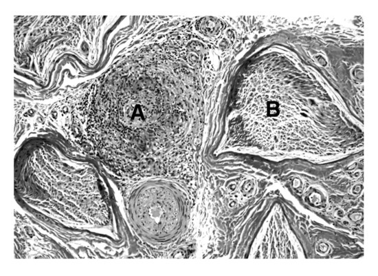
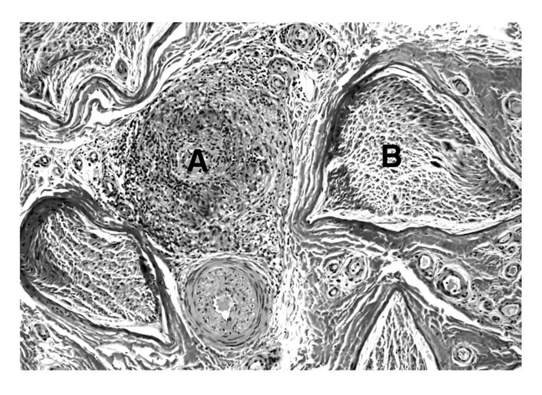
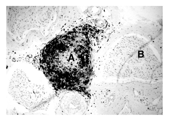
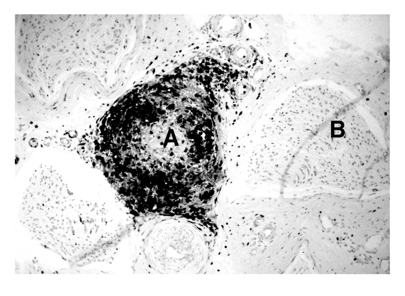
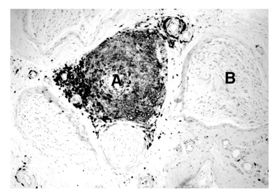
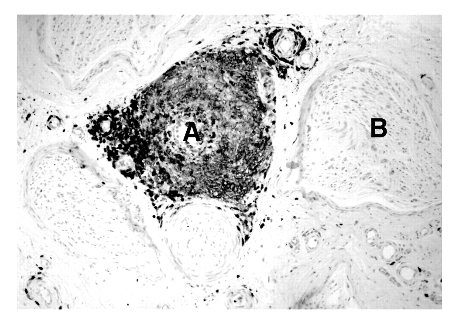
A sural nerve biopsy sample taken in the manner described by Bevilacqua et al[3] was sent to the Peripheral Nerve Center at Mayo Clinic (Rochester, Minnesota) for review. The histopathology report noted severely decreased myelinated fiber density in all of the fascicles. In addition, there was perivascular epineurial inflammation of medium and large vessels (Figure 1). Immunohistochemical analysis showed an infiltrate that was positive for CD68 cells (macrophages) (Figure 2) and CD45 cells (lymphocytes) (Figure 3), which was diagnostic of a granulomatous vasculitis.
Figure 1.
A section of sural nerve. A medium-sized arteriole (A) is visibly infiltrated with inflammatory cells and rests between nerve fascicles (B). (H&E, ×100).
Figure 2.
KP-1 immunohistochemical stain for CD68 cells (neutrophils) that are infiltrating the wall of the arteriole (A) and nearly occluding the lumen, evidence of necrotizing vasculitis. A neighboring nerve fascicle (B) is also visible.
Figure 3.
Leukocyte common antigen immunohistochemical stain for CD45 cells (lymphocytes) reveals these cells surrounding the macrophages in the arteriole wall (A). This is diagnostic of granulomatous vasculitis. The same nerve fascicle (B) is visible on the right.
Discussion
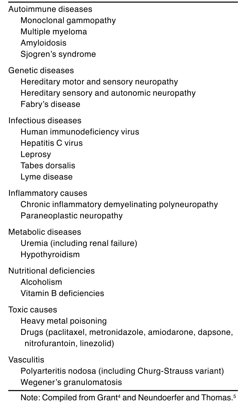
A key lesson herein is the need to take a clear and thorough history, which can reveal inconsistencies in the timeline of neuropathy in a diabetic patient suggestive of an alternate cause. The neurologic examination should include the upper and lower extremities, including assessment for cranial nerve deficits and autonomic dysfunction. A list of etiologies for neuropathies that may have signs and symptoms resembling diabetic peripheral neuropathy is given in Table 1.[4,5] Typically, the onset of neuropathic symptoms occurs 8 to 12 years after the presence of diabetes.[6] Occasionally, foot ulcers can be the presenting complaint in those with undiagnosed diabetes. However, the present patient’s presentation did not fit a protracted course of delayed diagnosis of diabetes. Furthermore, his father’s history of toe amputation in the absence of diabetes, despite a heightened sense of suspicion and a detailed work-up, triggered the decision for nerve biopsy.
Table 1.
Differential Diagnosis of Diabetic Neuropathy.
Vasculitis may be systemic or localized and can occur as a primary autoimmune disorder or secondary to infection, drugs, malignancy, or systemic lupus erythematosus.[7] It is usually classified by the size of the vessels involved (small, medium, or large). The neurologic symptoms of necrotizing vasculitis are caused by occlusion of the epineurial arterioles.[8,9] Vasculitic neuropathy of medium-sized arterioles (as in the present patient) is suggestive of polyarteritis nodosa or Wegener’s granulomatosis.[9,10] In a large review[11] of patients with Wegener’s granulomatosis, 34% had neurologic involvement, half of whom had peripheral neuropathy. Neurologic manifestations in Wegener’s granulomatosis generally present as mononeuritis multiplex, but they can also present as a distal symmetrical neuropathy, as in the present patient.[7,11,12] Peripheral neuropathic symptoms in patients with granulomatous vasculitis can be treated with highdose corticosteroids, immunosuppression, or plasmapheresis, which can reverse symptoms and signs and improve the prognosis.[10] The present patient illustrates this point because his amputations might have been prevented by earlier diagnosis and appropriate treatment, for which he was referred to the rheumatology department after pathologic diagnosis.
A diabetic patient with unusual neuropathic symptoms and a history of autoimmune disease, a family history of nondiabetic neuropathies, or a pattern other than a length-dependent one should be reviewed by a neurologist or specialist in peripheral neuropathy. Treatable causes of peripheral neuropathies should be excluded in these patients to avoid sequelae.
Funding
None reported.
Acknowledgments
Peter James Dyck, MD, and P. James B. Dyck, MD, of the Peripheral Nerve Center at The Mayo Clinic, Rochester, Minnesota for their help with the histology description in the preparation of this manuscript.
Conflicts of Interest
None reported.
References
- ADLER, A.I.; BOYKO, E.J.; AHRONI, J.H.; et al. Risk factors for diabetic peripheral sensory neuropathy: results of the Seattle Prospective Diabetic Foot Study. Diabetes Care 1997, 20, 1162. [Google Scholar] [CrossRef] [Scilit] [PubMed]
- DYCK, P.J.; KRATZ, K.M.; KARNES, J.L.; ET, A.L. The prevalence by staged severity of various types of diabetic neuropathy, retinopathy, and nephropathy in a population-based cohort: the Rochester Diabetic Neuropathy Study. Neurology 1993, 43, 817. [Google Scholar] [CrossRef] [Scilit] [PubMed]
- BEVILACQUA, N.J.; ROGERS, L.C.; MALIK, R.A. ET AL: Technique of the sural nerve biopsy. J Foot Ankle Surg 2007, 46, 139. [Google Scholar] [CrossRef] [Scilit]
- GRANT IA: “Differential Diagnosis of Diabetic Neuropathies,” in Diabetic Neuropathy, 2nd Ed, ed by PJ Dyck, PK Thomas, p 415, WB Saunders, Philadelphia, 1999. Dyck, P.J.; Thomas, P.K. (Eds.) .
- NEUNDOERFER, B.; THOMAS, P.K. Diabetic Sensorimotor Neuropathy: Clinical Features; Thieme: Stuttgart, 2003. [Google Scholar]
- PARTANEN, J.; NISKANEN, L.; LEHTINEN, J.; et al. Natural history of peripheral neuropathy in patients with non-insulin-dependent diabetes mellitus. N Engl J Med 1995, 333, 89. [Google Scholar] [CrossRef] [Scilit] [PubMed]
- ROANE, D.W.; GRIGER, D.R. An approach to diagnosis and initial management of systemic vasculitis. Am Fam Physician 1999, 60, 1421. [Google Scholar] [PubMed]
- SAID G: Necrotizing peripheral nerve vasculitis. Neurol Clin 1997, 15, 835. [CrossRef] [Scilit] [PubMed]
- OHKOSHI, N.; MIZUSAWA, H.; OGUNI, E.; et al. Sural nerve biopsy in vasculitic neuropathies: morphometric analysis of the caliber of involved vessels. J Med 1996, 27, 153. [Google Scholar] [PubMed]
- HAWKE, S.H.; DAVIES, L.; PAMPHLETT, R. ET AL: Vasculitic neuropathy: a clinical and pathological study. Brain 1991, 114(pt 5), 2175. [Google Scholar] [PubMed]
- NISHINO, H.; DEREMEE, R.A.; RUBINO, F.A.; et al. Wegener’s granulomatosis associated with vasculitis of the temporal artery: report of five cases. Mayo Clin Proc 1993, 68, 115. [Google Scholar] [CrossRef] [Scilit] [PubMed]
- NISHINO, H.; RUBINO, F.A.; PARISI, J.E. The spectrum of neurologic involvement in Wegener’s granulomatosis. Neurology 1993, 43, 1334. [Google Scholar] [CrossRef] [Scilit] [PubMed]

© 2008 American Podiatric Medical Association