


Abstract
Background: The plain film forefoot axial radiographic view is a specialized view in podiatric imaging. In everyday office practice, an axial orthoposer is used, and the study is nonweightbearing. Largely useless for imaging functional pathology, the forefoot axial view’s main use centers around imaging of the plantar metatarsal heads and the hallucal sesamoid complex. To this end, the name “forefoot” is somewhat of a misnomer, and the view is also known as the sesamoid axial study.
Methods: We report on the initial use of oblique sesamoid or “skyline” axial studies for juxtacortical lesions of the sesamoid bones and more proximal dorsolateral tarsal structures, an area traditionally difficult to assess accurately with plain film imaging.
Results: Rather simple, empiric modifications in the forefoot axial technique allowed for 1) “skyline” imaging of exostotic surface lesions involving the dorsal, medial, and lateral midfoot bones, and 2) significant improvement in imaging hallucal sesamoid pathology.
Conclusions: Although the proposed oblique forefoot axial modifications empirically add two additional plain film radiographic studies to a standard forefoot axial study, our initial study suggests that the imaging benefits far outweigh the liabilities. This is especially true when one considers the difficulties encountered when employing standard plain film pedal in an effort to clarify or delineate pathologic processes involving the midfoot bones or metatarsal bases. It is hoped that this study will spawn further use and research of these “novel” views.
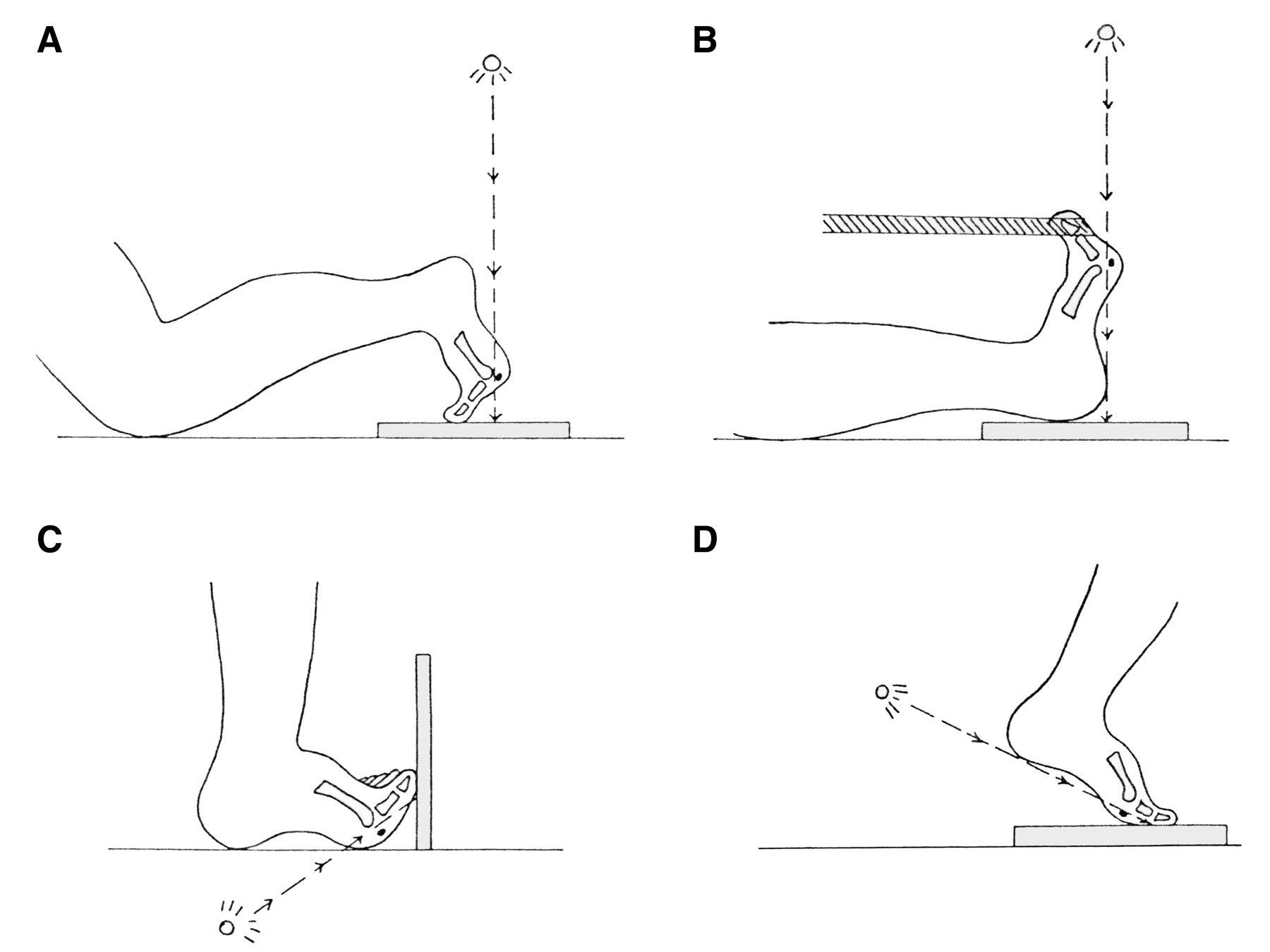
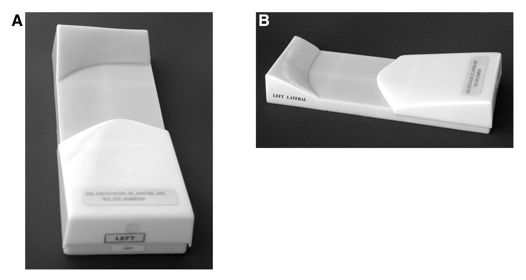
The first use of the axial sesamoid roentgenographic technique is ascribed to Muller. [1] Since then, nonweightbearing methods have been described by Lewis, [2] Holly, [3] and Chang et al, [4] along with the Viladot [5] (Puerta) modification of the Lewis method (Fig. 1). All of these views are “qualitative” methods for “skyline” axial sesamoid evaluation in orthopedic medicine. Whereas hospital radiographers typically acquire these views without the use of ready-made positioning devices, modern-day podiatric sesamoid axial imaging typically uses a prefabricated plastic orthoposer (Gill Podiatry Supply & Equipment Co, Middleburg Heights, Ohio) (Fig. 2). This “posed” method for obtaining the axial sesamoid (often called the “forefoot axial”) view was first described by Downey and Dorothy [6] in 1969. Largely developed for coronal plane imaging of the plantar forefoot, the technique specified that balsa wood wedges be placed beneath the heel and toes, thereby creating a prominent plantar “apex” at the level of the metatarsophalangeal joints. By elevating the heel and extending the digits via the aforementioned wedges, superimposition of the phalanges is all but eliminated (Fig. 3). Gamble and Yale [7] describe several other frontal plane imaging methods that seem to be modifications of the method of Downey and Dorothy. [6] If just the heel wedge is used, this special axial modification can clarify the more proximal first and second metatarsocuneiform segments. If only a forefoot wedge is used (thereby positioning the ankle in a calcaneus position), the view then profiles the plantar calcaneus. In 1987, Dreeben et al [8] proposed a new weightbearing method for axial radiography of the metatarsal heads using a specially designed platform. Although the technique showed promise for accurate “quantitative” evaluation of coronal plane structural (and perhaps functional) variations of the metatarsal heads, the technique has not experienced wide acceptance. [8]
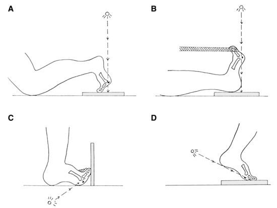
Figure 1.
Illustrations of Lewis (A), Holly (B), Viladot (C), and Meschan (D) sesamoid (forefoot) axial techniques. (Reprinted with permission from Chang et al. [4])
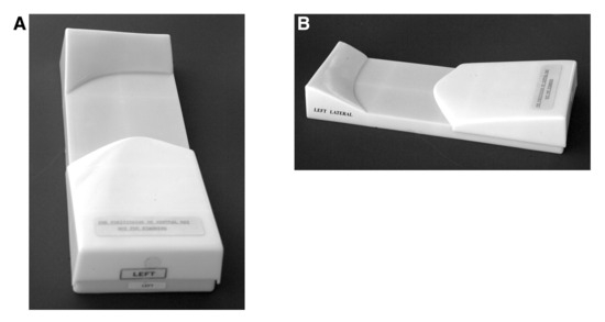
Figure 2.
Top rear (A) and side (B) views of the left forefoot axial orthoposer.
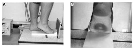
Figure 3.
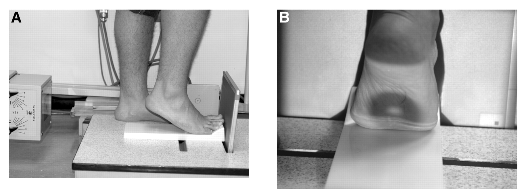
Side (A) and posterior (B) views demonstrating patient positioning and the optimal central ray target location for the modified “rectus” forefoot axial study. Note that the collimation is wide and that the heel elevation is increased to approximately 40°.
We discovered that some simple modifications of the forefoot axial study not only facilitate viewing of the hallucal sesamoid bones but can also enhance coronal plane skyline imaging of the more proximal forefoot and midfoot osseous structures. Between May 1, 2002, and June 30, 2005, these new views were successfully used on a variety of patients at the Cleveland Foot and Ankle Clinic, as illustrated by the following four case presentations. Note that in one case the modified plain film radiographic study “outperformed” preoperative magnetic resonance imaging (MRI).
Methods
Radiographic views were obtained with a fixed-pivot-arm (28-inch SID) radiography machine (X-Cel X-Ray Corp, Crystal Lake, Illinois), with kilovoltage and milliamperage settings of 65 kV(p) and 15 mA, respectively, and with an exposure time of 21/60th of a second. These settings may be adjusted per the discretion of the technician depending on variables such as bandages, edema, age, and foot size. A standard view is taken first, followed by two additional views that require patient repositioning for oblique forefoot axial pedal imaging.
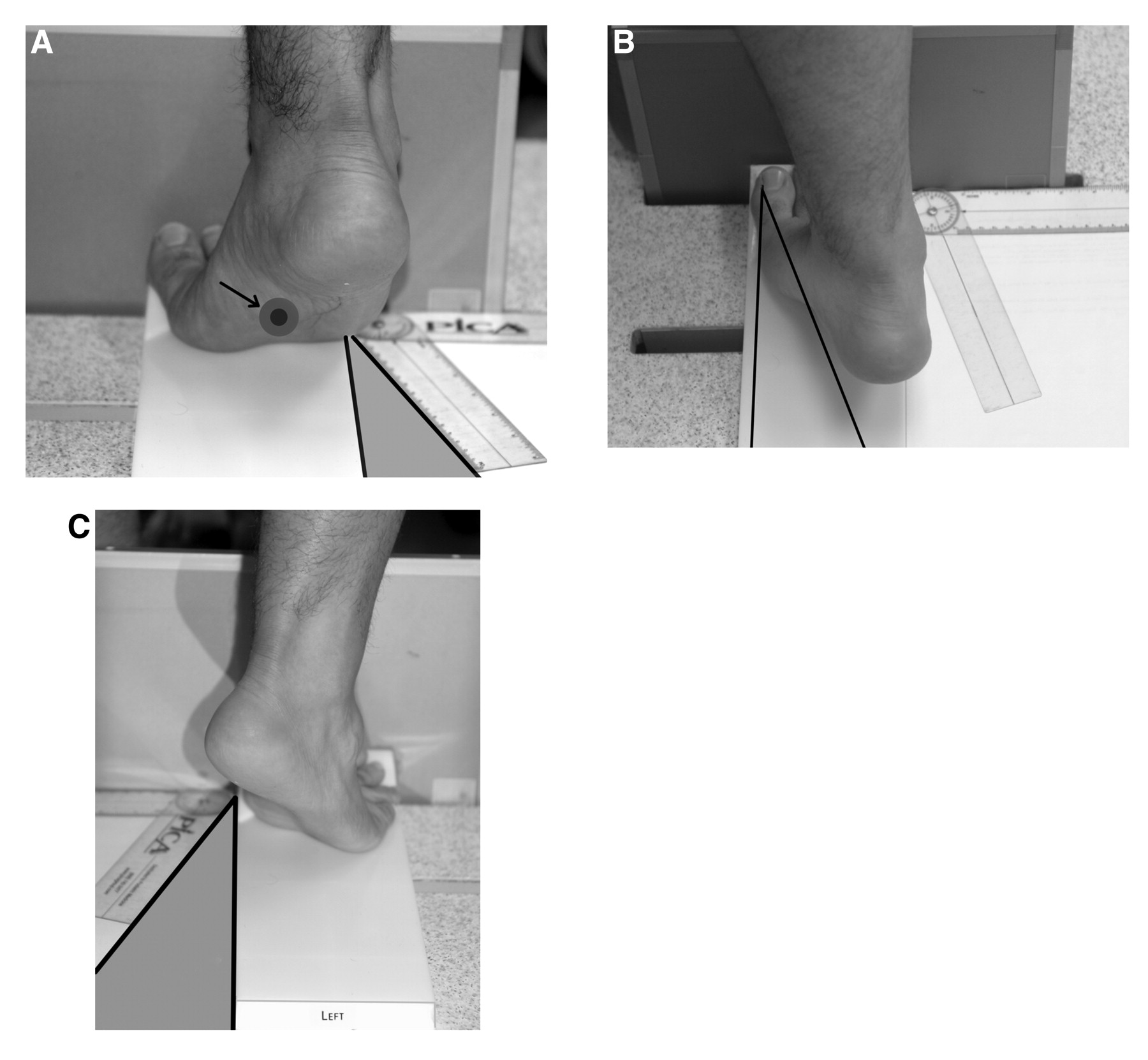
The initial setup for the three views is not unlike the method described by Downey and Dorothy [6] in that the film cassette is vertically oriented in the orthoposer slot, and the foot is placed on the forefoot positioner (Fig. 1) with the toes gently pushed up against the film. The orthoposer naturally extends the toes at the metatarsophalangeal joints to preset angles (Fig. 3). The tube head is then oriented at 90° to the film plane and is positioned posterior (caudad) to the patient, with the central ray coursing inferiorly from posterior to anterior. The plantar second metatarsal head is an optimal central ray target. However, unlike in the method of Downey and Dorothy, [6] the collimated field of view is widened to include the ankle, and the heel elevation is increased to a mean ± SD of 40° ± 5° (Fig. 4). The first view is acquired with the patient in this position.
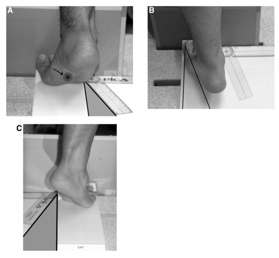
Figure 4.
A, Postero-plantar and B, posterior views of the foot. The arrow points to the optimal central ray target of the x-ray beam. Positioning for the adducted oblique forefoot axial study. Note the angle indicated by the tractograph. C, Positioning for the abducted oblique forefoot axial study.
The two additional oblique forefoot axial/sesamoid axial studies require internal (adduction) and external (abduction) transverse plane rotation of the foot by a mean ± SD of 30° ± 5°. For either view, the patient is asked to internally or externally rotate the foot approximately 30° while maintaining the same degree of heel elevation. The amount of rotation can be guided by using a tractograph (Fig. 4) or by using premade wedge templates that can be added to the front of the orthoposer. As can be noted from the figure, the abducted study is facilitated by using the orthoposer designed for imaging the contralateral foot. The central ray target is directed centrally toward either the ball of the foot or the midfoot, depending on the area of interest. In our practice, we do not use a heel wedge for forefoot axial views. Instead, the patient is asked to elevate the heel 35° to 45°. If hallux limitus is present and the sesamoid bones compose the area of interest, the patient is asked to elevate the heel until contact of the plantar first metatarsophalangeal joint with the orthoposer is lost. However, for midfoot imaging, patients with restricted dorsiflexion of the first metatarsophalangeal joint may lift the ball of the foot off the weightbearing surface while maintaining distal hallux (interphalangeal joint) contact.
Case Reports
Case 1
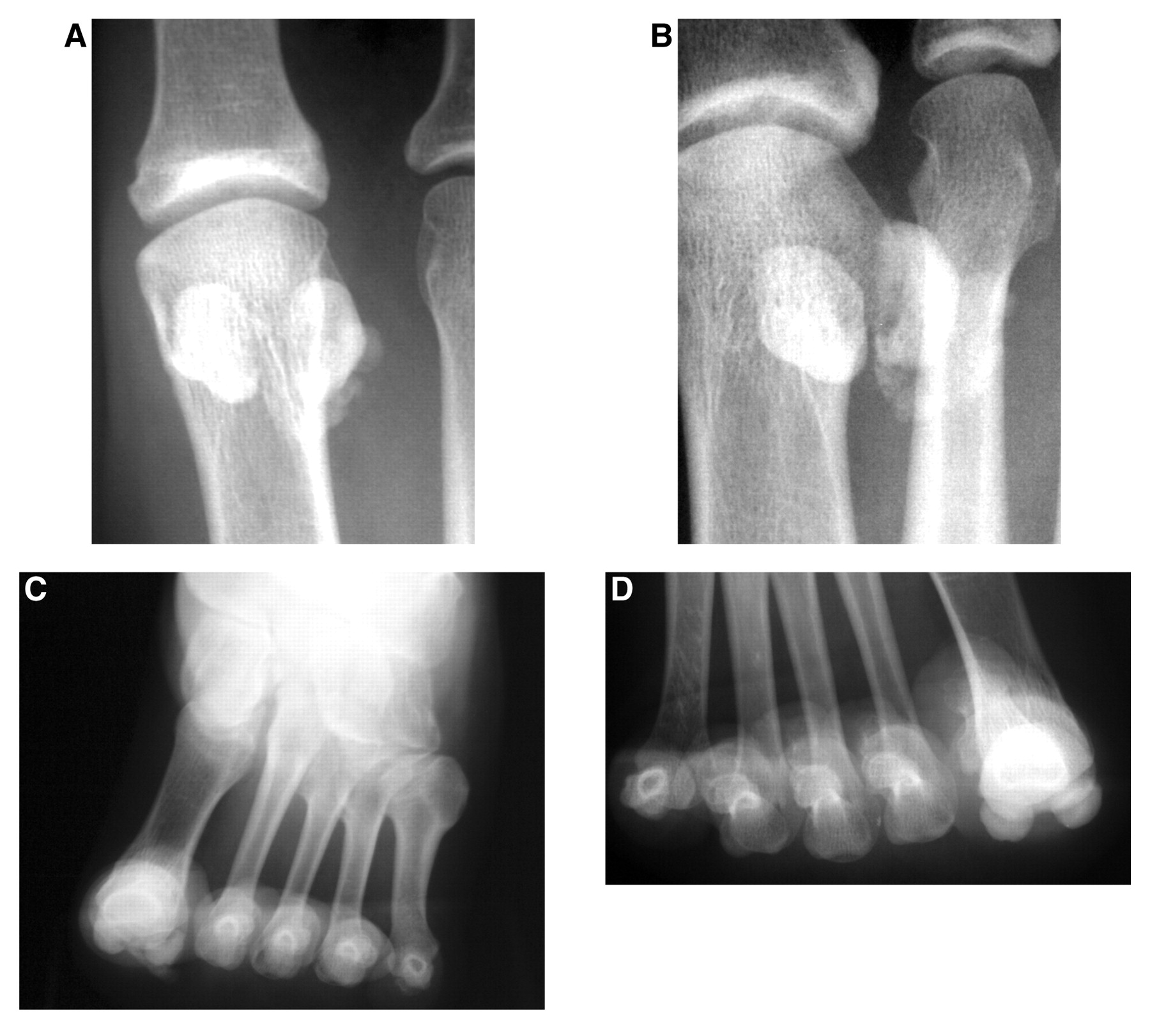
A 48-year-old nondiabetic woman presented to the Cleveland Foot and Ankle Clinic of the Ohio College of Podiatric Medicine with pain on the plantar aspect of her right hallux, the apex of which was centered over the plantar first metatarsophalangeal joint. The onset was traced to a treadmill injury approximately 7 months earlier, at which time acute pain and bruising along the medial arch were apparent. Swelling had been an inconsistent finding, however, and the quality of the pain had recently been of a burning nature, aggravated by walking or bearing weight for long periods. Although certain types of footwear (especially Birkenstock sandals) provided some relief, the pain “beneath her big toe” had recently become much worse. For financial reasons (lack of health insurance), the condition had not yet been medically managed. Standard anteroposterior and low-angle medial oblique radiographs of the right foot were obtained along with bilateral conventional forefoot axial studies (Fig. 5). The anteroposterior radiographic findings are generally suggestive of fibular sesamoid fragmentation and are reminiscent of Treves’/Renander’s disease (osteochondrosis). [9,10] However, the oblique and forefoot axial studies suggested the possibility of ossific or even calcific matrix formation in the soft tissues (capsular) adjacent to the fibular sesamoid. In the absence of any other information, the findings are best described as proliferative ossification along with fragmentation surrounding the fibular sesamoid, and without evidence of luxation or erosive changes. Differential diagnostic considerations should include avascular necrosis, osteochondrosis or osteochondritis, [11,12] post-traumatic heterotopic new bone formation, capsulosynovial calcifications (eg, synovial chondromatosis), and, remotely, neoplasm.
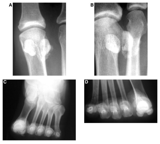
Figure 5.
Anteroposterior (A), medial oblique (B), and conventional forefoot axial (C) studies. D, Contralateral comparison study of the normal foot.
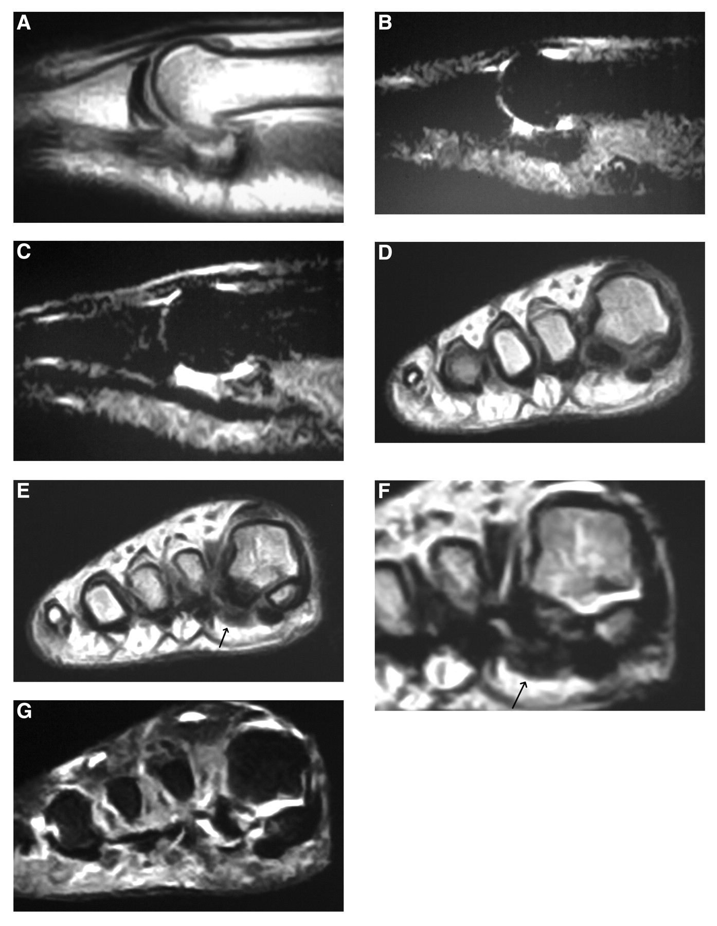
Because we anticipated future surgical intervention and were unable to clearly delineate the pathologic process, ancillary computed tomography (CT) or MRI was considered prudent. Although CT was preferred, for reasons of availability, cost, or both, only the MRI could be acquired (Fig. 6). The MRI was initially interpreted by an outside radiologist as demonstrating only mild, nonspecific periarticular soft-tissue edema with mild joint effusion (Fig. 6 A and B).
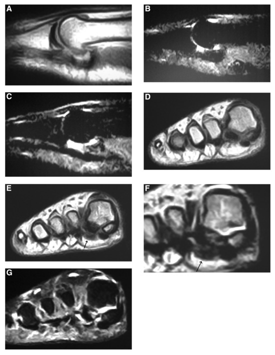
Figure 6.
A, Sagittal T1-weighted image of the first metatarsophalangeal joint. B and C, Sagittal short tau inversion recovery (STIR) images of the first metatarsophalangeal joint demonstrate mild to moderate periarticular/capsular edema. D and E, Coronal T1-weighted images at the level of the first metatarsal sesamoid complex. Lesion demonstrates a conspicuous low–signal intensity center with intermediate signal periphery (arrow). Note the absence of marrow fat/low–signal intensity fibular sesamoid. F, Close-up view of corresponding coronal T2-weighted image, with magnification. The lesion (arrow) and the contiguous fibular sesamoid somewhat similarly demonstrate heterogenous intermediate– and low–signal intensity areas, although a central low–signal intensity area dominates the lesion. G, Coronal STIR image. The low–signal intensity fibular sesamoid persists. See the “Case 1” subsection of the text for a full description.
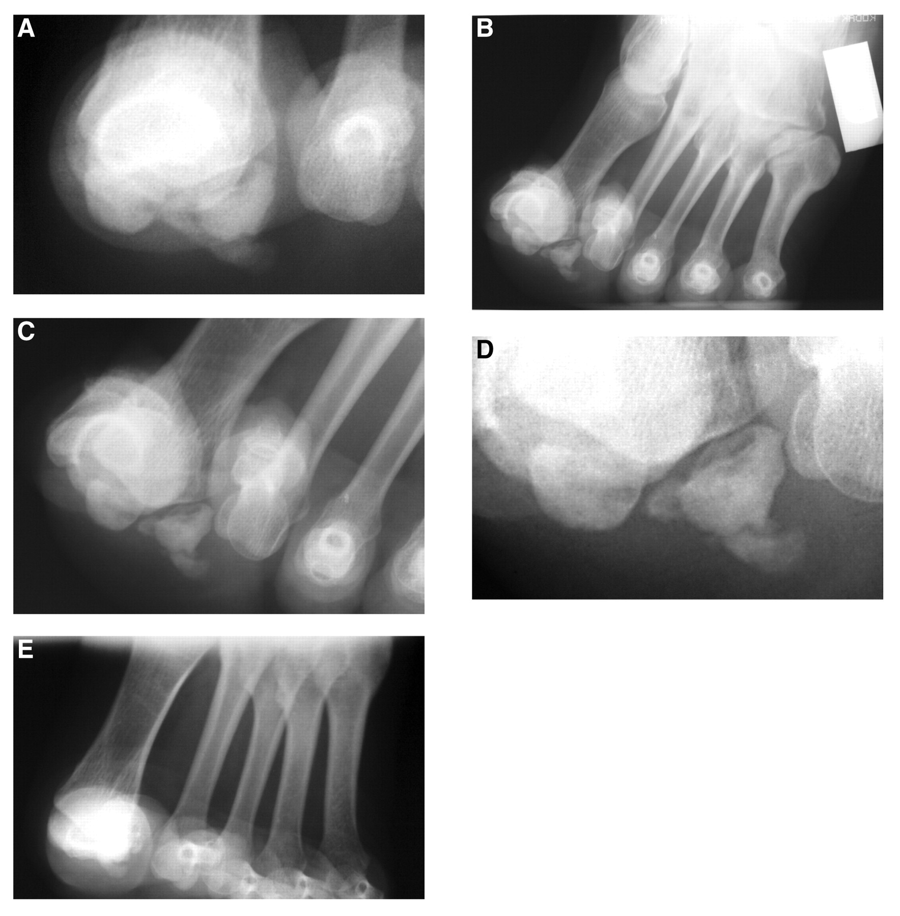
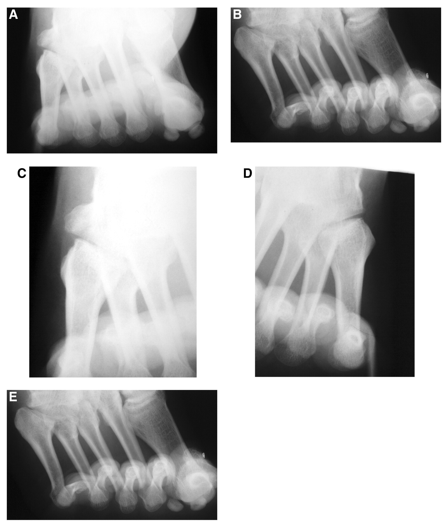
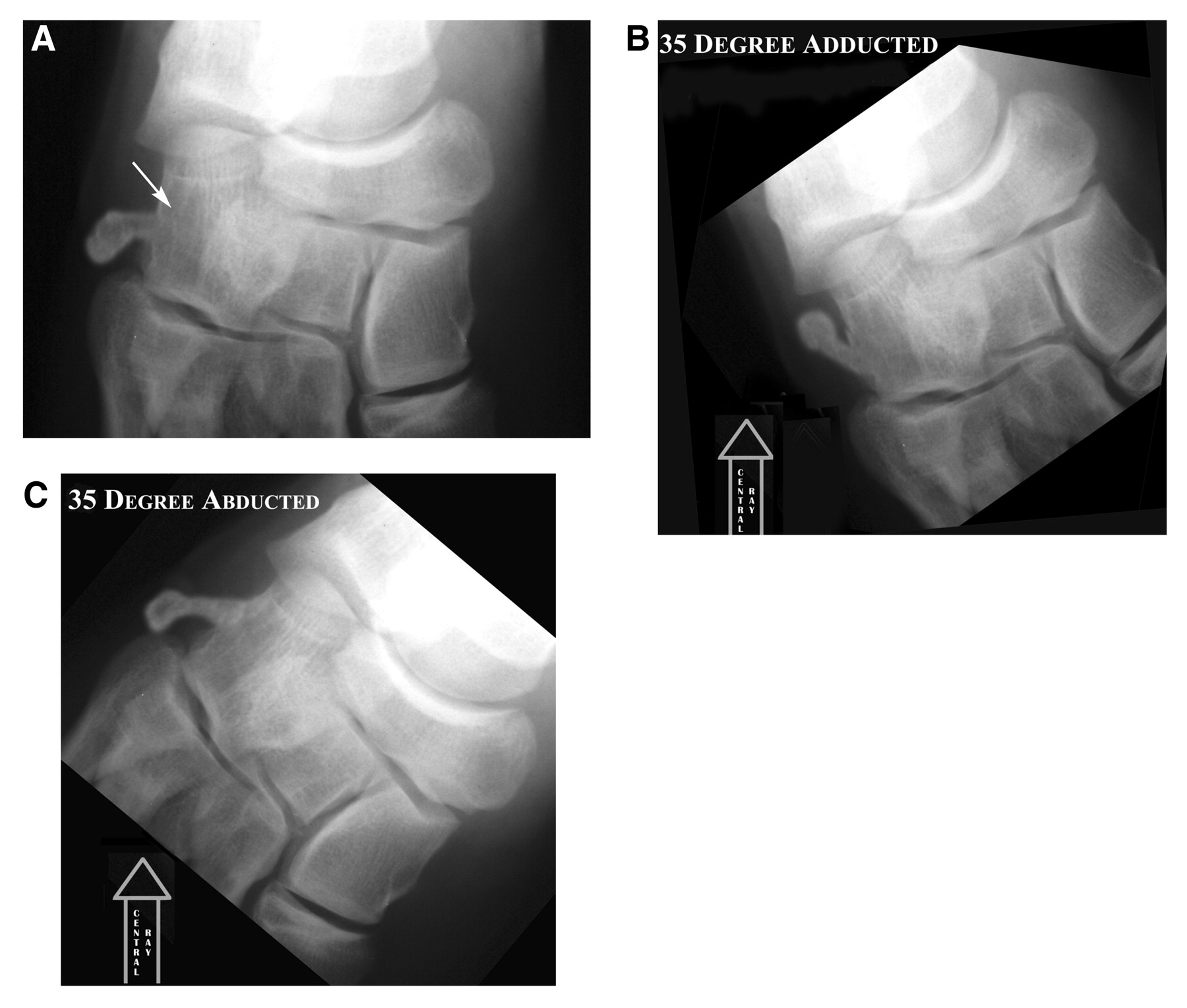
The MRIs were not available at the patient’s follow-up office visit. Because the diagnosis was still in question, three wide-field forefoot axial studies were then empirically acquired: the conventional forefoot axial study and two modified oblique forefoot axial views, one in abduction and one in adduction. The results of these modified oblique-coronal sesamoid axial studies are shown in Figure 7. Whereas the abducted oblique coronal view (Fig. 7E) did not add any useful information, the adducted views (Fig. 7 A–D) significantly clarified the radiographic picture and demonstrated the presence of a pedunculated exostotic lesion emanating from the apex of the plantar surface of the fibular sesamoid. A transverse fracture line could be appreciated through the base of its stalk, and, therefore, corticomedullary confluence could not be ascertained with certainty. In addition, collapse with irregularity of the superior subchondral cortex (articular surface) of the fibular sesamoid was noted. As with the other views, several discrete osseous fragments are apparent, without evidence of de novo calcific or ossific matrix formation. These findings were most suggestive of a fractured exostotic lesion emanating from the inferior fibular sesamoid surface, with possible avascular necrosis (possibly secondary) of the sesamoid. Prime differential diagnostic considerations then centered around osteochondroma and bizarre parosteal osteochondromatous proliferation (Nora’s lesion). Although rare, both lesions have been described as involving the sesamoid bones. [13-15] Post-traumatic heterotopic new bone formation is commonly included in this differential diagnosis, but it was believed that the older patient age, the radiographic absence of the characteristic “zoning” phenomena, and the appearance of a joining pedicle to the adjacent sesamoid ruled against this diagnosis.
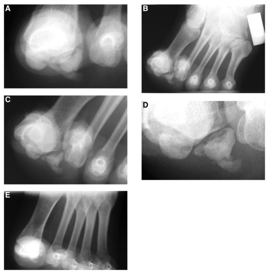
Figure 7.
A, Close-up view of Figure 5C does not resolve the details of the pathologic process involving the fibular sesamoid complex. B–D, Adducted oblique forefoot axial views at progressively close-up views of the hallucal sesamoidal complex. Note the clarification of the pathologic process in part D. E, Abducted oblique forefoot axial view does not clarify the pathologic process.
Once the MRIs were obtained, we compared the coronal studies (Fig. 6 D–G) with the adducted plain film forefoot axial images (Fig. 7 B–D). As can be seen, the coronal T1-weighted images demonstrate the presence of a spatially corroborative low–signal intensity area applied against the inferior pole of the fibular sesamoid (Fig. 6 D and E). This low–signal intensity zone seems to be contained by a thin, homogenous intermediate–signal intensity (gray) zone largely in circumference of the entire low–signal intensity zone. In the corresponding coronal T2-weighted image, the lesion (arrow) and the contiguous fibular sesamoid are somewhat similar in that they demonstrate heterogenous intermediate– and low–signal intensity areas, although the adjacent lesion demonstrates more of a central low–signal intensity area (Fig. 6F). The heavily water-weighted short tau inversion recovery (STIR) coronal images (Fig. 6G) do not clearly demonstrate a high signal perimeter that would constitute a cartilaginous cap, although the technique somewhat incompletely “sectioned” the suspected lesion. However, in concert with the T2-weighted images, a low–signal intensity medulla (stroma) is rather obvious. It was believed that these features were, nonetheless, consistent with an osteochondroma in an older adult.
In comparing the MRI features of osteochondroma and Nora’s lesion, the normally high water content in nonmineralized portions of the cartilage cap of an osteochondroma is typically of intermediate– to low–signal intensity on T1-weighted images and of high–signal intensity on T2-weighted images. However, the thickness of the cap is variable and generally correlates with the age of the patient; it can be quite thick (up to 3.0 cm) in children and adolescents and often thin or even absent in adult patients (similar to the present patient). In addition, progressive mineralization of cartilaginous areas in the cap will correspond with low–signal intensity with all MRI pulse sequences. As maturation of the cap proceeds, enchondral ossification can occur, and intermediate high– to high–signal T1-weighted “yellow marrow” signal may be apparent. [16] Conversely, in bizarre parosteal osteochondromatous proliferation, lesions typically demonstrate low–signal intensity T1 and high–signal intensity T2 characteristics, and the MRI findings in this patient were, therefore, deemed to be inconsistent with a diagnosis of Nora’s lesion. [17] Heterogenous high T2 signal intensity also characterizes the lesions of myositis ossficans, [18] and, therefore, this was deemed unlikely as well.
Correlation of the MRI (Fig. 6E) with the plain film radiographic image (Fig. 7C) suggests the diagnosis of fractured osteochondroma. In addition, on all pulse sequences, approximately 50% of the normal marrow fat signal within the fibular sesamoid has been subjected to heterogenous low–signal intensity replacement. These changes are suggestive of previous avascular necrosis with some reconstitution. [19,20] Once again, keep in mind that none of these details were noted by the interpreting radiologist. Only close correlation with the adducted forefoot axial plain film radiographic study allowed us to improve on the accuracy of the MRI interpretation.
Case 2
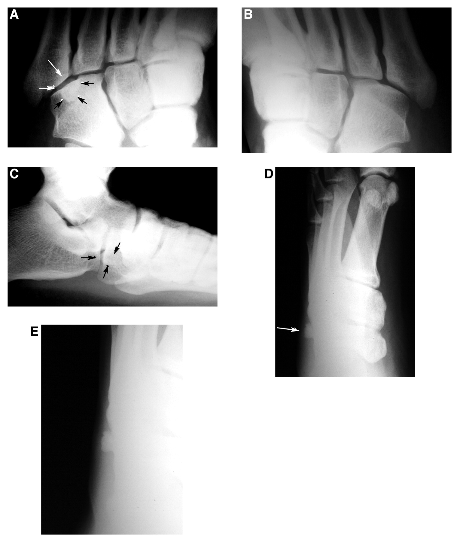
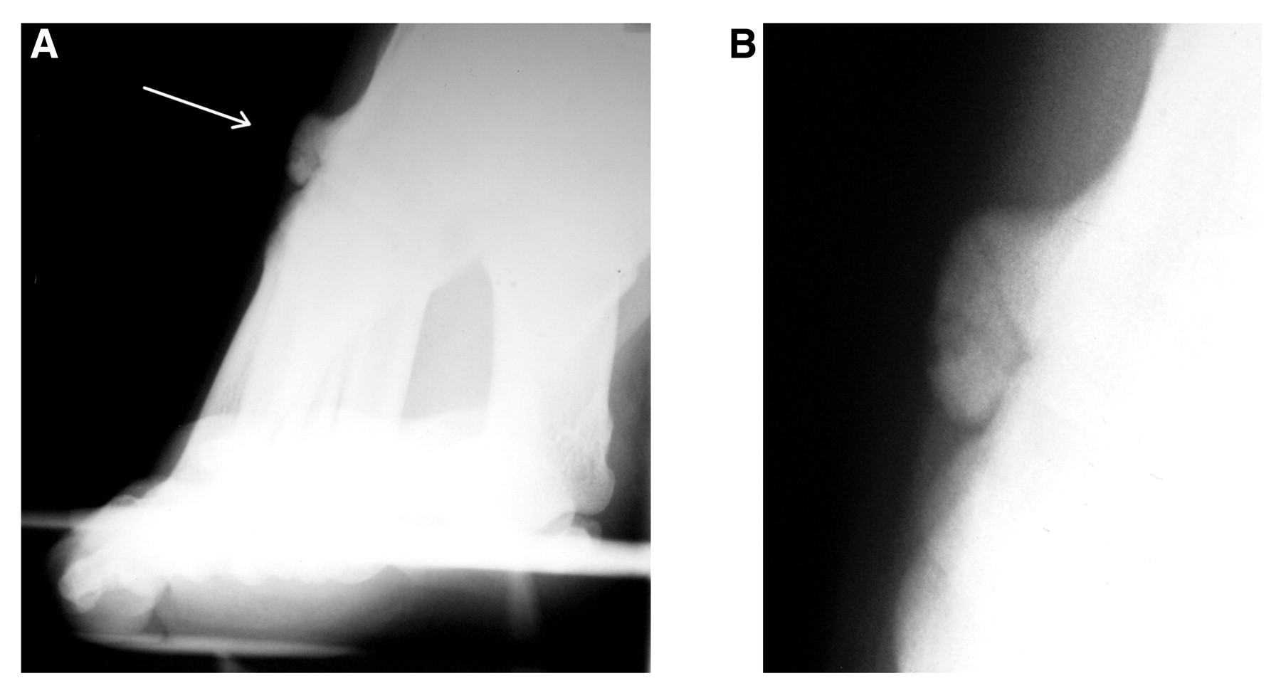
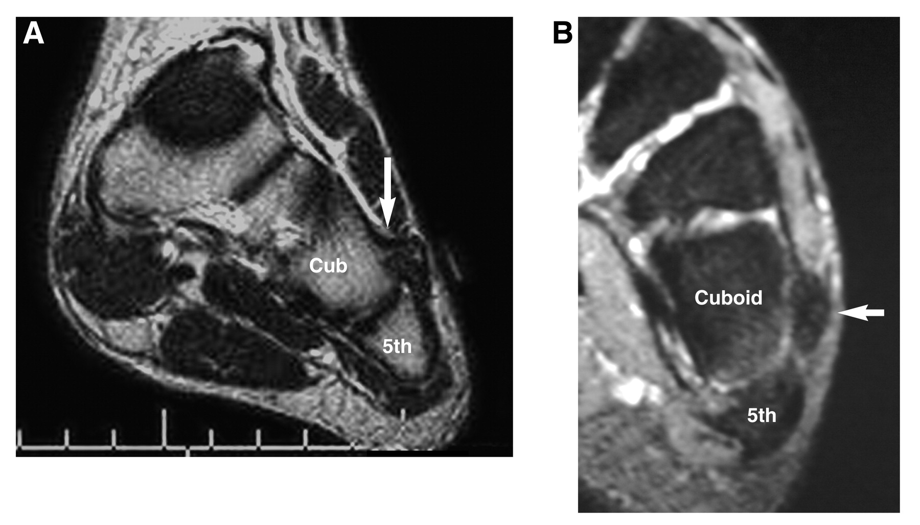
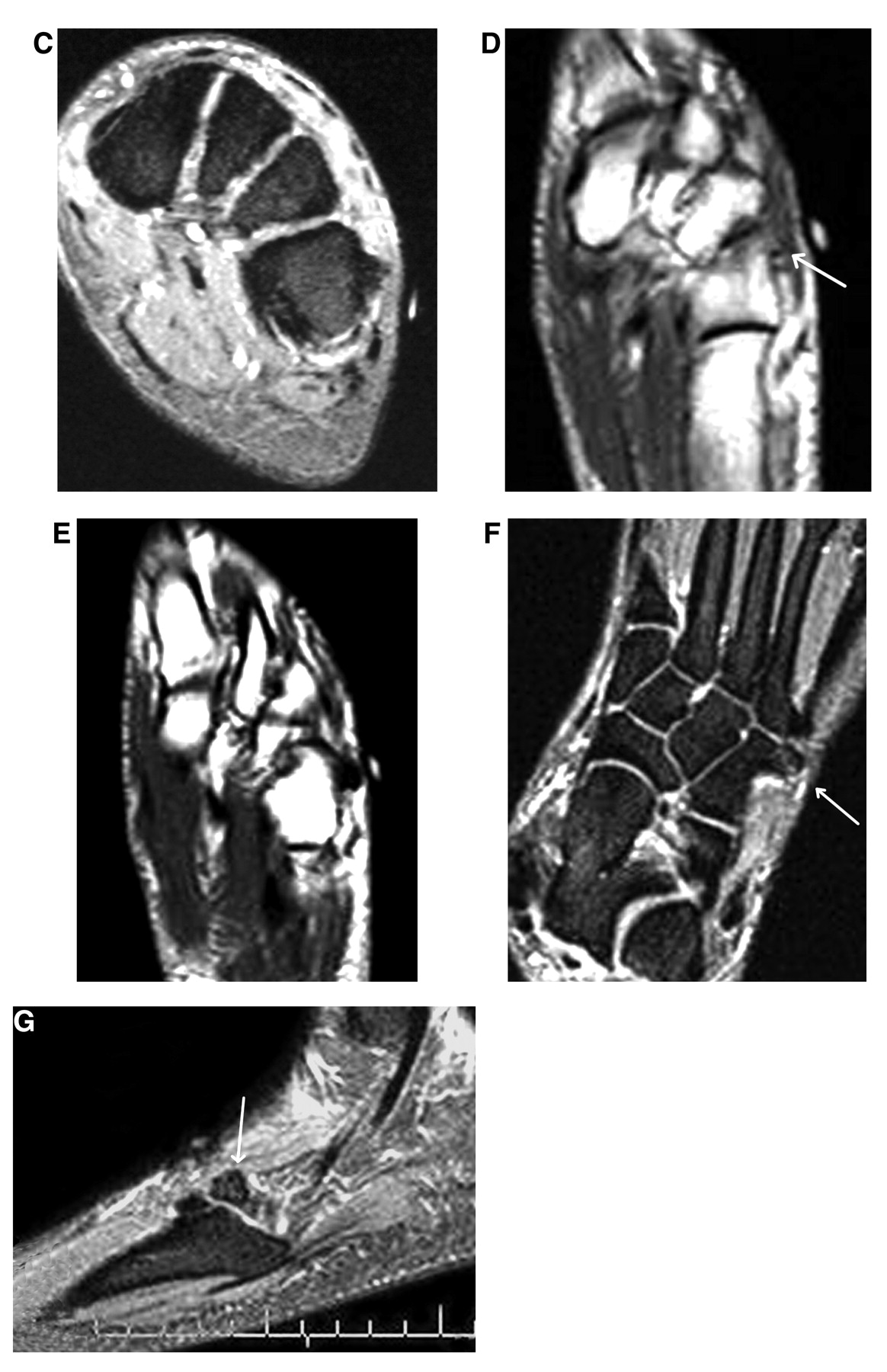
A 32-year-old man was experiencing mild pain in his left midfoot that began somewhat insidiously after a motorcycle accident 3 months earlier. Although swelling was initially noted, this resolved and has not persisted. The patient related that the pain was largely aggravated with weightbearing activities. His medical history was unremarkable, as were the dermatologic and neurovascular lower-extremity examinations. The musculoskeletal examination, however, demonstrated a palpable, hard, deep protuberance in the area of the dorsolateral tarsometatarsal joint. Despite the osseous “end feel,” precise localization was not clinically possible. The lesion generally approximated the dorsal cuboid–fourth metatarsal area. Anteroposterior plain film radiographic studies were unremarkable (not pictured), but the lateral and medial oblique views indicated the presence of a focal osteosclerotic zone superimposed over the fifth metatarsocuboid joint area (Fig. 8 A–C). The interposition over several bones was suggestive of periarticular soft-tissue ossification, but the high-angle lateral oblique studies improved specificity and indicated the presence of a flat-topped exostosis, most likely emanating from the dorsal cuboid bone (Fig. 8 D and E). Based on these studies, an alternative origin from the proximal fifth metatarsal base cannot be ruled out. Pursuant to these studies, standard and oblique forefoot axial studies were ordered and evaluated. Although the standard and adducted radiographic studies were unremarkable (not pictured), the abducted forefoot axial studies (30° abducted with 40° of heel elevation) nevertheless demonstrated the presence of a pedunculated (and not flat-topped) exostotic lesion projecting off of the dorsolateral surface of the cuboid bone (Fig. 9). A rounded cap could easily be noted angling downward, and (as with case 1) a pedicle fracture was now visible. Preoperative MRIs were ordered, and the results concurred with the plain film forefoot axial study interpretation (Fig. 10).
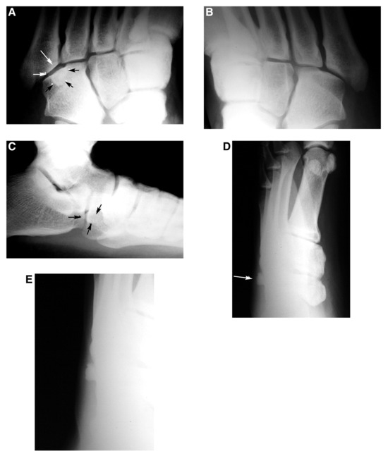
Figure 8.
Left (A) and right (B) high-angle medial oblique studies. The arrows outline the suspected lesion. C, Left lateral study. The arrows point to the lesion. D, High-angle lateral oblique studies demonstrate a flat-topped exostosis, likely arising from the cuboid surface. E, A close-up view of the lesion shown in D.
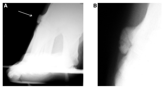
Figure 9.
A, Abducted oblique forefoot axial studies. B, A close-up view of the exostotic lesion designated by the arrow in A. The morphologic features of the lesion are considerably different, and it now appears pedunculated. Osteochondroma is a likely diagnosis, and the pedicle appears fractured.
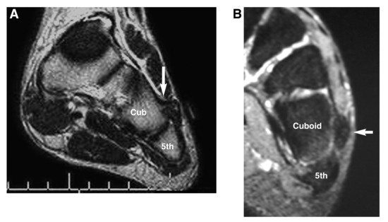
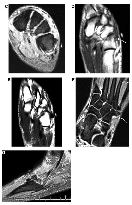
Figure 10.
Coronal T1-weighted (A) and short tau inversion recovery (STIR) images (B and C). A, The T1-weighted image clearly demonstrates the exostotic projection off of the dorsal surface of the cuboid (Cub) (arrow) and overt corticomedullary confluence. Note the high–signal intensity fat in the pedicle of the lesion. This favors true osteochondroma versus a post-traumatic reactive parosteal or juxtacortical surface lesion. B, An apparent low–signal intensity, oval pedunculated cap of the lesion is seen (arrow). This closely resembles the plain film oblique forefoot axial image in Figure 9B. C, This is a more proximal cut, which does not demonstrate the cap. This suggests that the stalk projects in a somewhat posterior to anterior direction. D, Axial T1-weighted image. Overt corticomedullary confluence is noted in the stalk of the lesion, with probable fracture at the junction with the cap. E, Slightly more plantaraxial slice than D. Oblique axial (F) and sagittal (G) short tau inversion recovery (STIR) images. The true anteroposterior diameter of the cap is better appreciated, consistent with the plain film medial oblique image (Figure 8A). All arrows point to the exostotic lesion that appears to emanate from the distal lateral cuboid bone.
Case 3
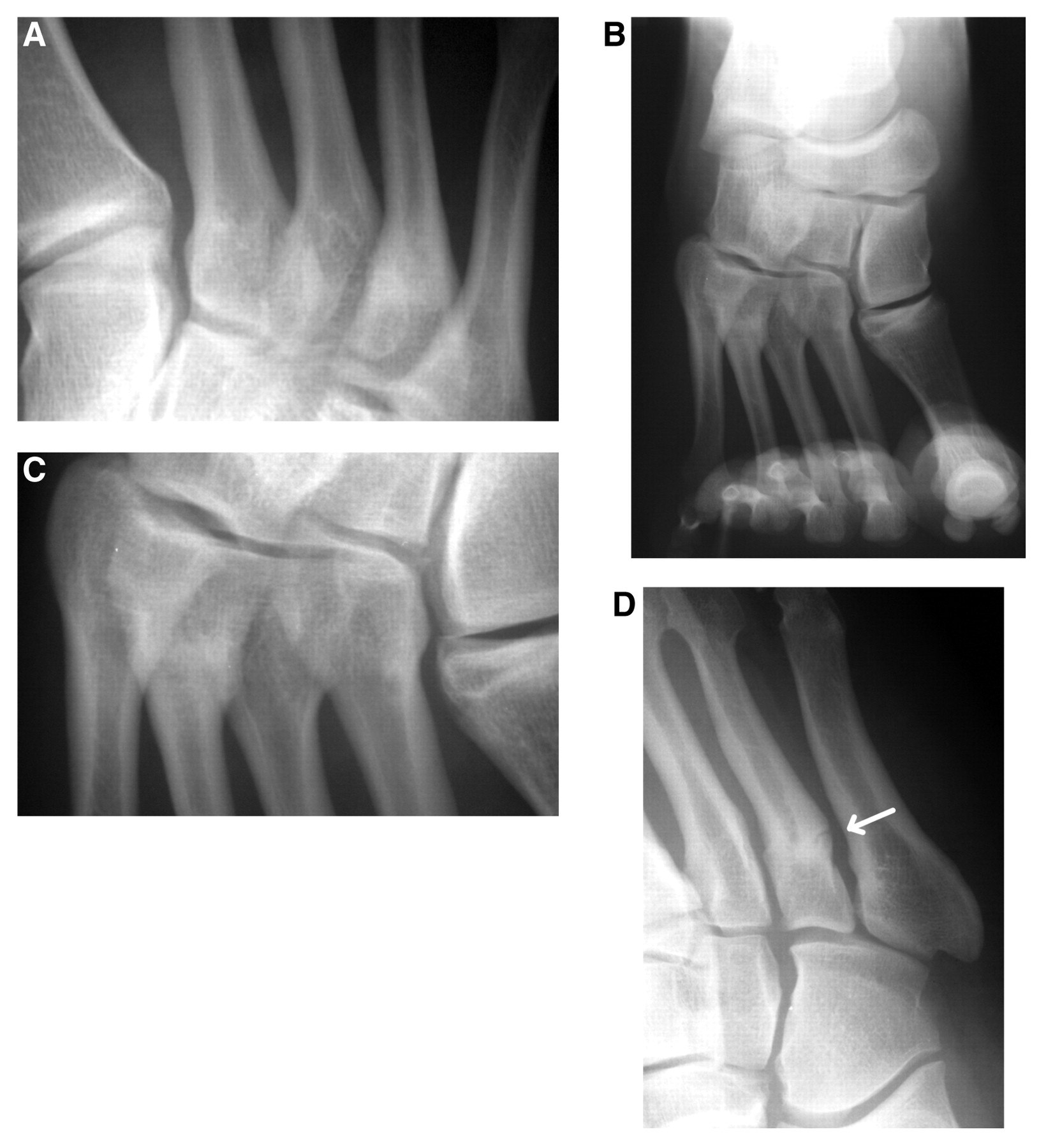
A 28-year-old man presented to the Cleveland Foot and Ankle Clinic with a long history of periodic left foot pain. The pain was localized to an area just proximal to the fifth metatarsal styloid process, and the patient attributes the onset to an injury incurred while playing college basketball approximately 10 years earlier. The foot was generally asymptomatic with daily weightbearing activities; however, with prolonged activities or sports, especially with lateral movement, foot pain occurred. Swelling was inconsistently noted but was primarily absent, and simple rest alleviated his symptoms. The history and pedal examinations were generally unremarkable, except for a palpable lump in the area of interest that was best appreciated with the left foot in a plantarflexed and inverted attitude. As can be seen from standard plain film pedal images (Fig. 11), a prominent distal lateral “corner” of the cuboid bone can be noted on the anteroposterior view (Fig. 11A). Beyond this, however, the medial oblique and lateral views did not clarify the nature of this pathologic entity (Fig. 11B–D). Lateral oblique studies were likewise unremarkable (not pictured).
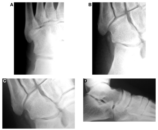
Figure 11.
Anteroposterior (A), medial oblique (B), close-up view of medial oblique (C), and lateral plain film (D) images. Only the anteroposterior view demonstrates bizarre hypertrophic enlargement of the anterolateral corner of the cuboid. The medial oblique and lateral views are unremarkable, and a specific diagnosis is lacking.
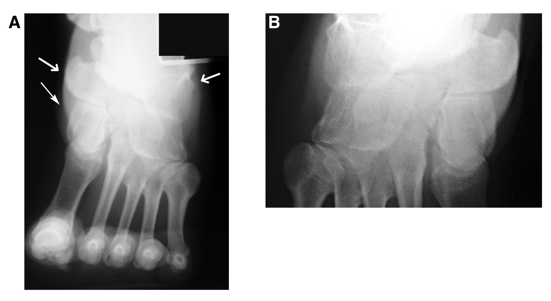
Rather than move to CT or MRI, we prospectively ordered a set of modified forefoot axial studies for the “off-label” use of midfoot imaging. Specifically, bilateral forefoot axial studies were performed (modified via 40° of heel elevation and wide collimation) along with the two oblique forefoot axial studies of only the affected foot (Fig. 12). As can be noted by comparison with the contralateral modified neutral (no abduction or adduction) forefoot axial image (wide field, increased heel elevation), a large, exostotic lesion is conspicuous on the left (affected) side. The magnified comparison images (Fig. 12 C and D) demonstrate the presence of an exostotic lesion originating from the cuboid, with broad-based deformation of the distal lateral bone. The lesion appears moderately large and plateau shaped, with extension in a lateral-to-horizontal direction. Although this process seems to involve the distal one-half of the bone, it extends to the marginal area of the lateral fifth metatarsocuboid joint. In consideration of the patient’s history, simple post-traumatic periarticular exostosis was favored, with sessile osteochondroma as the prime differential diagnosis. Note that imaging obtained by using the adducted oblique forefoot axial study was suboptimal (Fig. 12E), and the lesion was inconspicuous on the abducted study (not pictured).
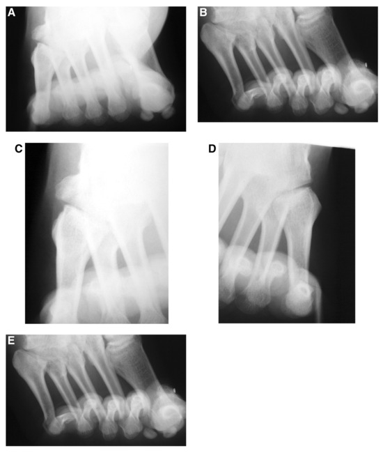
Figure 12.
Neutral wide-field forefoot axial with increased heel elevation standard (A) and close-up view (C) images of the affected left foot. Conspicuity of the lesion is optimal, and a short list of differential diagnoses can be generated (see the “Case 3” subsection for a description). Contralateral comparison standard (B) and close-up view (D) images of the unaffected right foot. E, Adducted oblique forefoot axial study. The lesion can be seen, but it is not as clearly defined as in parts A and C.
Case 4
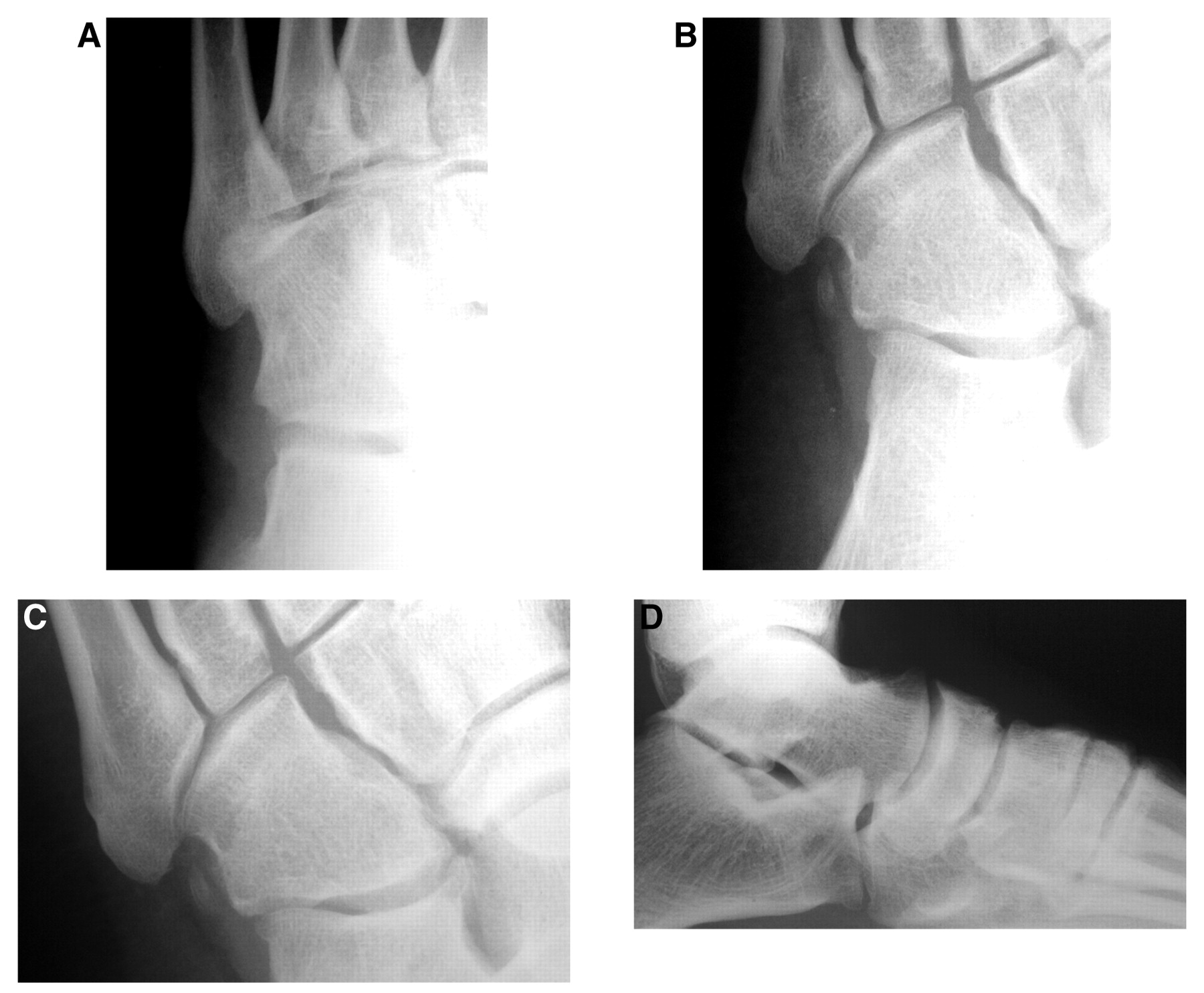
A 14-year-old boy felt a “pop” and the sudden onset of pain and swelling in his right lateral midfoot while landing on the mat during a high school wrestling match. Plain film radiographic anteroposterior, lateral, and oblique studies were originally read as negative in a local hospital emergency department, but later that evening the patient was called by the hospital radiologist and was told that he had a fracture. The patient presented to the Cleveland Foot and Ankle Clinic the next day, where standard anteroposterior, medial oblique, and lateral views were ordered along with a standard-position wide-field forefoot axial study with the heel elevated to 40° (Fig. 13). Whereas the anteroposterior view is unremarkable, the wide-field axial coronal study demonstrates loss of the concave proximal medial contour (now convex) and apparent “sclerosis” of the fourth metatarsal base, which, in actuality, is related to local brightening of the thickened cortices. The medial oblique study clearly indicates cortical thickening with interruption of the proximal lateral fourth metatarsal diametaphyseal cortex, consistent with an infraction. However, combining the two studies strongly suggested that this fracture is, indeed, “thru and thru” and not just an infraction. Not only did the modified forefoot axial study outperform the standard anteroposterior study, but it yielded unique information regarding imaging of the medial cortex. Owing to osseous overlap, this area is difficult to image with conventional anteroposterior, medial oblique, and lateral oblique studies.
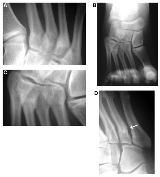
Figure 13.
A, Anteroposterior view of the foot does not demonstrate any pathologic abnormalities. B, Neutral wide-field forefoot axial study (with 40° of heel elevation). Circumferential enlargement/loss of concave contours of the proximal metaphysis of the fourth metatarsal bone is conspicuous and is consistent with a healing fracture. C, Close-up view of part B. Note the overt convexity of the medial and lateral proximal metaphyseal contours of the fourth metatarsal bone. D, Medial oblique study. Obvious lateral cortical discontinuity of the fourth metatarsal bone is seen at the level of the proximal diametaphysis.
Discussion
We examined the imaging capabilities of wide-field, oblique (abducted and adducted) forefoot axial studies for a limited range of pathologic abnormalities involving the proximal lateral forefoot bones and the hallucal sesamoid bones. In several cases, these oblique studies were used in lieu of more expensive imaging modalities for pathologic abnormalities initially deemed outside the imaging capabilities of standard pedal views. In positioning the foot for these modified forefoot axial studies, the foot is aligned at angles oblique to the cardinal frontal plane (of the foot), thereby increasing the percentage of sagittal plane information. Angles greater than 45° become sagittal plane dominant and, therefore, were not used. For clinically apparent promontories or suspected exostotic lesions emanating from the dorsolateral midfoot or anterior calcaneus, we initially attempted to palpate the lesion’s point of maximum prominence and then rotate the foot to skyline the lesion (alignment of the lesion perpendicular to the central ray). This is generally assumed when the point of maximum prominence is most conspicuous in the shadow of the foot in the lit radiographic field. In theory, this should provide the optimal pedal angular position for visualizing an exostotic lesion. In practice, however, results can be mixed. In some cases, it is simply not possible to accurately determine the point of maximum prominence. Other times, despite accurate determination of the point of maximum prominence, an exostotic lesion may not be straight or uniform in shape. For example, osteochondromas are commonly directed away from joints or have an angulation (angular deformity) between the pedicle and the cap. In addition, clinically prominent osteoproliferative lesions may be amorphous, and determination of the point of maximum prominence is of limited use. Finally, although further study is needed, we surmise that these oblique axial studies will be of use in imaging lytic or erosive surface defects of the dorsolateral foot bones.
In light of these issues, we prefer an initial empiric imaging approach that uses all three views (one neutral and two oblique studies) at the mid/medium angle of 30°. Additional angular “tweaks” can then be easily made as needed pending the results of these studies. However, for suspected exostotic lesions, estimation of the point of maximum prominence is certainly a reasonable optional approach when attempting to optimize the pedal angle of obliquity (abduction or adduction). In none of the presented case studies, however, was this necessary.
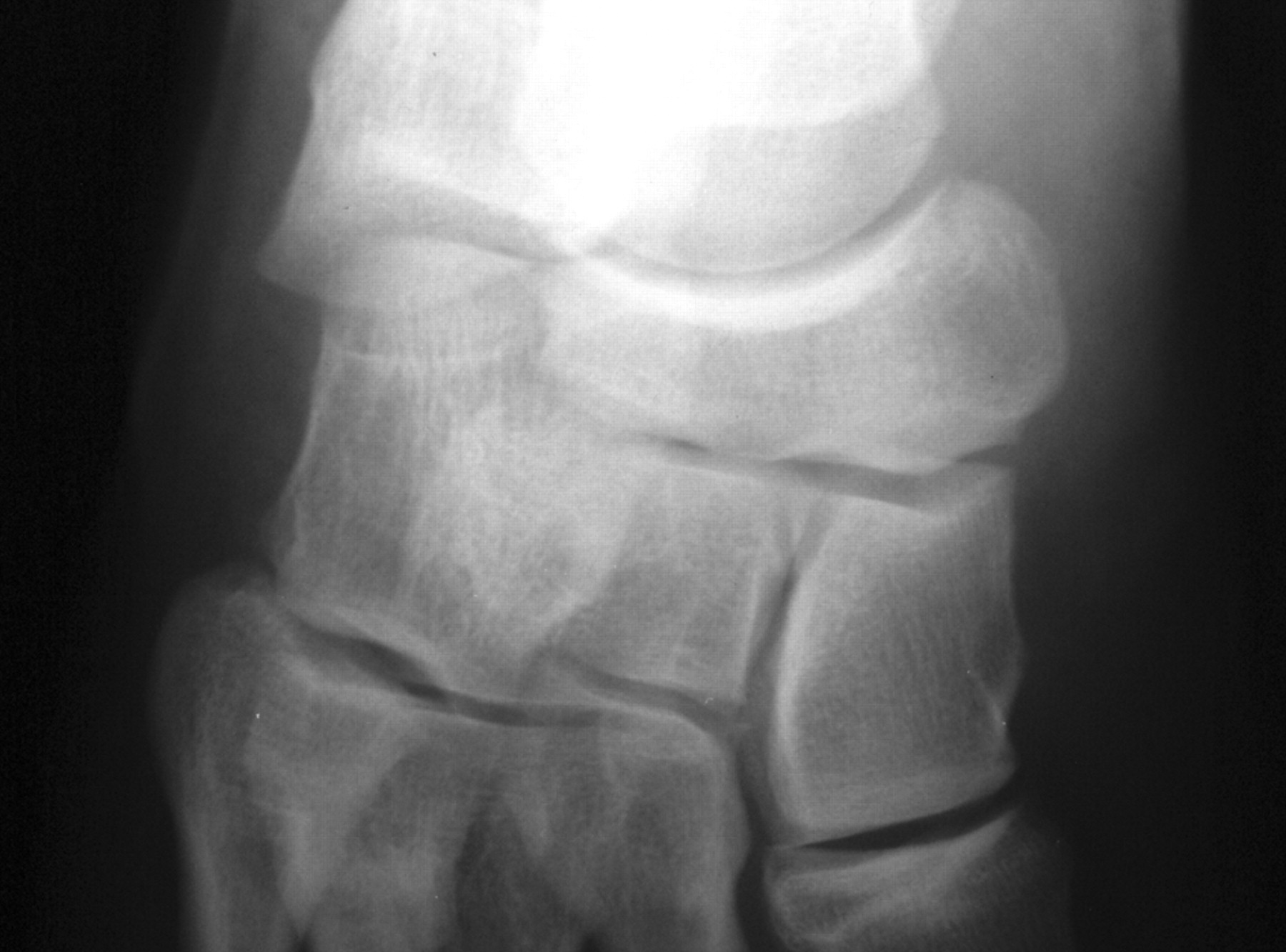
Rather than elevate the heel just enough to get it out of way of the x-ray central beam (conventional forefoot axial study), we increased the angle of heel elevation (in the sagittal plane). This increases the percentage of transverse plane information in the final study and places the metatarsals in a position that is less likely to result in overlap with more proximal osseous elements. Values greater than 45° were avoided, however, because they become transverse plane dominant and largely mirror the detail of the posterior-anterior projection. On the other hand, angles approaching 45° can provide excellent coronal plane midfoot imaging (Fig. 14).
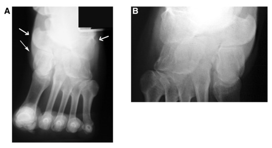
Figure 14.
Neutral wide-field forefoot axial study (with 35° of heel elevation) (A), close-up view of the midfoot (B). The arrows indicate the medial and lateral contours of the navicular and cuboid bones. A cornuate navicular (type III os tibial externum) is suggested by this study.
In essence, these forefoot axial modifications are significantly oblique to all of the cardinal body planes. The benefits of this are 1) enhanced imaging of osseous surfaces, contours, and shapes; 2) the ability to rotate cortical surface projections (exostotic lesions) into an improved or optimal imaging position (at a right angle to the central beam, providing skyline detail); and 3) ancillary hallucal sesamoid imaging. When imaging the hallucal sesamoid bones, the abducted forefoot axial views provide enhanced tibial sesamoid detail, whereas the adducted forefoot axial studies can aid in imaging fibular sesamoid pathologic abnormalities. Regarding the more proximal structures, abducted oblique forefoot axial studies generally skyline lateral midfoot structures (eg, the cuboid), whereas adducted oblique forefoot axial studies skyline medial structures. Simple wide-field forefoot axial views with increased foot plantarflexion (increased heel elevation) provided for excellent skyline or contour details of the medial and lateral midfoot bones, especially the fifth metatarsal cuboid area on the lateral side (Fig. 14A). We believe that imaging of the proximal medial tarsal bones is typically adequate when conventional anteroposterior and lateral oblique views are used. However, as illustrated in cases 2 and 3, the modified forefoot axial imaging sets were clearly superior to standard anteroposterior, lateral, and medial oblique foot studies for lateral midfoot imaging.
Plain film imaging of the proximal lateral lesser tarsal bony surfaces to identify fragmentation, exostoses, fractures, and cortical lysis is problematic at best. Lateral and anteroposterior pedal studies are simply inadequate owing to osseous overlap. Likewise, medial oblique pedal studies commonly rotate dorsolateral exostotic lesions inwardly into positions of increased osseous overlap and are likewise inadequate. Conversely, medium- to higher-angle lateral oblique studies can generally aid in imaging dorsolateral surface pathologic findings, such as erosive changes and exostoses in this locale provided that the long axis of the exostosis is directed either dorsally or plantarly. This is best illustrated in case 2, where the fractured cap of the cuboid osteochondroma is directed plantarly despite the stem emanating in a dorsolateral direction. As the figures demonstrate, the lateral oblique view identifies a rectangular, plateau-shaped base but does not image the cap. With wide-field oblique forefoot axial imaging, adduction of the foot rotates a posteriorly directed (lateral midfoot) cap into skyline position. Conversely, abduction of the foot likewise rotates an anteriorly directed exostotic lesion into the coronal plane. This is illustrated with the simulated cuboid exostosis in Figure 15. Finally, note that using a single lateral oblique foot study to obtain clear views of pathologic processes involving the dorsolateral tarsal bones is subject to a significant degree of chance. Medial oblique ankle studies are occasionally used for skyline imaging (eg, for extensor digitorum brevis avulsive injuries of the os calcis), but their use is highly limited, and the findings of this study suggest that consideration be given to the routine addition of these modified forefoot axial views to the imaging armamentarium.
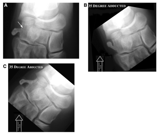
Figure 15.
Simulated pedunculated osteochondroma emanating from the lateral surface of the cuboid bone. A slight angular deformity between the cap and the pedicle has been created. All of the views are with 35° of heel elevation. A, The axis of the lesion without a significant anterior or posterior direction is adequately imaged by using the wide-open forefoot axial study in the standard position. B, When the axis of the lesion is directed posteriorly at an oblique angle, the adducted oblique forefoot axial study will best demonstrate the lesion. Note that an empiric value of 35° of adduction should adequately demonstrate the pedicle and the cap of the lesion. C, When the axis of the lesion is directed anteriorly, the abducted studies will best demonstrate the pedicle and the cap.
Conclusion
These case studies demonstrate how oblique forefoot axial views can be used to resolve equivocal or negative radiographic studies in light of clinically palpable exostoses located in difficult-to-image pedal areas, such as the proximal lateral foot. In addition, sesamoidal imaging can be enhanced. In these case studies, there was little to no benefit gained from MRI, and in one case, MRIs were even outperformed by oblique forefoot axial studies. Obtaining forefoot axial views with the foot abducted and adducted 30° along with 35° to 45° of heel elevation can greatly facilitate plain film imaging of the hallucal sesamoids and lateral midfoot bones. As noted previously herein, increasing heel elevation angles beyond 45° becomes more transverse (axial or posterior-anterior projection) than coronal plane, with the same imaging limitations (Fig. 16). The present experience with using these techniques indicates that abducted forefoot axial views generally provide enhanced imaging of the tibial sesamoid and medial midfoot structures (eg, navicular), whereas adducted forefoot axial views tend to illuminate fibular sesamoid and lateral midfoot structures (cuboid). Nevertheless, we must warn against trying to predict which view is best for midfoot imaging, as an anteriorly directed exostosis of the lateral midfoot is likely to be imaged by pedal abduction and vice versa. This cannot be predicted clinically, however, and we suggest that practitioners empirically obtain all three forefoot axial views when their use is indicated. These findings suggest that there may, indeed, be a place for these studies as part of the “routine” pedal radiographic regimen, and it is hoped that further work with these views will bear this out. Adding these views to the physician’s plain film pedal radiographic imaging armamentarium may, in some cases, obviate the need for more expensive ancillary imaging studies.
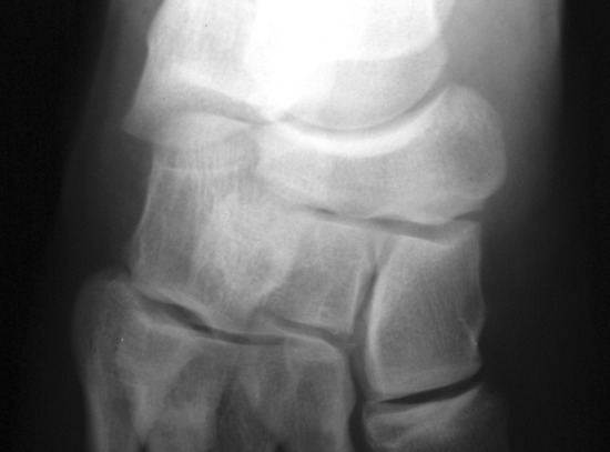
Figure 16.
Neutral wide-field anteroposterior forefoot axial study with approximately 65° of heel elevation. Note that the midfoot more closely approximates the transverse plane.
Financial Disclosure
None reported.
Conflict of Interest
None reported.
References
- Muller W: Malacie des sesamknochens des I metatarsole, ein typesches krank-heitsbild: beitr. 2. Klin Chir 134: 308, 1925.
- Lewis RW: Non-routine views in roentgen examination of the extremities. Surg Gynecol Obstet 67: 38, 1938.
- Holly EW: Radography of the tarsal sesamoid bones. Med Radiogr Photogr 31: 73, 1955.
- Chang JW, Griffiths H, Chan DPK: A new radiological technique for the forefoot. Foot Ankle 5: 77, 1984.
- Viladot A: “The Metatarsals,” in Disorders of the Foot, ed by M Jahss, p 667, WB Saunders, Philadelphia, 1982.
- Downey MA, Dorothy WL: A radiographic technique to demonstrate the plantar aspect of the forefoot in stance. JAPA 59: 140, 1969.
- Gamble FO, Yale J: Clinical Foot Roentgenology, 2nd Ed, Robert E. Kreiger Publishing Co Inc, Huntington, NY, 1975.
- Dreeben S, Thomas PBM, Noble PC, et al: A new method for radiography of weightbearing metatarsal heads. Clin Orthop 224: 260, 1987.
- Treves A: Un cas de sesamoidite interne du gros orteil. Rev d’Orthop 15: 411, 1928.
- Renander A: Two cases of typical osteochondropathy of the medial sesamoid bone of the first metatarsal. Acta Radiol 3: 521, 1924.
- Ilfeld FW, Rosen V: Osteochondritis of the first metatarsal sesamoid. Clin Orthop 85: 38, 1972.
- Kliman ME, Gross AE, Pritzker KP, et al: Osteochondritis of the hallux sesamoid bones. Foot Ankle 3: 220, 1983.
- Noguchi M, Ikoma K, Matsumoto N, et al: Bizarre parosteal osteochondromatous proliferation of the sesamoid: an unusual hallux valgus deformity. Foot ankle Int 25: 503, 2004.
- Harty JA, Kelly P, Niall D, et al: Bizarre parosteal osteochondromatous proliferation (Nora’s lesion) of the sesamoid: a case report. Foot Ankle Int 21: 408, 2000.
- Mowad SC Sr, Zichichi S, Mullin R: Osteochondroma of the tibial sesamoid. JAPMA 85: 765, 1995.
- Murphey MD, Choi JJ, Kransdorf MJ, et al: Imaging of osteochondroma: variants and complications with radiologic-pathologic correlation. RadioGraphics 20: 1407, 2000.
- Torreggiani WC, Munk PL, Al-Ismail K, et al: MR imaging features of bizarre parosteal osteochondromatous proliferation of bone (Nora’s lesion). Eur J Radiol 40: 224, 2001.
- Kransdorf MJ, Meis JM, Jelinek JS: Myositis ossificans: MR appearance with radiologic-pathologic correlation. AJR Am J Roentgenol 157: 1243, 1991.
- Karasick D, Schweitzer ME: Disorders of the hallux sesamoid complex: MR features. Skeletal Radiol 27: 411, 1998.
- Toussirot E, Jeunet L, Michel F, et al: Avascular necrosis of the hallucal sesamoids update with reference to two case-reports. Joint Bone Spine 70: 307, 2003.
© 2008 American Podiatric Medical Association