


Abstract
Reconstructive surgery for hindfoot, ankle, and leg deformities is facilitated by proper radiographic analysis. The long leg calcaneal axial and hindfoot alignment views have been proved to be useful in deformity planning at The Foot and Ankle Institute at The Western Pennsylvania Hospital. These radiographic views can be attained in an office setting or in any hospital radiology department. The details provided herein of this radiographic technique will be useful to physicians, office staff, and radiology technicians to facilitate proper imaging of hindfoot, ankle, and leg deformities.
Frontal plane radiographs, such as the long leg calcaneal axial and hindfoot alignment views, are critical in the preoperative assessment and management of hindfoot and ankle deformities. Developed by Kleiger and Mankin,[1] the long leg calcaneal axial view is a modification of the Harris and Beath[2] view, which was originally described for evaluation of subtalar joint coalitions. Johnson et al[3] devised a modified radiographic measurement method for determining an angular measurement of hindfoot coronal alignment based on a cadaveric study.
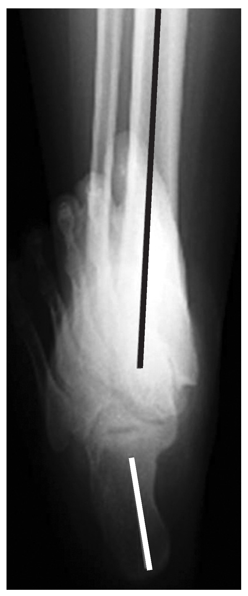
The long leg calcaneal axial view displays the relationship of the calcaneus to the leg and allows for adequate visualization of the subtalar joint. Evaluation of the subtalar joint (varus, valgus, and coalition assessments), the relationship of the calcaneus to the distal one-third of the tibia, and any deformity that may exist in the calcaneus itself can be visualized using the long leg calcaneal axial view (Fig. 1).
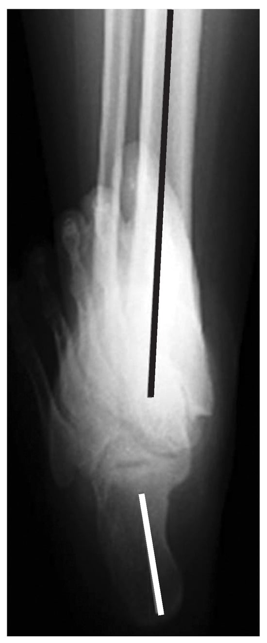
Figure 1.
The long leg calcaneal axial view provides visualization of the distal tibia and its relationship to the subtalar joint and calcaneal bisection.
The hindfoot alignment view demonstrates the relationship of the hindfoot to the leg but, in comparison, the long leg calcaneal axial view provides better visualization of the ankle joint. This view allows for assessment of deformities in or adjacent to the ankle joint (Fig. 2). These radiographic views can be attained in an office setting or in any hospital radiology department. For both views, proximal incorporation of at least the distal one-third of the tibia is crucial to accurately determine angular relationships among the foot, ankle, and leg. Thus, a radiology department may be better suited to perform these useful views owing to the availability of larger radiographic cassettes to allow inclusion of the tibia in the radiographic view. A special Plexiglas (Rohm, Darmstadt, Germany) platform is necessary to perform the hindfoot alignment view, and it may need to be fabricated or purchased. The optimal cassette sizes, materials, positioning, angles, and settings are necessary for success and have been collaborated between our clinical practice and the institution’s radiology department.
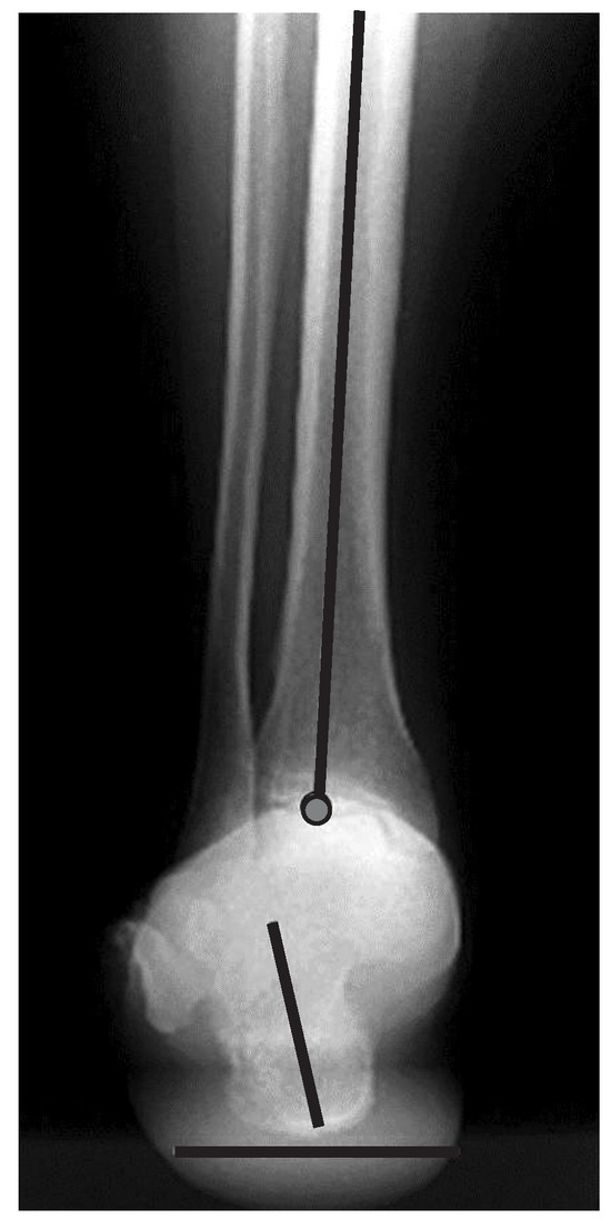
Figure 2.
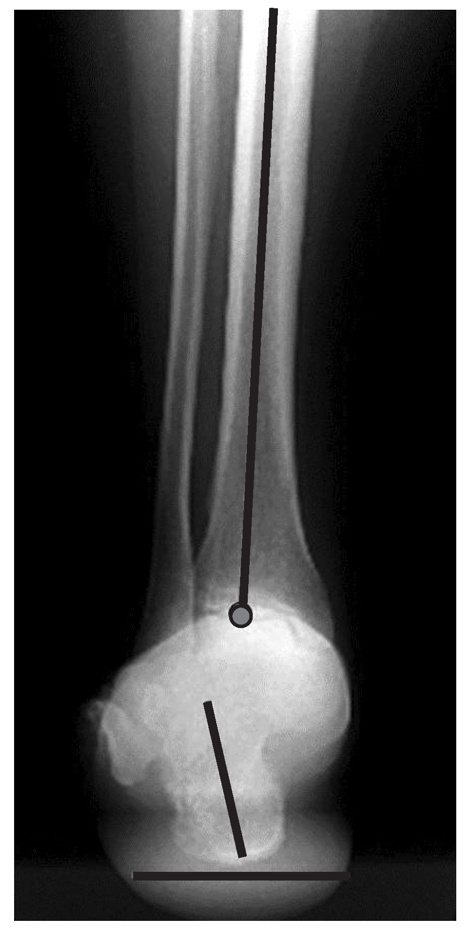
The hindfoot alignment view provides visualization of the tibia, ankle, and calcaneus. Malalignment in the tibia and ankle joint can be assessed.
Radiographic Technique
Long Leg Calcaneal Axial View
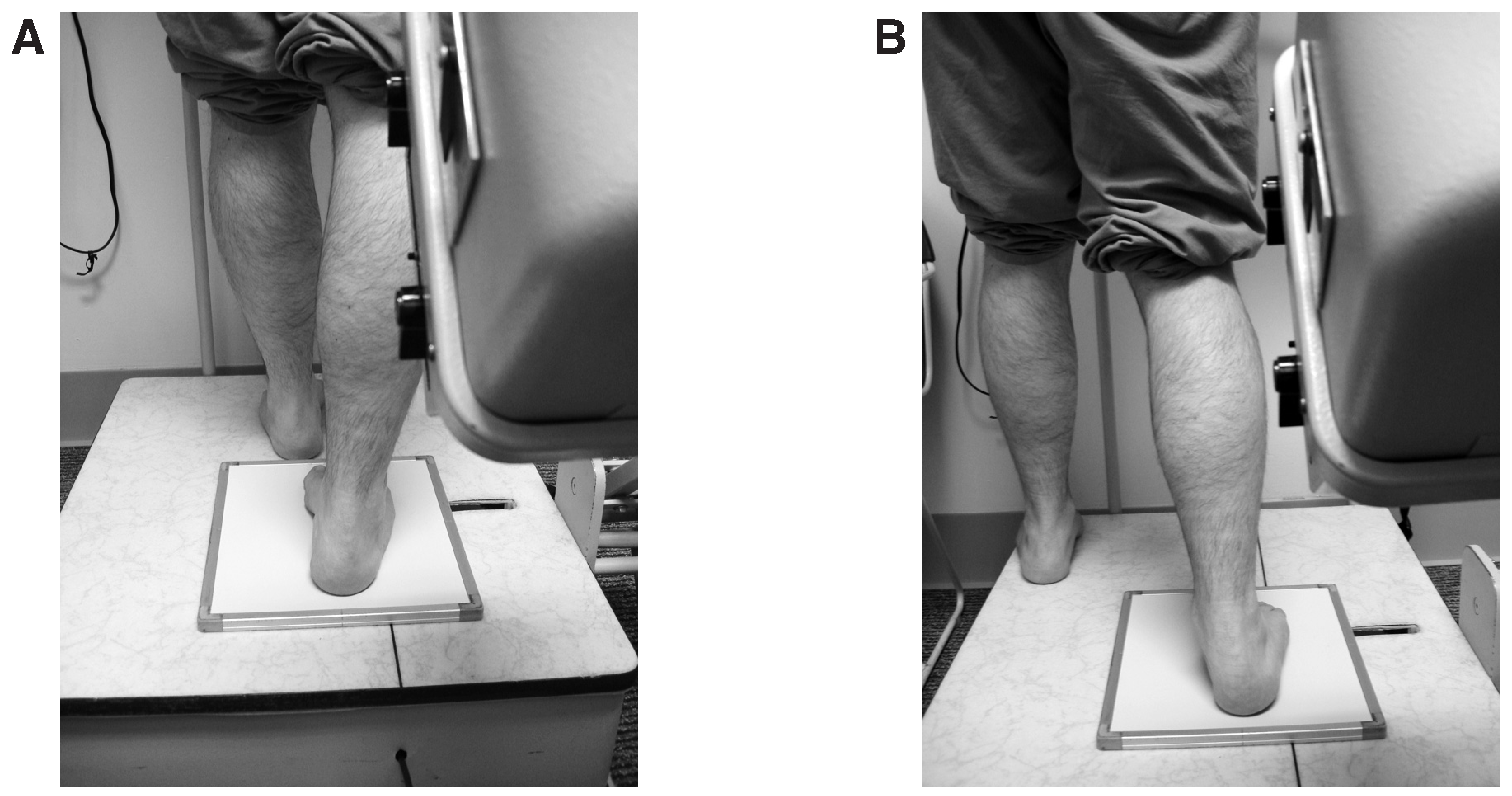
The long leg calcaneal axial radiograph is a weightbearing view. The cassette is placed on the floor, and the tube head is angled 45° to the film (Fig. 3). A 14 × 17-inch x-ray cassette is preferred to a 10 × 12-inch cassette, although either may be used. The x-ray beam is centered at the subtalar joint. The imaged foot should be plantigrade on the cassette and in 10° of dorsiflexion at the ankle, and the knee should be in an extended position. Error can occur with supination or pronation of the foot. When placing the patient in the correct stance position, the technician should make sure that the patient is properly positioned so as to prevent such pronated or supinated positions (Fig. 4). The nonimaged foot is placed in front of the patient for balance. The radiographic settings should be 6.4 mA sec and 65 kVp.
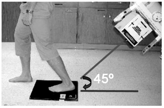
Figure 3.
The long leg calcaneal axial view is taken with the patient in the “ski jump” position. The involved limb is centered on a 14 × 17-inch cassette with the heel placed at the cassette edge closest to the tube head. The involved foot is plantigrade on the cassette, with the ankle dorsiflexed approximately 10° in relation to the leg (ski jump position). The involved knee should be held in extension. The contralateral limb should be placed in front of the film. The tube head is positioned approximately 40 inches posterior to the involved heel, with a declination of 45° to the vertical and the central beam centered at the subtalar joint.
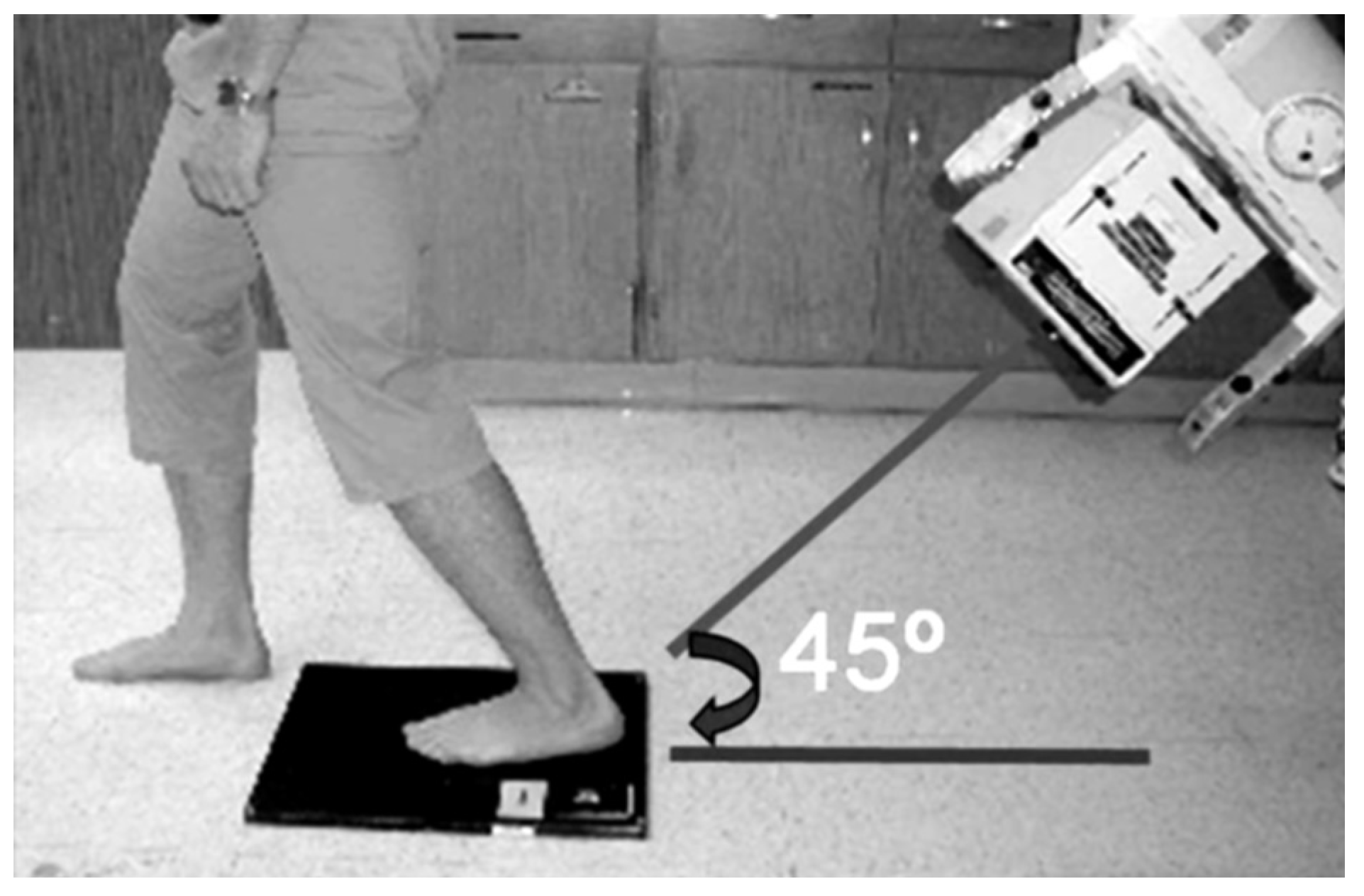
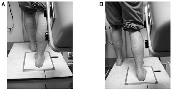
Figure 4.
When positioning for the “ski jump” alignment, the technician should take caution to avoid pronating (A) or supinating (B) the patient, which will result in malalignment.
Intraoperative acquisition of the long leg calcaneal axial view can be achieved with a fluoroscopy unit and with the radiographic beam positioned to pass from distal superior to proximal posterior through the plantar aspect of the foot while being at an angle of 45° to the operative table.
Hindfoot Alignment View
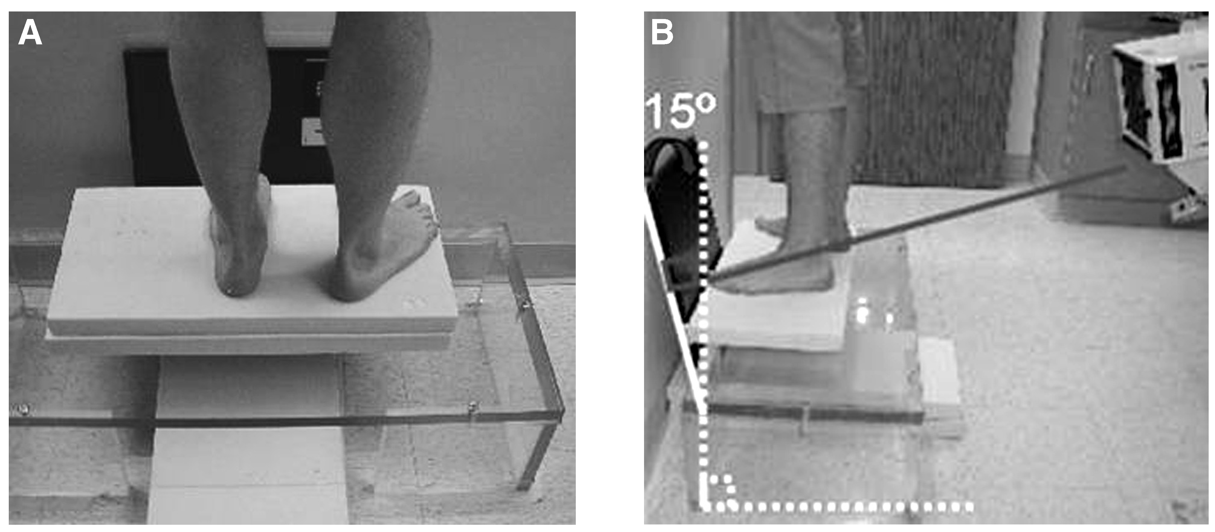
The hindfoot alignment view is a weightbearing view as well. Unlike the long leg calcaneal axial view, this view is taken in the resting calcaneal stance position. Another difference is the use of a Plexiglas platform. The film is placed anterior to the box angled 15° from vertical. The 14 × 17-inch film cassette is ideal. The patient stands on the special Plexiglas platform. The tube head should be at 90° in relation to the position of the cassette, and the radiographic beam is centered on the ankle joint. The film cassette is angled 15° from the vertical (Fig. 5). The patient’s toes are placed against the cassette in the angle and base of gait. The midline of the foot should be perpendicular to the cassette. The radiographic settings should be 6.4 mA sec and 65 kVp.
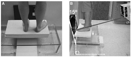
Figure 5.
Positioning for the hindfoot alignment view. The patient stands on the Plexiglas platform in the angle and base of gait with the involved limb perpendicular to the film. The 14 × 17-inch cassette is placed at the distal aspect of the foot on the platform at a 15° angle to the vertical. The hallux should rest against the cassette. The tube head is positioned approximately 40 inches posterior to the involved limb, with a declination of 15° to the vertical and the central beam centered at the ankle joint.
Discussion
Reconstructive hindfoot, ankle, and leg surgical success hinges on preoperative, intraoperative, and postoperative radiographic alignment. The long leg calcaneal axial and hindfoot alignment views are useful adjuncts to frontal plane deformity assessment in the ankle and leg. These radiographic measurements have been shown to correlate well with the clinical measurement of the resting calcaneal stance position.[4] We found these views to be useful in preoperative and postoperative evaluations and in intraoperative fluoroscopic assessment of major hindfoot, ankle, and leg procedures. It is our intent that the procedures and figures supplied herein will assist office staff, radiology technicians, and hospital radiology departments in reproducing the appropriate techniques and views described.
Financial Disclosure
None reported.
Conflicts of Interest
None reported.
References
- Kleiger B, Mankin HJ: A roentgenographic study of development of the calcaneus by means of the posterior tangential view. J Bone Joint Surg Am43:961, 1961.
- Harris RI, Beath T: Hypermobile flat-foot with short tendo Achilles. J Bone Joint Surg Am78:116, 1948.
- Johnson JE, Lamdan R, Granberry WF, et al: Hindfoot coronal alignment: a modified radiographic method. Foot Ankle Int20:818, 1999.
- Lamm BM, Mendicino RW, Catanzariti AR, et al: Static rearfoot alignment: a comparison of clinical and radiographic measures. JAPMA95:26, 2005.
© 2008 American Podiatric Medical Association