


Abstract
Background: Sophisticated methods of cryotherapy, such as application of a water-circulating device, have recently been popularized to provide a constant or intermittent therapeutic source in the foot and ankle postoperative setting. In this study, the efficacy and safety of three selected cryotherapy devices (Iceman, EBIce, and Ankle Cryo/Cuff) were investigated. Methods: Each cryotherapy unit, in the coldest setting, was applied over standard surgical dressings in group I, over one layer of Jones compression bandage in group II, and over two layers of Jones compression bandage in group III on four individuals in excellent overall health. The skin temperature was then recorded every 15 min for 180 min in each trial. Results: In group I, the Iceman was the only device that required discontinuation in one subject, and the EBIce and Cryo/Cuff were tolerated in all of the subjects. However, the temperatures in all of the devices continued to decrease at the end of the trials. In group II, all of the cryotherapy devices controlled temperatures between 20°C (68°F) and 30°C (86°F). In group III, no device significantly lowered the initial surface skin temperature. Conclusions: We achieved the safe and effective temperature range when the cryotherapy devices were applied over one layer of Jones compression dressing. The cryotherapy devices resulted in less predicable temperature declination when applied over the thinner surgical dressing. When the devices were applied over two layers of Jones compression dressing, surface skin temperature declination was minimal.
Cryotherapy—an application of cold to an injured area—is one of the most valuable and efficacious postoperative management modalities because it affects multiple physiologic processes and leads to better patient recovery and comfort. Nerve conduction is gradually suppressed starting at 25°C (77°F),[1] and analgesia is accomplished when the temperature is less than 10°C (50°F) to 15°C (59°F).[2,3] By a similar mechanism, cold therapy can also reduce muscle spasticity when the temperature is less than 25°C (<77°F).[1,4] Cooling can also lower oxygen requirements in the tissue to prevent tissue hypoxia at a microscopic level[2,4,5] and to prevent hematoma formation by triggering vasoconstriction, decreasing vessel wall permeability, and increasing blood viscosity.[2,5]
On the other hand, application of cold can result in many devastating sequelae if not used properly. Long durations of cold exposure, even above-freezing temperatures of 1.6°C (34.9°F) to 4.4°C (39.9°F), could result in frostbite,[6] although the most damaging phase, the freeze-thaw phase, has been shown to occur at temperatures of −15°C (5°F) to −6.7°C (20°F).[7] During the prefreeze phase of frostbite, which occurs at temperatures of 2.7°C (36.9°F) to 10°C (50°F), an affected part becomes edematous. McMaster and Liddle[8] used a New Zealand white rabbit model in which a group of injured limbs submerged in 30°C (86°F) water had greater edema reduction than limbs submerged in 20°C (68°F) water and limbs at ambient room temperature. Matsen et al[9] also showed increased swelling after fracture in rabbits’ hind limbs when the temperature was 5°C (41°F) to 15°C (59°F).
Cold therapy may entail many modalities to help cool an injured part. It includes application of ice packs or ice cubes directly onto the skin or immersion of the injured part into cold water. Recently, more sophisticated methods of cryotherapy (application of a water-circulating device to a body part of interest) have been popularized to provide a constant or intermittent therapeutic source. Its cooler contains ice water, which is delivered through the hose to the bladder. The bladder is applied to the area of interest or is incorporated into the dressing. There are many commercially available cryotherapy devices that can be used in foot and ankle surgeries; however, each device is unique in its temperature regulation mechanism, bladder structure, and water pump cycle pattern (Table 1).

Table 1.
Commercially Available Cryotherapy Devices Differ in Their Water Flow Pattern, Bladder-Filling Mechanism, and Temperature Regulation
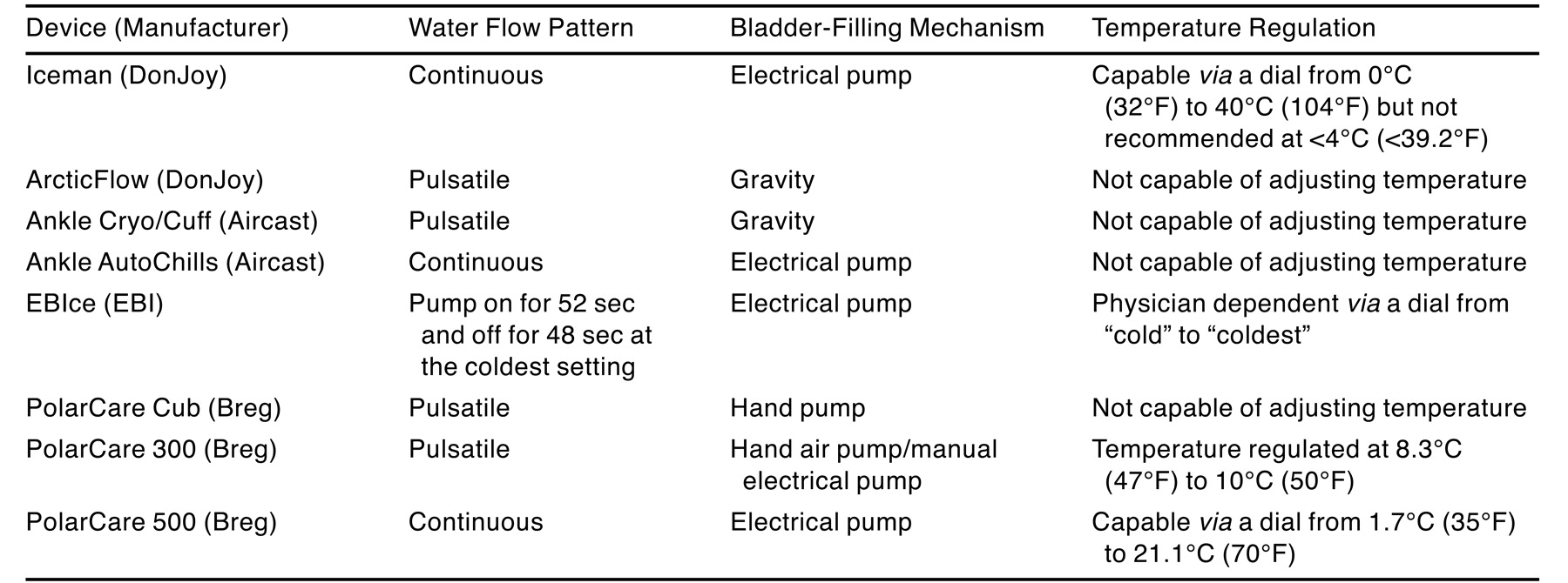
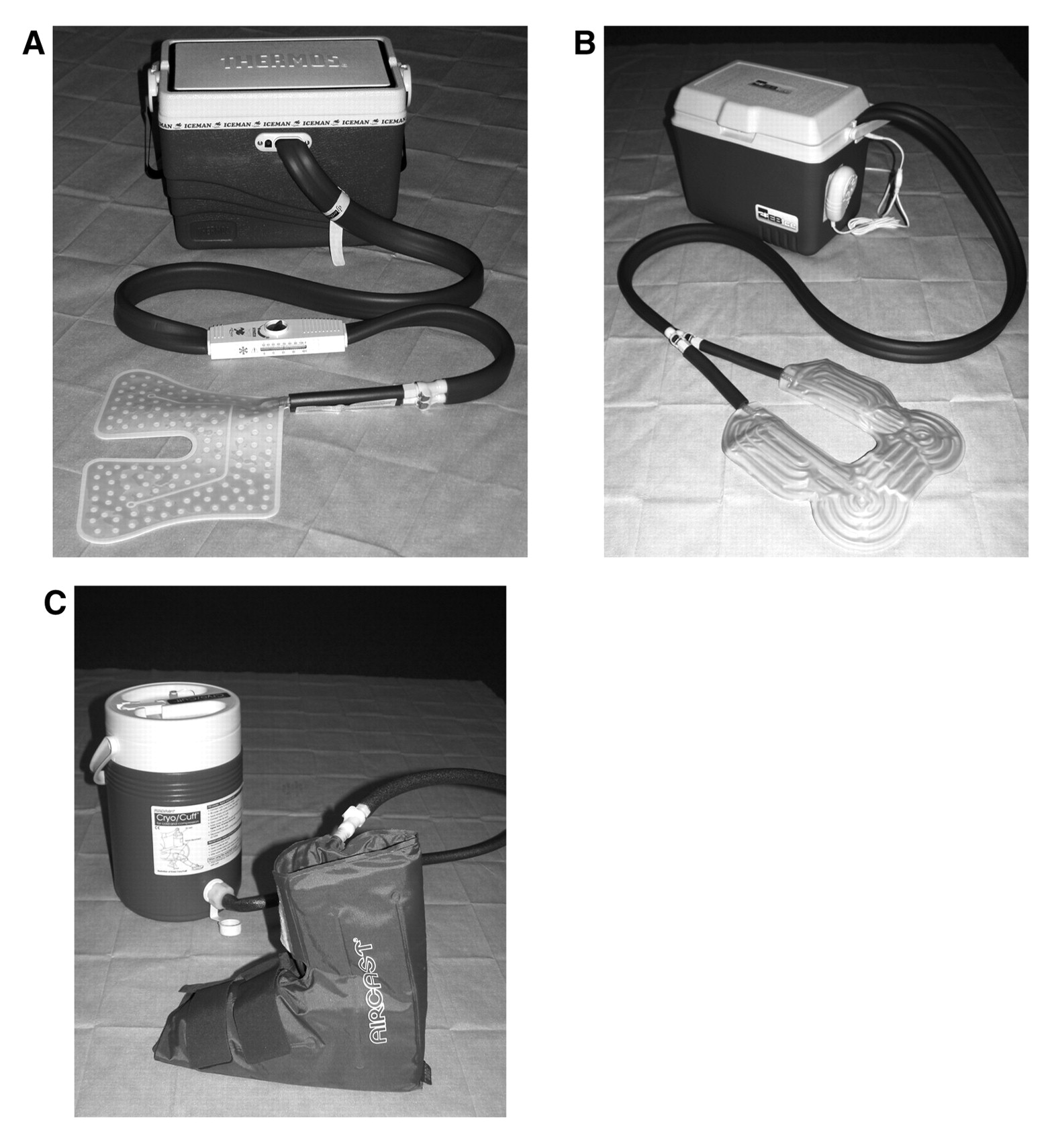
In this study, three representative cryotherapy devices were investigated for their efficacy and potential implications. Iceman Model 1100 Cold Therapy System (DonJoy Orthopedics, Vista, California) is a continuous-flow cryotherapy device that has an electrical pump that mechanically delivers water continuously through the hose and into the bladder (Fig. 1A). The unit’s temperature-adjusting dial regulates the velocity of the cold water circulating through the bladder to adjust the temperature from 0°C (32°F) to 40°C (104°F). EBIce (EBI Medical Systems, Parsippany, New Jersey) also delivers water in a continuous manner via an electrical pump. Similarly, it has a temperature control dial ranging from “cold” to “coldest” to determine the interval of cold water circulation (Fig. 1B). At the least cold setting, the unit pumps water for 9 of 100 sec, whereas at the coldest setting, water circulates for 52 of every 100 sec. Ankle Cryo/Cuff (Aircast Inc, Summit, New Jersey) delivers cold water into its bladder using gravitational force (Fig. 1C). Manual elevation of the cooler above the bladder draws the cold water into the bladder; therefore, the temperature regulation is patient dependent. The company’s recommendation is to repeat this cycle every 2 to 3 hours. The device is also capable of providing 30 to 35 mm Hg of compression to the affected area.[10,11]
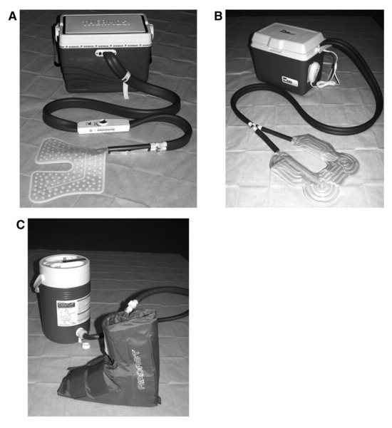
Figure 1.
Three commercially available cryotherapy devices are investigated in this study: Iceman (Don-Joy) (A), EBIce (EBI) (B), and Cryo/Cuff (Aircast) (C).
These devices were applied over standard postsurgical dressings and over different layers of Jones compression bandages to mimic various postsurgical situations at St. Vincent Charity Hospital, Cleveland, Ohio, and in suspect for many of our colleagues, and surface skin temperature was measured in four healthy individuals.
Materials and Methods
Four individuals (two men and two women aged 26–35 years) in excellent overall health with no medical history were selected for this study. They had no signs of neurovascular compromise—normal pedal pulses, capillary fill time, and epicritic sensation. The dorsalis pedis and posterior tibial pulses of all of the subjects were normal (2 out of 4). They had no risk factors for peripheral vascular or vasoconstrictive disease, such as a history of cardiac diseases, hyperlipidemia, diabetes, coagulopathy, obesity, or smoking.
To measure skin temperature, we chose the HH508 Handheld Digital Thermometer (Omega Engineering Inc, Stamford, Connecticut) based on its ability to precisely measure surface temperature with minimal variance (Fig. 2). The unit is capable of being operated at an ambient temperature of 0°C (32°F) to 50°C (122°F) and of measuring temperature ranges from −200°C (–328°F) to 1,093°C (1,999°F) with accuracy of ±0.1% of the reading +1°C. This temperature-measuring device has wire probes that can easily be inserted under the dressings.
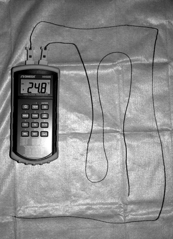
Figure 2.
The HH508 Handheld Digital Thermometer (Omega) was used to measure surface skin temperatures.
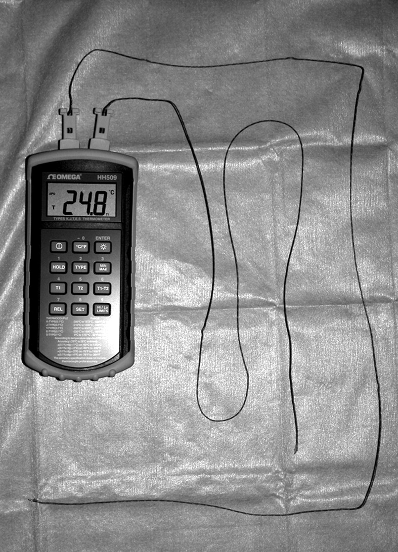
For each subject, the progression of surface skin temperature was recorded after initiation of cryotherapy using all three cryotherapy devices in three surgical dressing patterns (groups I–III). As a control, the skin temperature was also measured under all of the dressing patterns without any cryotherapy device applied.
Subjects were placed in the supine position with their extremities elevated with two pillows. The temperature-measuring probe was then placed on the midfoot dorsally in the area of the lateral cuneiform and secured with paper tape. To mimic the postsurgical scenario, three layers of eight-ply, 4 × 4-inch gauze sponges (#2318; Johnson & Johnson Medical, Arlington, Texas) were placed over the midfoot above the temperature-measuring probe. One roll of 3 × 75-inch Sof-Kling conforming gauze bandage (#6993; Johnson & Johnson Medical) followed by one 4.5-inch × 4.1-yard Kling fluff roll (#6930; Johnson & Johnson Medical) were applied uniformly to cover the entire foot from the proximal digits to the ankle.
In group I, the Iceman and EBIce devices were applied over the dressings mentioned previously herein and were held in place using a Swift-Wrap Elastic Bandage (Medline Industries Inc, Mundelein, Illinois). The Ankle Cryo/Cuff was placed over the top of the dressings and the Swift-Wrap Elastic Bandage. The skin temperature was obtained immediately after application of the dressings. Ice cubes were put in the cooler of each cryotherapy device to the level recommended by the manufacturer, and tap water was poured into the cooler. The water was then cooled for 10 min, and the units were set at the lowest temperature before starting the experiment. The Cryo/Cuff was manually recirculated every 15 min to achieve the coldest possible temperature, against the manufacturer’s recommendation (which was to recirculate the water every 2–3 hours), just before each reading. The skin temperature was then recorded every 15 min for 180 min starting at the initiation of cryotherapy.
In group II, one layer of Jones compression bandage, which consisted of two 4-inch × 4-yard Specialist cast paddings (#9044; Johnson & Johnson Orthopaedics, Raynham, Massachusetts) and a 6-inch Swift-Wrap Elastic Bandage, was applied uniformly from the digits to the tibial tuberosity over the dressings used in group I. The Iceman and EBIce devices were held in place over the Specialist cast paddings underneath the 6-inch Swift-Wrap Elastic Bandage, whereas the Cryo/Cuff was placed over the 6-inch Swift-Wrap Elastic Bandage, and skin temperature was recorded in the manner described for group I.
In group III, a second layer of Jones compression bandage was applied over the dressing model of group II in the same way, and the devices were applied on top of this layer using the same method as described for group II. The temperatures were again recorded in the same manner.
On collection of data, the mean and lowest temperatures were determined from the recordings of all four subjects. The standard deviation was also calculated. At the end of the trials, the subjects were asked if they had any discomfort from each device. If they did, they were asked at what temperature they started to feel the discomfort.
Results
A summary of the mean and lowest temperatures of each recording is shown in Table 2, and temperature progression curves are shown in Figure 3, Figure 4 and Figure 5. In group I, use of the Iceman continuous-flow cryotherapy device was discontinued in one subject owing to intolerable pain and paresthesia experienced at 105 min in the trial. The skin temperature reading at this point was 14.2°C (57.6°F). Extensive forefoot erythema and delayed capillary fill time of the digits (11 sec) was noted before discontinuation of the Iceman unit. In this particular group, the final temperature (14.2°C or 57.6°F) was recorded as the lowest temperature and the mean temperature was calculated from the available data. EBIce and Cryo/Cuff were tolerated for the full 180 min in all four subjects. Temperature progression on the mean was similar for all of the cryotherapy devices, although the standard deviations in group I were generally higher than those in the other groups. Temperature was still declining at the end of each trial (180 min) in all of the devices.
Table 2.
Summary of the Surface Skin Temperature Progression With Application of Three Cryotherapy Devices Over Different Layers of Dressings
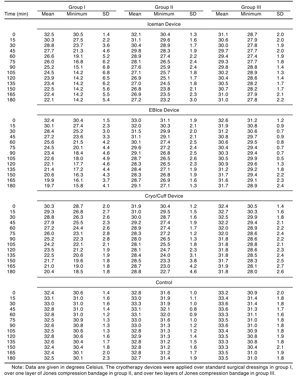
Figure 3.
Mean (A) and minimum (B) surface skin temperature progression curves under no Jones compression bandage (group I).
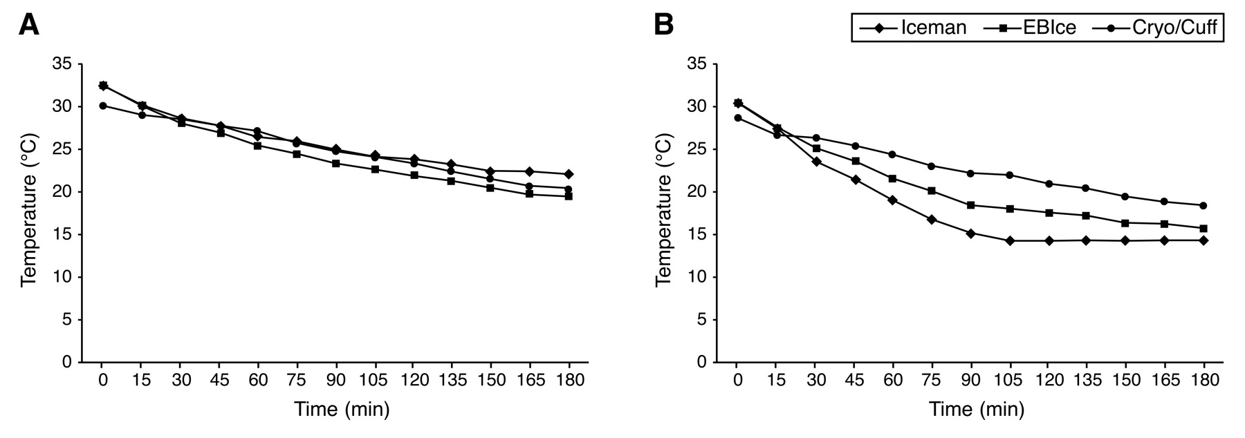
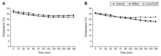
Figure 4.
Mean (A) and minimum (B) surface skin temperature progression curves under one layer of Jones compression dressing (group II).
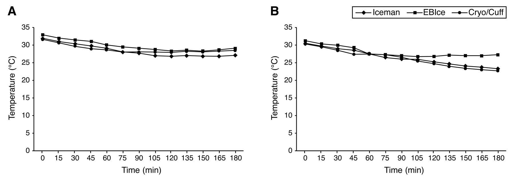
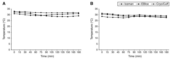
Figure 5.
Mean (A) and minimum (B) surface skin temperature progression curves under two layers of Jones compression dressing (group III).
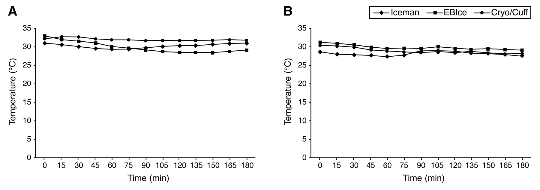
In group II, all of the subjects were pain free throughout all of the trials, and temperature depression leveled off before the end of the trials in all three units. All of the devices kept the skin temperature below 30°C (86°F) for the length of the cryotherapy trials. The lowest temperature achieved by each device was not far from its mean value.
In group III, skin temperatures in all three cryotherapy units were not significantly different from the initial readings. Temperatures achieved by each unit throughout the trial were slightly lower than those of the control. The difference in temperature depression between units was minimal.
The subjective information received from the test subjects throughout the study indicated that the most comfortable period of testing (the temperatures at which the subject felt coolness from the cryotherapy units without pain or discomfort) was between 23°C (73°F) and 30°C (86°F). Once the skin temperature rose above 30°C (86°F), subjects relayed that they felt extremity “hot” and actually began to perspire. When the temperature fell below 23°C (73°F) using the Iceman device, the subjects began to feel discomfort, and it became intolerable at 14.2°C (57.6°F) and required discontinuation of the trial in one subject. The subjects did not experience discomfort with the EBIce and Cryo/Cuff devices until the temperatures reached approximately 18°C (64°F). The subjects relayed that discomfort began at a higher temperature with the Iceman simply because the continuous flow of the unit resulted in an earlier intolerance to cold, whereas the intermittent-flow devices were easier to tolerate and gave them a “break” between pulses of cold infiltration.
Discussion
In this study, the Iceman was the only device that required discontinuation. In group I, the lowest surface skin temperature (14.2°C or 57.6°F) in one subject reached by the Iceman was theoretically capable of complete interruption of nerve conduction or increased local tissue edema as suggested by the studies of Matsen et al[9] and McMaster and Liddle.[8] Because of the high standard deviation and skewed distribution of the data in this group, mean temperature progression among all of the subjects should not be overemphasized, and neither is it relevant. This only suggests that the efficacy and safety of these cryotherapy devices were less predictable when they were applied over a thinner postsurgical dressing. Moreover, although none of these devices reached the temperature that may cause frostbite during the 180-min trial, note that the temperature was still declining at the end of the trial. Therefore, we cannot simply rule out the possibility of such complications when a longer duration of cryotherapy is used in a thin surgical dressing.
Over one layer of Jones compression bandage, all of the cryotherapy devices maintained the temperatures between 20°C (68°F) and 30°C (86°F). This is still within the temperature range at which edema reduction is thought to be attained. Some devices even reached less than 25°C (<77°F), which is the level at which nerve conduction starts to be suppressed, resulting in reduction of muscle spasm and pain signal transmission. However, more than 90 min of cryotherapy was required to reach this level. This finding is consistent with a study by Weresh et al,[1] who demonstrated that temperatures less than 25°C (<77°F) were not achieved by the application of ice packs over one layer of Jones dressing within 90 min. All of the cryotherapy devices were tolerated throughout the experiment in this group. Temperature progression also leveled off at the end of the trials.
Over two layers of Jones compression dressing, no device significantly lowered the surface skin temperature from the initial reading. Therefore, it is likely that nerve conduction would not be affected by these devices, and neither would reduction of metabolism in the tissue. Jones compressive bandages are generally poor conductors of cold,[1] and the efficacy of the cryotherapy device is questionable if two layers are applied. However, edema reduction may still take place at this temperature range. Compared with the control group, the skin temperature in this group was slightly lower and closer to the 30°C (86°F) point, where edema reduction was best achieved in the study by McMaster and Liddle.[8]
Wilke and Weiner[4] studied continuous-flow cryotherapy units in foot and ankle postoperative settings and concluded that they controlled pain, inflammation, and blood loss. However, they also noted that caution should be taken because rare complications can be devastating. Mora et al[11] discovered that pulsatile cold compression achieved significant ankle circumference reduction in patients after ankle fracture compared with patients treated with splinting and elevation alone. A similar result was obtained by Scheffler et al[12] in human subjects with compressive cryotherapy using the Cryo/Cuff. Furthermore, Stockle et al[13] suggested that continuous-flow cryotherapy devices reduced tissue swelling in the lower extremity more effectively than the simple ice pack application.
Based on the subjective and objective analyses from this study and previous animal and clinical studies, we feel comfortable providing cryotherapy in the temperature range of 20°C (68°F) to 30°C (86°F) for intermittent-flow cryotherapy units and 25°C (77°F) to 30°C (86°F) for continuous-flow units in a healthy individual. However, one needs to keep in mind that a patient with peripheral vascular disease, vasospastic disease, or neuropathy, or a patient in the postoperative period with a local anesthetic nerve block or taking oral narcotic analgesics is more susceptible to complications with cryotherapy. External validity, especially in a high-risk patient population, cannot be justified from this small study conducted in healthy individuals. Because of the small sample size and the nature of the study design, this study was more susceptible to sampling bias, and the power of the study may not be adequate to draw significant conclusions. Case-controlled clinical studies are needed to determine the true therapeutic temperature range of cryotherapy in the postoperative setting, and this experiment may serve as a framework for such future studies.
Conclusion
Newer cryotherapy devices can be very effective at minimizing postoperative pain, swelling, muscle spasm, and discomfort when used appropriately. In this study, we achieved the safe and effective temperature range when the cryotherapy devices were applied over one layer of Jones compression dressing. The cryotherapy devices resulted in less predicable temperature declination when applied over the thinner surgical dressing. When the devices were applied over two layers of Jones compression dressing, surface skin temperature declination was minimal.
Although statistically significant data were not derived from this experiment, the subjective analysis showed that the intermittent-flow devices were more comfortable to the subjects than the continuous-flow device even in the same temperature range. The continuous-flow device required discontinuation in one subject, whereas the intermittent-flow devices were tolerated in all of the subjects for at least 180 min from initiation of cryotherapy in all of the surgical dressing types tested.
Financial Disclosures
None reported.
Conflict of Interest
None reported.
References
- Weresh MJ, Bennett GL, Njus G: Analysis of cryotherapy penetration: a comparison of the plaster cast, synthetic cast, Ace wrap dressing, and Robert-Jones dressing. Foot Ankle Int 17: 37, 1996.
- Olson JE, Stravino VD: A review of cryotherapy. Phys Ther 52: 840, 1972.
- Bugaj R: The cooling, analgesic, and rewarming effects of ice massage on localized skin. Phys Ther 55: 11, 1975.
- Wilke B, Weiner RD: Postoperative cryotherapy: risks versus benefits of continuous-flow cryotherapy units. Clin Podiatr Med Surg 20: 307, 2003.
- Ciolek JJ: Cryotherapy: review of physiological effects and clinical application. Cleve Clin Q 52: 193, 1985.
- Murphy JV, Banwell PE, Roberts AH, et al: Frostbite: pathogenesis and treatment. J Trauma 48: 171, 2000.
- Yu GV, Vincent AL, Hudson JR: Inside insight for treating frostbite. Podiatry Today 14: 46, 2001.
- McMaster WC, Liddle S: Cryotherapy influence on posttraumatic limb edema. Clin Orthop Relat Res 150: 283, 1980.
- Matsen FA III, Questad K, Matsen AL: The effect of local cooling on postfracture swelling: a controlled study. Clin Orthop Relat Res 109: 201, 1975.
- Mindrebo N, Shelbourne D: Knee pressure dressings and their effects on lower extremity venous capacitance and venous outflow. Orthop Int Ed 2: 273, 1994.
- Mora S, Zalavras CG, Wang L, et al: The role of pulsatile cold compression in edema resolution following ankle fractures: a randomized clinical trial. Foot Ankle Int 23: 999, 2002.
- Scheffler NM, Sheitel PL, Lipton MN: Use of Cryo/Cuff for the control of postoperative pain and edema. J Foot Surg 31: 141, 1992.
- Stockle U, Hoffmann R, Schutz M, et al: Fastest reduction of posttraumatic edema: continuous cryotherapy or intermittent impulse compression? Foot Ankle Int 18: 432, 1997.
© 2007 American Podiatric Medical Association