


Abstract
A posterior bone-block operation is one of the few treatment options in cases of paralytic footdrop. A case with a flail ankle and no bony deformity is ideal for this approach. Two cases of acquired flail ankle with equinus deformity were treated using a new modification of the bone-block technique that does not interfere with subtalar joint motion. A bone block harvested from the iliac crest was fixed at the posterior talus after partial resection of the posterior tubercle. The graft was in contact with the posterior malleolus of the tibia. Satisfactory correction was achieved, and both patients could walk without the use of external splints.
A posterior bone-block operation for footdrop was first performed by Campbell in 1921, as a result of a report from a committee appointed by the American Orthopaedic Association to investigate the different treatment operations for footdrop.[1] Various modifications of his technique have been reported since the first description published in 1923.[2,3,4,5] Enthusiasm for this approach waned after the refinement of tendon transfer principles and techniques in the 1930s.
In some patients, all of the muscles distal to the knee are affected at a similar level, and tendon transfer is not an option. The disease is usually acquired after puberty; thus neuromuscular deficiency is not accompanied by a bony deformity. Footdrop is the main complaint, and lifelong use of orthoses is the most common solution. We report two such cases of footdrop that were operated on using a new modification of the posterior bone-block technique, aiming to allow an orthosis-free life.
Patients and Method
Surgical Technique
A lazy S–shaped longitudinal incision approximately 13 cm long is made ending at the posterior tubercle of the calcaneal bone. The Achilles tendon is lengthened using a Z-type cut. The length of the cut depends on the severity of the equinus contracture of the ankle joint. If a fixed contracture is not present, a cut of approximately 10 cm is adequate. The posterior tibiotalar and subtalar joints are released medially and laterally until dorsiflexion of approximately 10° is achieved. The posterior tubercle of the talus is cleared of soft tissues.
A tricortical iliac bone graft is harvested using standard surgical technique. The graft is approximately 4 cm wide and 3 cm deep. The anterior cut on the iliac bone is placed 5 cm proximal to the anterior superior iliac spine because the iliac crest is thicker at this level. The posterior talar tubercle is partially resected to prepare a sleeve for the graft (Fig. 1A). The anteroposterior dimension of this sleeve should be similar to the thickness of the iliac bone graft. The graft is placed such that the iliac crest side touches the posterior malleolus of the tibia when the ankle joint is in slight dorsiflexion of approximately 5°. The shapes of the host side and the graft and the position of the graft are adjusted after several trials. After completion of this step, the graft is fixed temporarily using two 1.5-mm Kirschner wires. A standard 6.5-mm AO screw is placed in a posterior-to-anterior direction through the graft into the talar body over a washer (Fig. 1B). After removal of the Kirschner wires, the stability of the fixation is checked. The passive range of motion of the ankle should be limited to 0° to 5° of dorsiflexion with moderate force applied by the surgeon. The Achilles tendon is repaired without undue tension, and the skin is closed over a suction drain. A short-leg cast is applied in slight dorsiflexion, which is changed the second week after surgery. The patient is not allowed to bear full weight until week 12, when the cast is removed.
Case 1
A 55-year-old woman was referred to the emergency department of Istanbul Medical Faculty with a diagnosis of acute compartment syndrome of the right lower leg that had occurred after an elective knee arthroscopy performed for osteoarthritic changes in the joint. The operation was performed 2 days before her referral. An injury to the popliteal artery was detected and repaired using a venous graft. A double-incision fasciotomy was also added. The wounds healed after split-thickness skin grafting. In month 6, no active motion was detected at the ankle. A weak toe flexion was the single active movement in the foot, and all of the muscles of all three calf compartments were fibrotic. The ankle was fixed in 35° of equinus, with a tibiocalcaneal angle of 53° (Fig. 2A).
A posterior bone block was placed at the ankle joint in month 65 after the iatrogenic vascular injury (Fig. 2B). The wound healed uneventfully. In month 3, the patient was bearing full weight without using crutches. Radiographic examination revealed partial resorption at the proximal part of the graft. In the first year, interphalangeal/proximal interphalangeal arthrodeses were performed for claw-toe deformities of the first through fourth toes. Displacement of the washer was seen on the radiograph. In the second year, the graft had healed, and the position of the washer had remained unchanged for the past year (Fig. 2C). The ankle was in 5° of equinus (Fig. 2D), and the patient was walking without using any aids. The tibiocalcaneal angle was measured as 83°.
Case 2
A 29-year-old woman presented to Istanbul Medical Faculty, Department of Orthopaedics and Traumatology, complaining of a nonhealing wound at the right fourth toe tip of 6 months’ duration and gait disturbances related to equinus deformity. She had a fall-related injury 12 years earlier and an irreversible neurologic deficit at the right lower leg due to a burst-type spinal fracture.
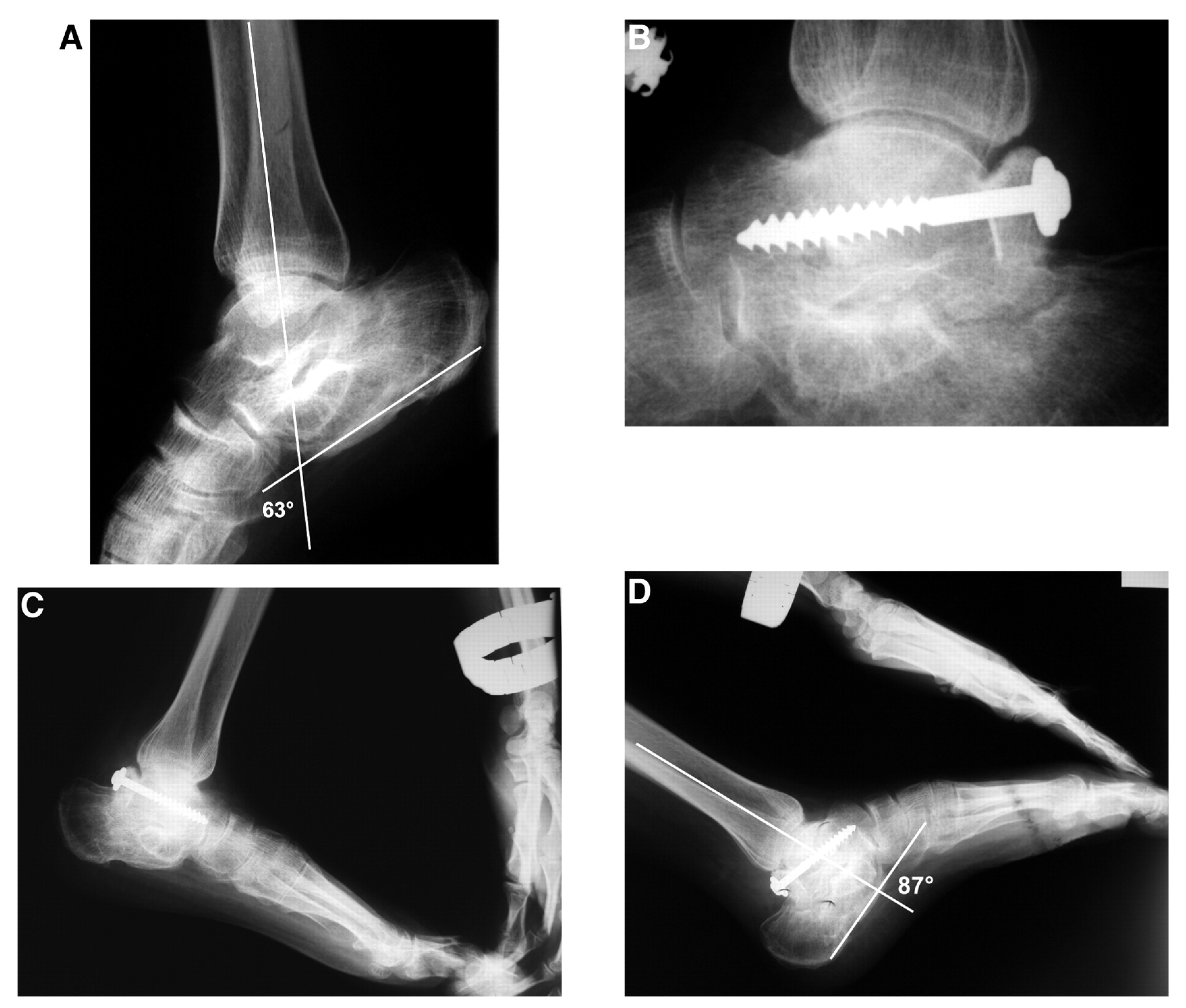
Passive range of motion of her right ankle was 20° to 30° of plantarflexion (Fig. 3A). Clawing of all of the toes was evident with passive ankle dorsiflexion, and no active motion was present at the ankle or foot. The subtalar joint was stiff in the neutral position, and the heel was not painful. The tibiocalcaneal angle was measured as 63°.
The patient was previously advised to undergo an arthrodesis operation involving the ankle joint. The patient strongly objected to this approach. Radiologic examination revealed malunion of the right calcaneus with ankylosis of the subtalar joint. An accompanying forefoot drop was also identified.
Posterior bone-block technique was applied after an Achilles tendon lengthening and posterior capsular release. A larger graft compared with the first case was used (Fig. 3B). The tricortical side (iliac crest side) was positioned proximally. Tenotomy of the long flexor tendon was also added in the four lesser toes.
A superficial wound infection occurred postoperatively and healed uneventfully after parenteral and oral antibiotic drug treatment. Proximal interphalangeal arthrodesis was performed for residual clawing deformities of the third through fifth toes in postoperative month 8. In postoperative month 13, she was walking without using an external support and was satisfied with the result of the ankle operation. A decrease of 24° was observed in the tibiocalcaneal angle (Fig. 3 C and D). The forefoot drop, which hindered a plantigrade barefoot gait, could be overcome with the use of a high-lace shoe.
Discussion
Posterior bone block was a popular technique for the treatment of footdrop in the 1920s and 1930s, with many reported modifications. In the original technique described by Campbell,[2] a large bone block was placed between the ankle joint and the Achilles tendon using autologous bone grafts. The mechanism was similar to that of hyperextension block of the olecranon. Gill[3] modified this approach by inserting a wedge of bone taken from the calcaneus into the posterior talus. Wagner[4] proposed the use of an intra-articular bone block placed posterior to the talus using a screw, similar to our technique. This relatively smaller bone block was placed without resecting the posterior tubercle of the talus and spanned the calcaneus and the tibia. It was still much larger than the graft we used. The graft was usually the talar head, which was resected during a simultaneous triple arthrodesis, or the superior part of the posterior calcaneus. Lewin[6] proposed turning down a flap from the posterior surface of the tibia. The main difference in the described technique is preservation of the subtalar joint. The graft is placed at the posterior talus, above the level of the subtalar joint, whereas all of the other reported techniques use grafts placed between the calcaneus and the talus.
Results of the previous modifications were inconsistent. A satisfying result was obtained in 44% to 91% of the cases in the reported series.[7,8,9,10,11] Ingram (a surgeon from Campbell’s clinic) and Hundley[7] reported on the long-term results (5–27 years) of 90 feet in 1951. The study group consisted of patients with various diagnoses, such as poliomyelitis, cerebral palsy, and peripheral nerve injury. Both Campbell’s original technique and Wagner’s modification were included in the study. Results were satisfying in 82% of the cases. Unsatisfactory results were mostly caused by hypertrophic arthritis of the tibiotalar joint and aseptic necrosis of the talus. These problems were related to the triple arthrodesis operation, performed simultaneously for the correction of foot deformities. The best results were obtained in a small group of patients with a flail foot or a balanced foot in which there is a discrepancy of less than two grades between the plantarflexor group and dorsiflexors. In patients with a satisfactory result, the mean degree of motion of the foot on the leg was 17°. Three-fourths of this range was in the tibiotalar joint, and the remaining one-fourth was in the intertarsal and tarsometatarsal joints. According to these results, both presented cases were good candidates for this type of operation, having a flail but balanced ankle joint and no bony deformity requiring arthrodesis of the foot bones.
Posterior bone-block techniques have a limited but unequaled role in footdrop treatment. The techniques of posterior bone-block placement should be refined and incorporated into advanced training programs.
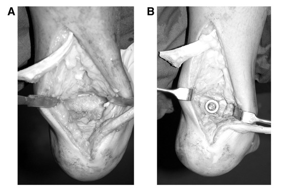
Figure 1.
A, A quadrangular portion of the posterior talus is removed. B, The tricortical iliac bone graft is fixed using a screw and washer.
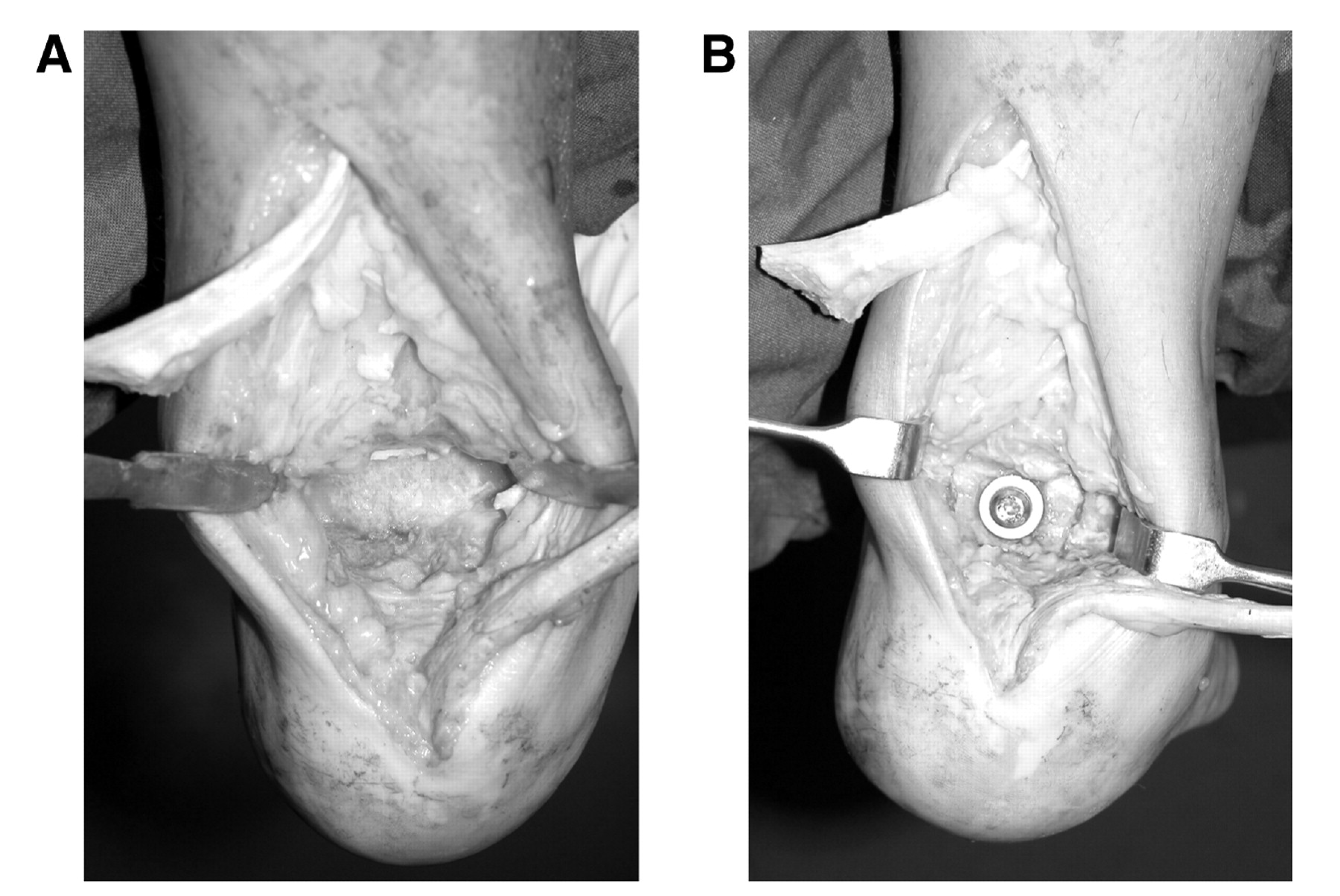
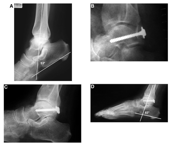
Figure 2.
A, Preoperative lateral radiograph of the ankle. The tibiocalcaneal angle was measured as 53°. B, Early postoperative radiograph showing the graft at the posterior talus. The tricortical side of the graft is facing plantarward. C, Follow-up radiograph in month 23. The displacement of the washer indicates partial resorption of the graft. D, Despite the resorption of the graft, the tibiocalcaneal angle was measured as 83°, 30° more than the preoperative value.
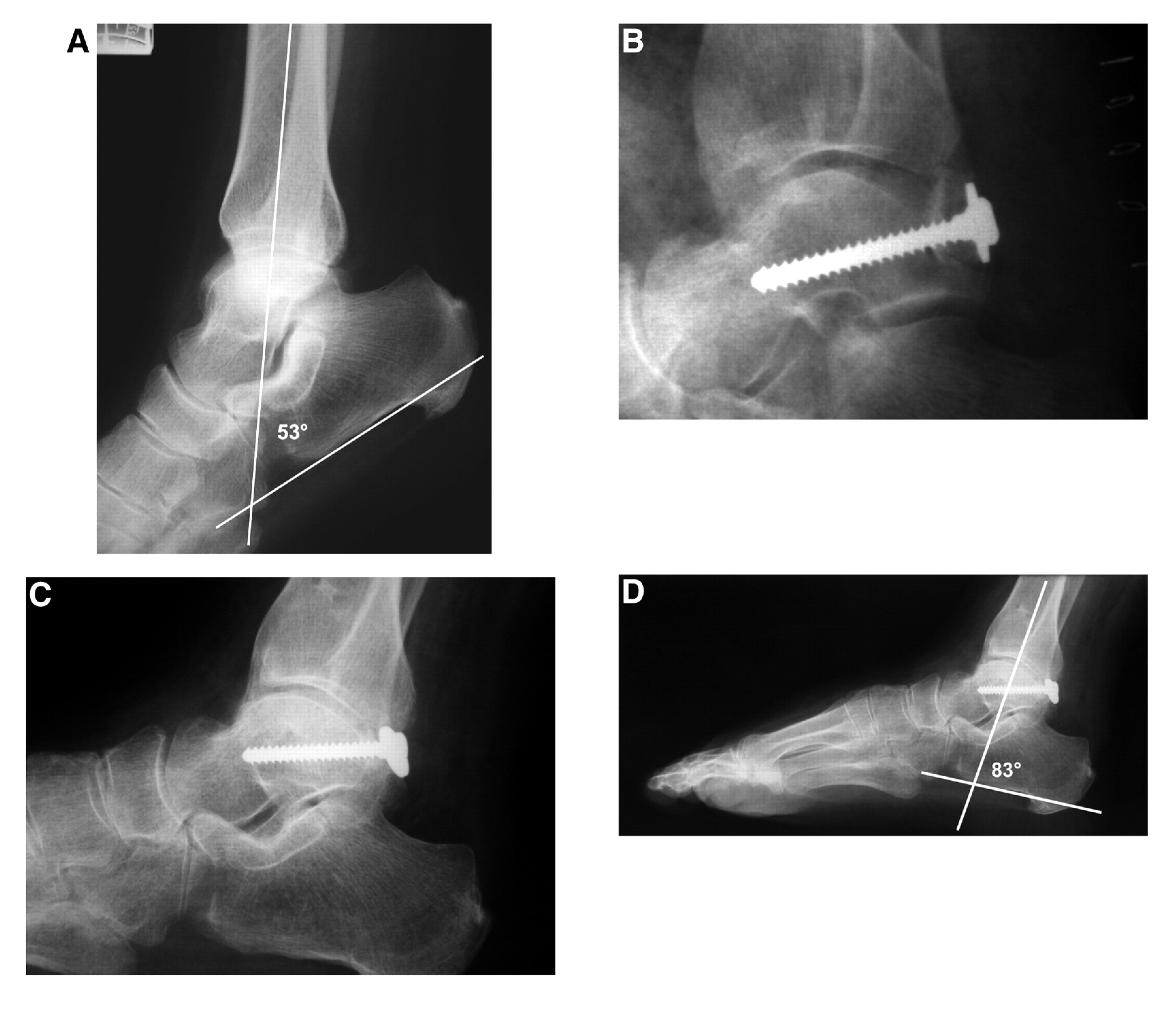
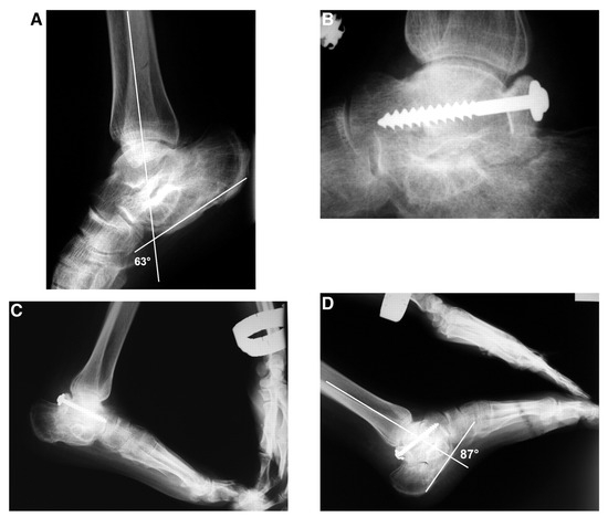
Figure 3.
A, Preoperative lateral view of the resting ankle. Narrowing and irregularities of the subtalar joint are seen. The tibiocalcaneal angle was measured as 63°. B, Lateral view in postoperative month 9. The position of the graft remains unchanged. C and D, Stress dorsiflexion (C) and plantarflexion (D) lateral radiographic examination of the ankle joint. The tibiocalcaneal angle at maximum passive plantarflexion was measured as 87°.
Financial Disclosure
None reported.
Conflict of Interest
None reported.
References
- Moore JR: Comment on J Bone Joint Surg Am 33: 679, 1951. J Bone Joint Surg Am 33: 690, 1951.
- Campbell WC: An operation for the correction of “drop-foot.”. J Bone Joint Surg 5: 815, 1923.
- Gill AB: An operation to make a posterior bone block at the ankle to limit foot-drop. J Bone Joint Surg 15: 166, 1933.
- Wagner LC: Modified bone block (Campbell) of ankle for paralytic drop-foot: with report of twenty-seven cases. J Bone Joint Surg 13: 142, 1931.
- Ingram AJ: “Paralytic Disorders,” in Campbell’s Operative Orthopaedics, Vol 4, ed by AH Crenshaw, p 2952, CV Mosby, St Louis. ,1987.
- Lewin P: “Anterior Poliomyelitis: Infantile Paralysis,” in The Foot and Ankle, p 550, Lea & Febiger, Philadelphia. ,1947.
- Ingram AJ, Hundley JM: Posterior bone block of the ankle for paralytic equinus: an end-result study. J Bone Joint Surg Am 33: 679, 1951.
- Inclan A: End results in physiological blocking of flail joints. J Bone Joint Surg Am 31: 748, 1949.
- Wheeldon TF, Clark MM: The Gill bone block operation for drop foot. JAMA 106: 447, 1936.
- Branch H: Drop-foot: end results of a series of bone-block operations. J Bone Joint Surg 21: 141, 1939.
- Carrell WB, Childress HM: Posterior bone block in talipes equinus: factors determining end results. South Med J 32: 528, 1939.
© 2007 American Podiatric Medical Association