


Abstract
Background: A study was undertaken to investigate the ability to predict dynamic foot posture from static measurements. Methods: Arch height and arch height ratio measurements were obtained from videotape images of the medial aspect of the foot during standing, walking, and jogging in 5 male and 13 female asymptomatic subjects. Results: Arch height and arch height ratio measurements taken in standing explained 66% to 83% of the variance associated with these measurements at midstance during walking and running. Arch height and arch height ratio demonstrated high reliability as static and dynamic measures. Conclusions: The results of this study support the use of arch height and arch height ratio measurements taken statically in the clinical assessment of the foot and may assist the clinician in estimating foot posture during dynamic activity in patients with lower-limb injuries.
Emerging evidence suggests that foot posture is linked to musculoskeletal pathology and overuse injuries, particularly in runners.[1,2] Foot posture or structure is often determined by measurement of the medial longitudinal arch. To detect abnormal foot structure in a patient, it is imperative to complete a comprehensive clinical examination. It is also considered best practice to evaluate dynamic motion in functional tasks that aggravate symptoms.[3] Measuring the quantity and quality of motion dynamically in a clinical setting is often difficult, time-consuming, and expensive. As a result, clinicians commonly use static measures, with an implicit understanding that “structure dictates function.”[4]
An issue for practitioners is that research is inconclusive when explaining the relationship between static and dynamic measures of foot posture. McPoil and Cornwall[5] investigated the ability of 17 static measurements of the lower extremity to predict maximum rearfoot pronation and time to maximum pronation during walking. This study found that only one static measure—difference in navicular height between resting and neutral standing postures—predicted maximum rearfoot pronation. However, this measure explained only 17% of the variability associated with maximum rearfoot pronation and led the authors to the conclusion that static measurements are poor predictors of dynamic foot function, as found in previous studies.[6]
Conventionally, subtalar joint motion has been measured using landmarks on the distal leg and calcaneum, yet Stacoff et al[7] suggest that when evaluating orthotic effects on foot motion, midfoot and forefoot movements may be more important than that of the calcaneus. Such thoughts have prompted the search for a more appropriate measure of foot posture and investigations into measurements obtained from the medial side of the foot, such as arch height ratio and longitudinal arch angle.[8,9]
Longitudinal arch angle was first defined by Dahle et al[10] as the angle formed by a line from the midpoint of the medial malleolus to the navicular tuberosity and another line from the midpoint of the first metatarsal head to the navicular tuberosity. Jonson and Gross[11] reported that measures of the longitudinal arch angle obtained in static foot positions are highly reliable, with intrarater and interrater intraclass correlation coefficients (ICCs) of 0.90 and 0.81, respectively.
McPoil and Cornwall[9] also reported longitudinal arch angle to be a reliable measure, with intrarater and interrater ICCs of 0.96 and 0.67, respectively, and investigated whether static measures of the longitudinal arch angle could be used to predict dynamic foot posture during walking. They reported that static measurements of the longitudinal arch angle were highly predictive of dynamic foot posture, with static longitudinal arch angle explaining more than 90% of the variance associated with the angle at midstance during walking. Although the longitudinal arch angle demonstrates high reliability as a static measure, its reliability dynamically is questionable, possibly because the measurement relies on the identification and skin marking of three bony landmarks. Skin markers can be a source of error because skin movement during dynamic activity could displace the skin markers being used to identify bony landmarks by 4.3 mm at the navicular tuberosity and 1.8 mm at the first metatarsal head.[12] This variance suggests the need for a measurement that can be used to assess foot structure statically and dynamically that does not require marking the skin over a bony landmark.
Williams et al[2,8,13] first described the arch height ratio as the arch height divided by the truncated foot length. Arch height was defined as the height of the dorsum of the foot from the floor at 50% of the foot length, and truncated foot length was defined as the length of the foot from the first metatarsophalangeal joint to the most posterior portion of the calcaneus. The arch height ratio measurement demonstrated the highest reliability and validity across the 10% and 90% weightbearing conditions compared with the six other measurements that were tested, with interrater reliability of 0.81 and 0.85 and intrarater reliability of 0.94 and 0.98, respectively. Although arch height ratio has been demonstrated to be a highly reliable static measure in several studies,[2,8] this measurement has not yet been tested dynamically. In addition, the 10% and 90% weightbearing conditions used by Williams and McClay[8] are potentially problematic because of the need of the patient to achieve a prespecified amount of body load through the foot. Having the patient with equal body weight on each foot (ie, 50% body weight) would facilitate easier and more efficient patient foot placement. Results of the study by McPoil and Cornwall[9] support the measurement of foot posture with equal body weight on each foot because they reported that the static longitudinal arch angle measure obtained in 50% body weight was highly predictive of the angle at midstance during walking.
Because the arch height ratio measure shows high reliability as a static measure, it could be of value in predicting dynamic foot posture, as with the longitudinal arch angle. It is tempting to suggest that because the arch height ratio relies on only one mark to be placed on the skin (ie, at the metatarsophalangeal joint), it may provide a more suitable measurement of dynamic foot posture. Furthermore, the arch height measure has not been tested during gait, and because it is less prone to skin marker movement (ie, no skin markers are required), it is highly likely that it could provide a more suitable measurement.
The purpose of this study was twofold. The first aim was to investigate the reliability of arch height and arch height ratio as dynamic measures of foot posture. The second aim was to evaluate the relationship between static and dynamic measurements of arch height and arch height ratio.
Methods
Participants
A convenience sample of 18 asymptomatic volunteers (36 feet), 5 males and 13 females with a mean ± SD age of 27 ± 5.8 years, participated in the study. Criteria for participant exclusion included coronary risk factors, orthotic device use, and a current lower-limb injury requiring a decrease in activity or consultation with a health-care professional. Volunteers who had severe foot deformities, such as pes cavus, severe pes planus, or hallux abducto valgus, as determined by visual examination, were also excluded from the study. All of the participants were recruited from the Northern Arizona University population, performed regular fitness or athletic activity, and were capable of completing the necessary controlled exercise program. The Northern Arizona University institutional review board provided ethical approval for the study, and all of the participants provided informed consent.
Technical Equipment
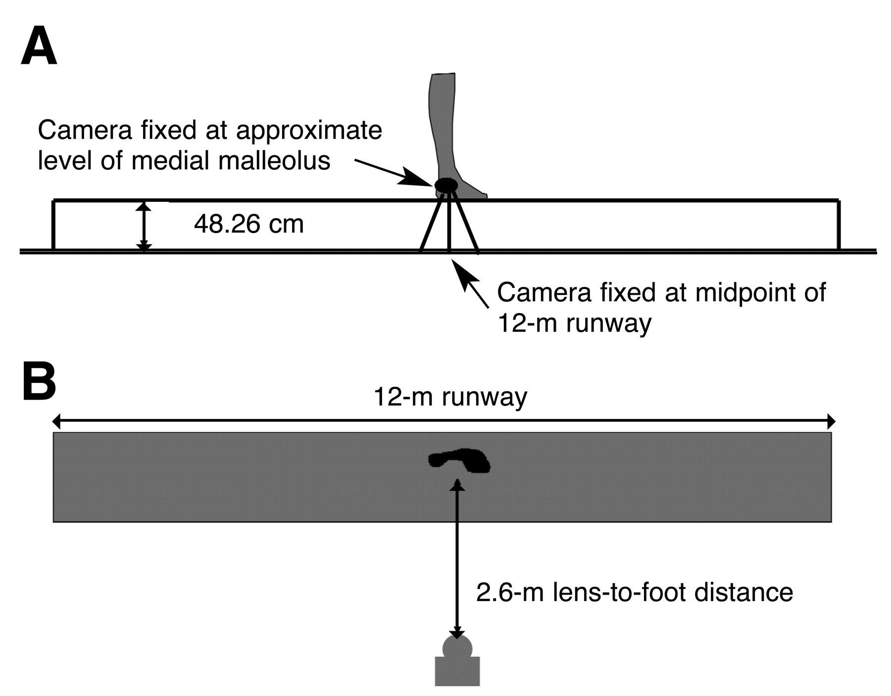
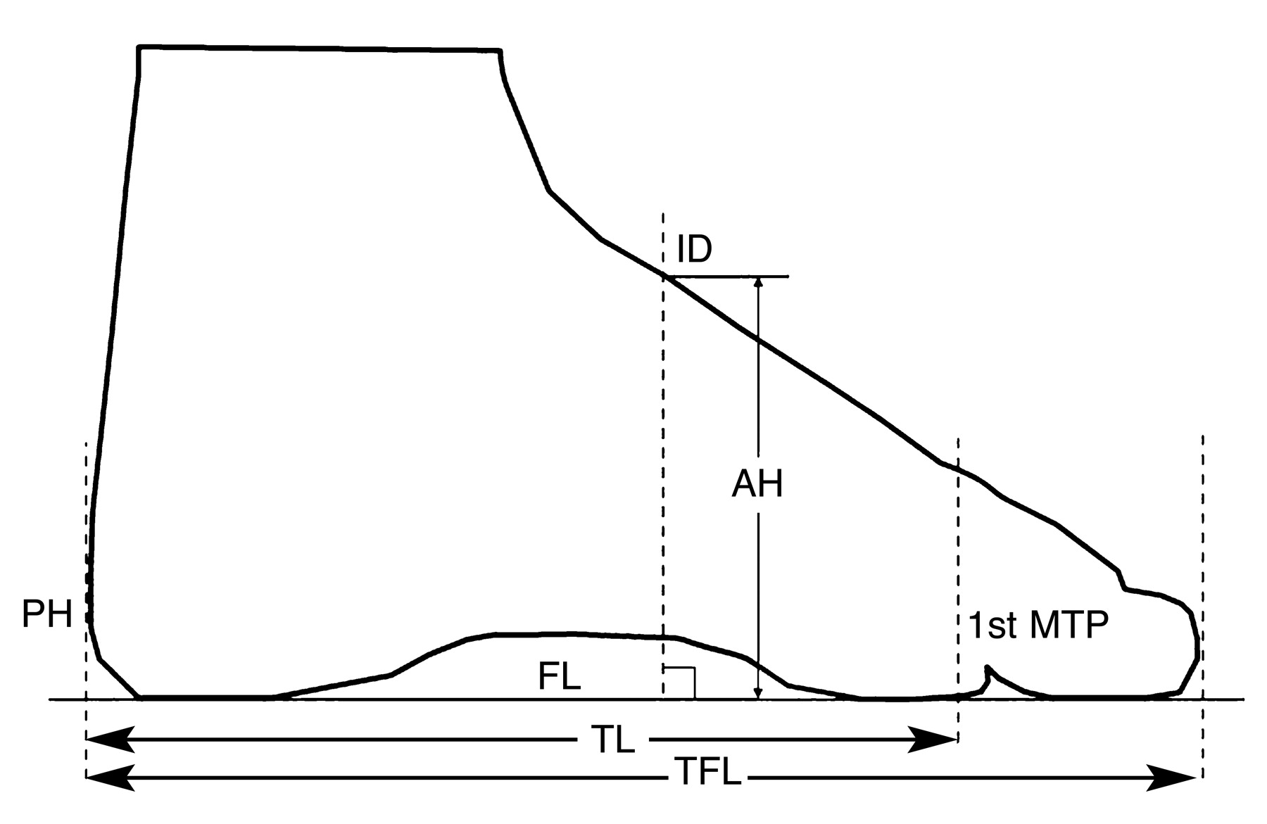
A digital videotape camera (model GR-DV2000; JVC, London, England) filming at 250 frames per second, with a resolution of 720 × 576 phase alternating line, was used to record images of the medial aspect of the foot in standing, walking, and jogging tasks. The videotape recordings were uploaded onto iMovie software (Apple Computer Inc, Cupertino, California) on a G3 Apple Mac Computer (Apple Computer Inc) with a capture rate of 30 frames per second. The camera was installed and fixed to the midpoint of an elevated 12-m runway and was not moved during data collection. The camera was positioned with the axis of the lens orthogonal to the runway and with a lens-to-foot distance of 2.6 m. The center of the lens was aligned approximately at the level of the medial malleolus in single-limb stance. Figure 1 provides a schematic representation of the experimental setup.
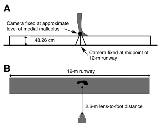
Figure 1.
Side (A) and top (B) views of the schematic representation of the experimental setup used for data collection.
Experimental Protocol
Subjects attended two assessment sessions, and a standardized protocol was followed at each session. The first session assessed the inclusion criteria of the subject, and then, where appropriate, information about the study was given and consent was obtained. During the second session, the foot used for this study was marked by the same investigator (B.V.) using an indelible pen at the site of the first metatarsophalangeal joint line (for the arch height ratio measure). For the standing task, the subject was positioned in the center of the 12-m runway in stride stance with the experimental foot forward and was told to look directly ahead. After completion of the three trials of static stance measurements, three trials of videotape footage were completed for the walking and jogging tasks. For the walking and jogging tasks, the subject was instructed to walk or jog across the 12-m runway at a self-selected speed that was monitored for consistency of foot placement on the platform. The same investigator (B.V.) conducted all of the trials, and subjects self-selected the interval between repeated trials.
Software Program
A software program, adapted by one of us (T.R.) from previous research in telerehabilitation and calibrated for this experimental setup, was used to calculate the two foot measurements. The software program performed linear and angular measurements on the basis of identified landmarks, such as the first metatarsophalangeal joint, posterior calcaneum, anterior hallux, and dorsum of the foot (Fig. 2). The software, described previously in the telemedicine literature,[14–16] has been demonstrated to produce accurate and reliable anthropometric measurements of the knee (type [2,2] ICC > 0.96),[15] shoulder (type [2,2] ICC > 0.97),[16] elbow (type [2,2] ICC > 0.93), and wrist joints (type [2,2] ICC > 0.96) (T.R., unpublished data, 2004). In addition, a study by two of us (T.R. and B.V., unpublished data, 2005) demonstrated high reliability (all interrater and intrarater type [2,1] ICCs > 0.97) for the measurements of foot length, truncated foot length, navicular height, and foot dorsum height.
Obtaining Foot Measurements from Videotape Footage
Using the method previously described by Vicenzino et al,[17] videotape footage was edited to obtain static frames for the midstance phase of gait. The midstance phase was identified from the videotape footage as the middle frame between heel contact and toe-off. The midstance phase was chosen because previous studies[18,19] analyzing rearfoot motion in walking and running have shown that the foot is maximally pronated at this point in the gait cycle. In addition, McPoil and Cornwall[9] reported that static measures of the longitudinal arch angle taken in standing were highly correlated to measures of the angle at midstance during walking. The arch height and arch height ratio measurements were then calculated from the digital images. Figure 2 explains how the arch height and arch height ratio were obtained from the static frames of midstance.
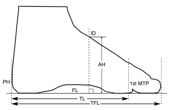
Figure 2.
The indices of foot posture defined. Arch height ratio was calculated by dividing arch height (AH) by truncated foot length (TL). The AH was the linear distance along a perpendicular line projected up from the floor line (FL) to its intersection with the dorsum of the foot (ID). The perpendicular line was placed halfway along the total length of the foot (TLF), which was measured from the most posterior aspect of the heel to the most anterior aspect of the hallux. The TL was measured from the posterior heel surface (PH) to the first metatarsophalangeal joint line (1st MTP).
Data Analysis
The ICCs and the standard errors of measurement (SEMs) were calculated from the data to determine the intrarater and interrater reliability of the two raters for the arch height and arch height ratio measurements. Before analysis, the data were averaged across the three trials. Pearson product moment correlations were performed using a software program (SPSS 11.0 for Windows; SPSS Inc, Chicago, Illinois) to evaluate the relationship between static and dynamic measures of arch height and arch height ratio.
Results
Reliability of Arch Height and Arch Height Ratio
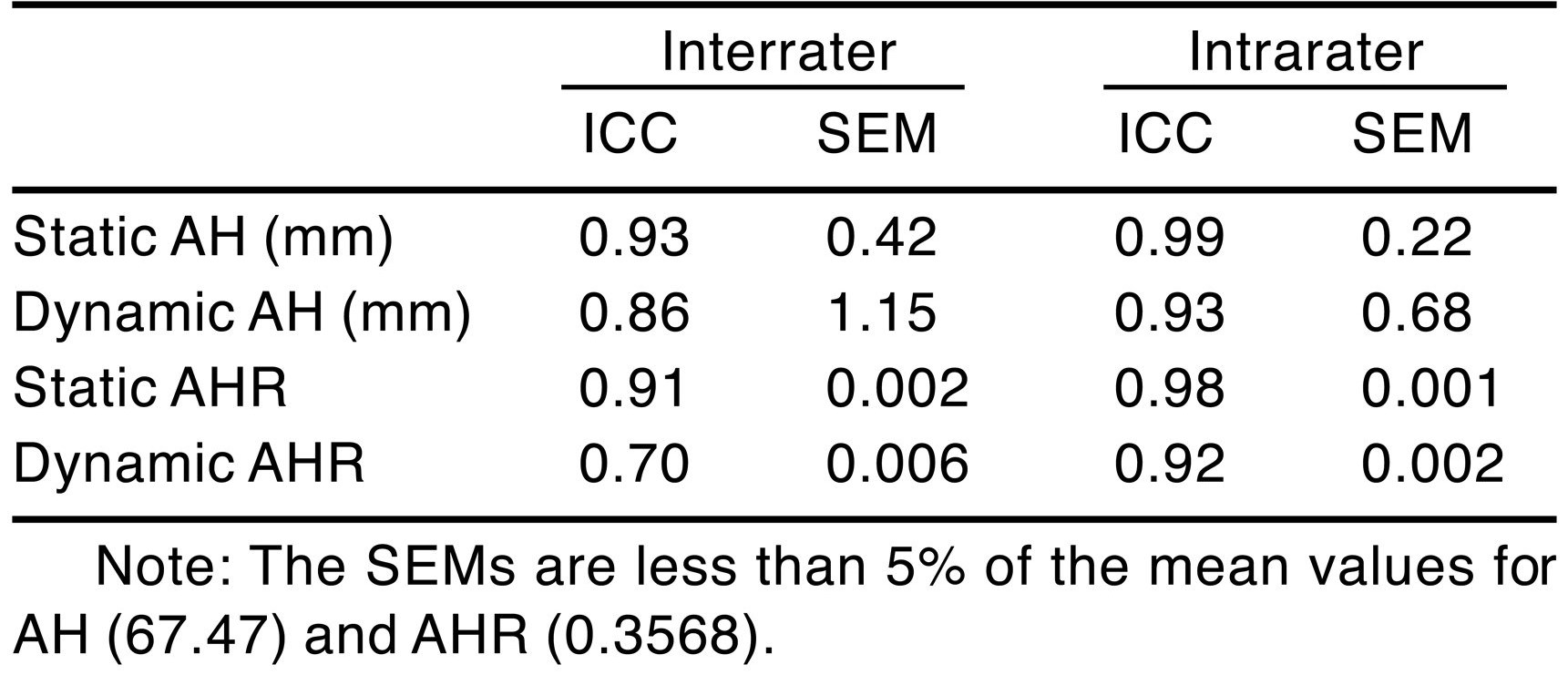
Table 1 displays the intrarater and interrater type (1,2) ICCs and SEMs for arch height and arch height ratio. For this study we used the classifications reported by Landis and Koch20 to determine the level of reliability for the ICCs. Except for the dynamic arch height ratio interrater ICC, all values of ICC would be classified as “almost perfect,” being greater than 0.86. Although both measures demonstrate high reliability, the arch height interrater and intrarater ICCs were higher and more consistent across static and dynamic conditions.

Table 1.
Intrarater and Interrater Type (1,2) Intraclass Correlation Coefficients (ICCs) and Standard Errors of Measurement (SEMs) for Arch Height (AH) and Arch Height Ratio (AHR).
Relationship of Arch Height and Arch Height Ratio Among the Standing, Walking, and Jogging Tasks
The Pearson product moment correlation coefficients for static arch height measured while standing in relation to dynamic arch height measured at midstance during walking and jogging were 0.81 (r2 = 0.66) and 0.91 (r2 = 0.83), respectively. The arch height ratio obtained in static standing was also highly correlated to the arch height ratio at midstance during walking and jogging, with Pearson correlation coefficients of 0.85 (r2 = 0.72) and 0.87 (r2 = 0.76), respectively.
Discussion
One objective of this study was to evaluate the reliability of arch height and arch height ratio measurements as static and dynamic measures of foot posture. The results of this study show arch height to be highly reliable as a static and dynamic measurement of foot posture, demonstrating “almost perfect” intrarater and interrater ICC values, based on the criteria described by Landis and Koch.[20] The arch height ratio also demonstrated high reliability as a static measure, with “almost perfect” intrarater and interrater ICC values. As a dynamic measurement, arch height ratio demonstrated “almost perfect” intrarater ICC values and “substantial” interrater ICC values. Overall, the arch height measure demonstrated consistently higher reliability as a static and dynamic measurement compared with the arch height ratio measure. The relatively poorer reliability of arch height ratio may be attributable to error inherent in identifying the metatarsophalangeal joint bony landmark.
The reliability of arch height ratio (interrater ICC = 0.91 and intrarater ICC = 0.98) and arch height (interrater ICC = 0.93 and intrarater ICC = 0.99) as static measures was favorably comparable with previous studies of arch height ratio (interrater ICC = 0.81–0.85 and intrarater ICC = 0.94–0.98) and arch height (interrater ICC = 0.79–0.77 and intrarater ICC = 0.94–0.98) by Williams and McClay.[8] This finding supports the premise that 50% weightbearing may be a more reliable method of obtaining the foot measurements required to calculate arch height ratio and arch height compared with the 10% and 90% weightbearing conditions previously described.[8] A possible explanation for this finding is that 50% weightbearing requires the subject to simply stand with equal weight on both feet, which can provide the clinician with a more efficient and reproducible foot position for measurement.
The excellent intrarater reliability of static arch height and arch height ratio (ICC = 0.99 and 0.98) is comparable with that of static longitudinal arch angle (ICC = 0.90–0.96) previously described.[9,11] However, the higher level of interrater reliability of arch height (ICC = 0.93) and arch height ratio (ICC = 0.91) seems to be superior to the static longitudinal arch angle, with previously reported interrater reliability ranging from 0.67 to 0.81.[9,11] A possible explanation for the lower interrater reliability of the static longitudinal arch angle measure is that it relies on the identification and skin marking by palpation of three bony landmarks (the medial malleolus, navicular tuberosity, and first metatarsophalangeal joint) for its calculation, which may increase the margin of error owing to skin movement.[12] The arch height ratio measure requires palpation of only one bony landmark in its calculation. In addition, Tranberg and Karlsson[12] reported that less skin movement occurred at distal sites such as the first metatarsophalangeal joint (1.8 mm) compared with proximal sites, such as the medial malleolus and navicular tuberosity (4.3 mm). The arch height measure requires no marks to be placed on the skin before videotape analysis, and this may explain its higher interrater reliability compared with arch height ratio and longitudinal arch angle.
The main aim of this study was to evaluate the relationship between static (standing) and dynamic (walking and jogging) measurements of arch height and arch height ratio. Pearson product moment correlation coefficients were used for this purpose. Results indicate that the standing measurement of arch height explained 66% of the variance associated with arch height measured at midstance during walking and 83% of the variance in arch height measured at midstance during jogging. The arch height ratio measured in standing explained 72% of the variance associated with arch height ratio measured at midstance during walking and 76% of the variance in arch height ratio measured at midstance during jogging. Results of the present study suggest that static measurements of arch height and arch height ratio may be used to provide the clinician with insight into the posture of the foot during dynamic activities.
Results of the study by McPoil and Cornwall[9] indicate that standing longitudinal arch angle explained more than 90% of the variance associated with the angle measured at midstance. This finding suggests that compared with arch height and arch height ratio, the longitudinal arch angle may have a greater ability to predict dynamic foot posture from static measures, as it explains a larger percentage of the variation.
In the process of determining whether arch height, arch height ratio, or longitudinal arch angle is the most suitable measure of foot posture, it is necessary to consider all of the factors that contribute to a measure’s suitability. We believe that these factors are threefold: reliability, validity, and practicality. Comparison suggests that longitudinal arch angle is the most valid measure in predicting dynamic foot posture from static measurement, yet it demonstrates substantially lower interrater reliability. On the other hand, arch height and arch height ratio demonstrate excellent interrater reliability but explain less of the variance between static measurements and dynamic foot posture. Thus it seems that longitudinal arch angle, arch height, and arch height ratio are suitable static measures of foot posture that can predict dynamic foot posture. If the ease of performing the measurement in the clinic is considered, we propose that arch height is the simplest and most efficient measure to obtain.
Conclusion
This study focused on the use of arch height and arch height ratio as part of the clinician’s assessment of the foot and their ability to improve the clinician’s understanding of foot posture during dynamic activity. Results of the present study indicate that arch height and arch height ratio are highly reliable measures of foot posture and that these static measurements can predict dynamic foot posture during walking and jogging. The results of this study support the clinician’s use of arch height and arch height ratio for assessment of the foot.
Funding
This study was partly funded
by a University of Queensland Travel Grant awarded to one of the authors (B.V.).
Conflicts of Interest
None reported.
References
- Willems TM, De Clercq D, Delbaere K, et al: A prospective study of gait related risk factors for exercise-related lower leg pain. Gait Posture 23: 91, 2005.
- Williams DS, McClay IS, Hamill J: Arch structure and injury patterns in runners. Clin Biomech 16: 341, 2001.
- Vicenzino B: Foot orthotics in the treatment of lower limb conditions: a musculoskeletal physiotherapy perspective. Man Ther 9: 185, 2004.
- Hlavac HF: “Examining Feet,” in The Foot Book: Advice for Athletes, ed by HF Hlavac, p 39, World Publications, Mountain View, CA, 1977.
- McPoil TG, Cornwall MW: The relationship between static lower extremity measurements and rearfoot motion during walking. J Orthop Sports Phys Ther 24: 309, 1996.
- Hamill J, Bates B, Knutzen KM, et al: Relationship between selected static and dynamic lower extremity measures. Clin Biomech 4: 217, 1989.
- Stacoff A, Reinschmidt C, Nigg BM, et al: Effects of foot orthoses on skeletal motion during running. Clin Biomech 15: 54, 2000.
- Williams DS, McClay IS: Measurements used to characterize the foot and medial longitudinal arch: reliability and validity. Phys Ther 80: 864, 2000.
- McPoil TG, Cornwall MW: Use of the longitudinal arch angle to predict dynamic foot posture in walking. JAPMA 95: 114, 2005.
- Dahle LK, Meuller M, Delitto A, et al: Visual assessment of foot type and relationship of foot type to lower extremity injury. J Orthop Sports Phys Ther 14: 70, 1991.
- Jonson SR, Gross MT: Intraexaminer reliability, interexaminer reliability, and mean values for nine lower extremity skeletal measures in healthy naval midshipmen. J Orthop Sports Phys Ther 25: 253, 1997.
- Tranberg R, Karlsson D: The relative skin movement of the foot: a 2-D roentgen photogrammetry study. Clin Biomech 13: 71, 1998.
- Williams DS, Davis IM, Scholz JD, et al: High-arched runners exhibit increased leg stiffness compared to low-arched runners. Gait Posture 19: 263, 2004.
- Russell T: Establishing the Efficacy of Telemedicine as a Clinical Tool for Physiotherapists: From Systems Design to Randomized Controlled Trial [PhD thesis], University of Queensland, Brisbane, 2004.
- Russell T, Jull G, Wootton R: Can the Internet be used as a medium to evaluate knee angle?. Man Ther 8: 242, 2003.
- Russell T, Wootton R, Jull G: Physical outcome measurements via the Internet: reliability at two Internet speeds. J Telemed Telecare 8: 50, 2002.
- Vicenzino B, Franettovich M, McPoil T, et al: Initial effects of anti-pronation tape on the medial longitudinal arch during walking and running. Br J Sports Med 39: 939, 2005.
- Cornwall MW, McPoil TG: Three-dimensional movement of the foot during the stance phase of walking. JAPMA 89: 56, 1999.
- McClay I, Manal K: A comparison of three-dimensional lower extremity kinematics during running between excessive pronators and normals. Clin Biomech 13: 195, 1998.
- Landis JR, Koch GG: The measurement of observer agreement for categorical data. Biometrics 33: 159, 1977.
© 2007 American Podiatric Medical Association