


Abstract
Achilles tendon injuries are among the three most frequent sports-related injuries of the foot and ankle. Proper function of the Achilles tendon is critical to performance in sports. A thorough knowledge of the anatomy and biomechanical function of this tendon is essential to the effective treatment of these injuries. Distinguishing among the various pathologies of the Achilles tendon is an important first step toward successful treatment and return of the athlete to sports activity. The term Achilles tendinitis is a nonspecific diagnosis that does not accurately describe an actual injury. This review is intended to provide the sports medicine physician with a means of classifying Achilles tendon injuries and, thus, arriving at an accurate diagnosis and treatment plan.
Greek mythology describes the infant Achilles (who would grow up to be the greatest Greek warrior) being held at his heel by his mother and dipped into the river Styx, an act that was supposed to give him an arrowproof layer. The warrior Achilles was brought down by a well-placed arrow shot by his nemesis, Paris, into his calcaneal tendon—the area that his mother’s hand covered and, therefore, the only part of his body that was not arrowproof.
In modern times, many high-profile collegiate and professional athletes, as well as “weekend-warrior” fitness athletes, have been sidelined by Achilles tendon injuries, some more devastating than others. Achilles tendon injuries account for 5% to 18% of all running injuries and are the most common overuse syndrome of the lower leg.[1–6] As the number of years of running increases, so does the risk of sustaining an Achilles tendon injury.[1] A long-term study[7] by the Centers for Disease Control and Prevention and the Cooper Institute for Aerobics Research found that men sustained Achilles tendon injuries at a rate 14.25% higher than women.
The scope and complexity of Achilles tendon sports and fitness injuries vary widely. Instead of diagnosing every athlete’s pain in the back of the foot or ankle as Achilles tendinitis (and thus treating all of these injuries alike), it is critical for the sports physician to distinguish among various types of Achilles tendon pathology. For example, differentiating an athlete’s paratenonitis from tendinosis or retrocalcaneal bursitis from Achilles insertional calcific tendinosis will affect treatment, rehabilitation, and return to activity. Ruptured Achilles tendons are season-ending injuries, whereas inflammatory Achilles tendon injuries are less severe but often cause an athlete to spend weeks or months on injured reserve.
Arriving at an accurate diagnosis and creating an effective treatment plan requires a detailed injury history, a thorough knowledge of anatomy and biomechanics, and, often, advanced diagnostic imaging. Early intervention is necessary to provide the athlete with the best chance of promptly returning to activity and avoiding repeated injury.[8]
Key Anatomical Considerations
From its most proximal origin on the femur to its distal insertion on the calcaneus, the Achilles tendon complex is subject to a variety of pathologies. Identifying which structure in this complex is causing an athlete’s pain is essential to a safe and rapid return to activity. The Achilles tendon complex crosses three joints and comprises three muscles that form a conjoined Achilles tendon, and it attaches to the calcaneal bone and deep fascia distally. Because the entire complex crosses three joints, range-of-motion assessment of the Achilles tendon and ankle joint must be evaluated with the knee joint in a flexed position and in a maximally extended position. A tight gastrocnemius muscle will result in limitation of ankle joint dorsiflexion with the knee extended but will not affect ankle joint dorsiflexion with the knee flexed. Saxena and Kim[9] recently found that ankle dorsiflexion in asymptomatic adolescent athletes is approximately 0° with the knee extended and just less than 5° with the knee flexed.
The triceps surae muscle includes the gastrocnemius medial and lateral heads, which originate from the posteromedial and posterolateral femoral condyles, respectively, and the soleus. Distally, the gastrocnemius aponeurosis merges with the aponeurosis of the soleus muscle to form the Achilles (calcaneal) tendon. The plantaris muscle and tendon course adjacent to the medial head of the gastrocnemius muscle and calcaneal tendon and insert into the medial one-third of the posterior calcaneus.
The Achilles (calcaneal) tendon is formed proximally by the conjoined aponeuroses of the gastrocnemius and soleus muscles. It is one of the largest and strongest tendons in the body, able to transmit up to 7,000 N of force during intense exercise.[10,11] Distally, its fibers rotate internally 90° as its proximal medial fibers become posterior and its posterior fibers become more lateral.[8] At its insertion at the middle one-third of the posterior calcaneus, retrocalcaneal and retro-Achilles bursae surround the tendon anteriorly and posteriorly, respectively.
The paratenon is an important sheath surrounding the Achilles tendon (Fig. 1). This structure is not a true synovial sheath but rather incorporates a loose, fatty, areolar network of tissue, including the mesotenon anteriorly, which supplies a vascular network to the Achilles tendon. The paratenon allows for the normal gliding function of the Achilles tendon in this encapsulating structure.
The “watershed area” is an avascular area with poor healing potential located 2 to 6 cm proximal to the Achilles tendon insertion and is a common site of pathology. A “watershed band” consisting of contiguous fascial bands from the deep posterior compartment to the Achilles tendon watershed region has been described recently and may be clinically significant.[12]
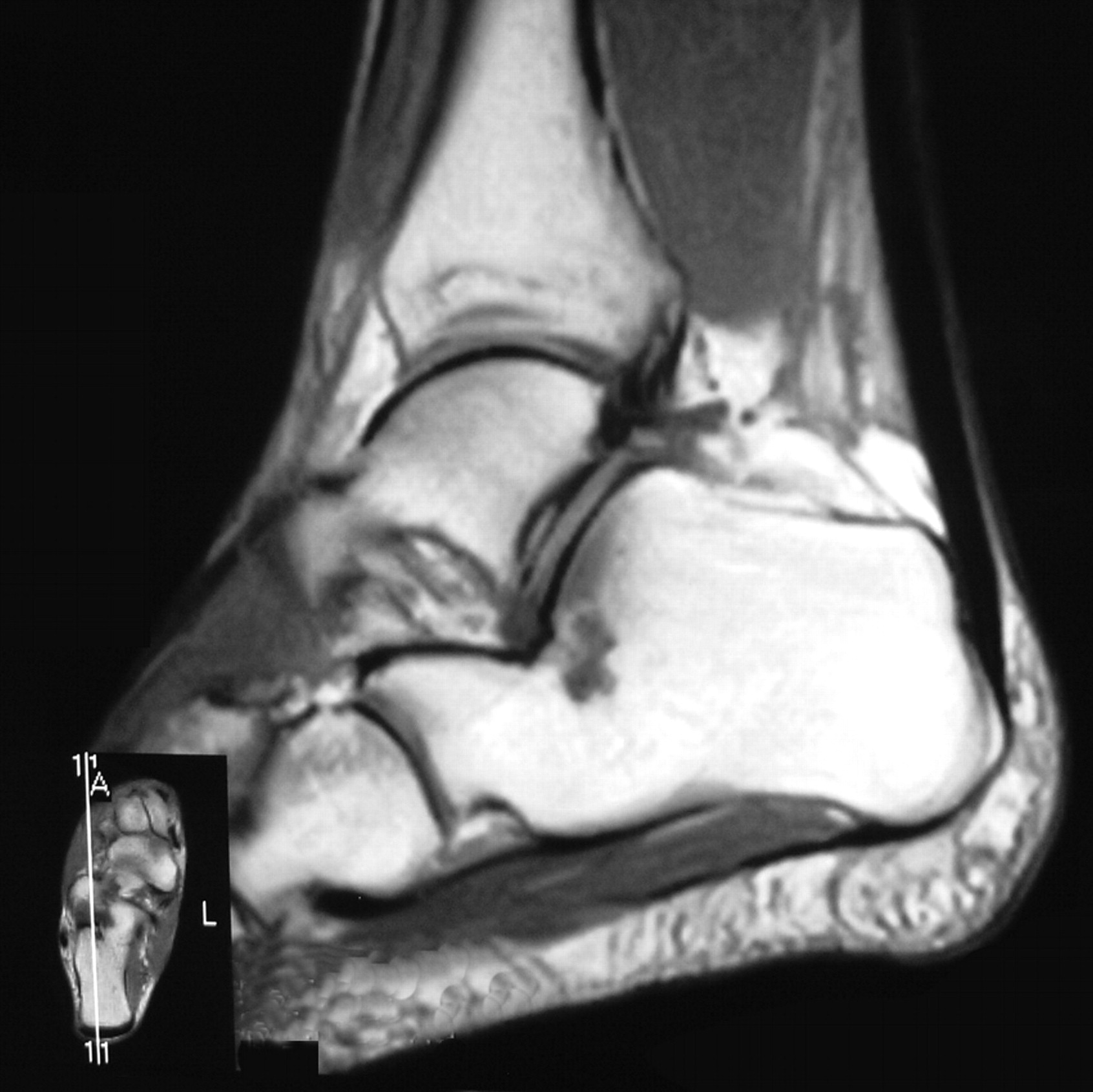
The distal Achilles tendo-fascia consists of deep fibers of the Achilles tendon that continue distally past the insertion and into the plantar aspect of the calcaneus (Fig. 2). These deep fibers are contiguous with plantar fascia fibers.
Two important bursal sacs are also associated with Achilles tendon sports injuries. The retro-Achilles bursa protects the posterior portion of the Achilles tendon at its insertion. External shoe pressure frequently causes this bursa to become inflamed. The retrocalcaneal bursa protects the anterior portion of the Achilles tendon from the posterior and superior aspect of the calcaneus. An enlargement of the calcaneus or pathology in the Achilles tendon can cause this bursa to become inflamed. Both of these structures can be visualized on the magnetic resonance image in Figure 2.
Figure 1.
A, The Achilles paratenon is held proximally by two Allis forceps during a reconstructive Achilles tendon repair. B, Note the covering the paratenon provides after it is coapted again over the proximal portion of the Achilles tendon, thereby encapsulating the Achilles tendon, providing nutrition, and preserving its gliding function.
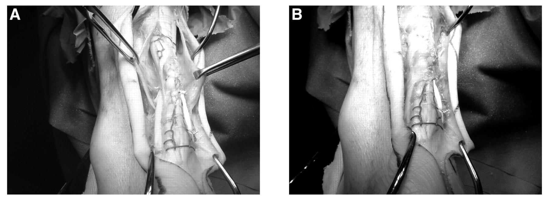
Figure 2.
Sagittal magnetic resonance image of the distal Achilles tendo-fascia, a continuum of deep connective tissue between the Achilles tendon and the plantar fascia.
Athletic Injuries of the Achilles Tendon
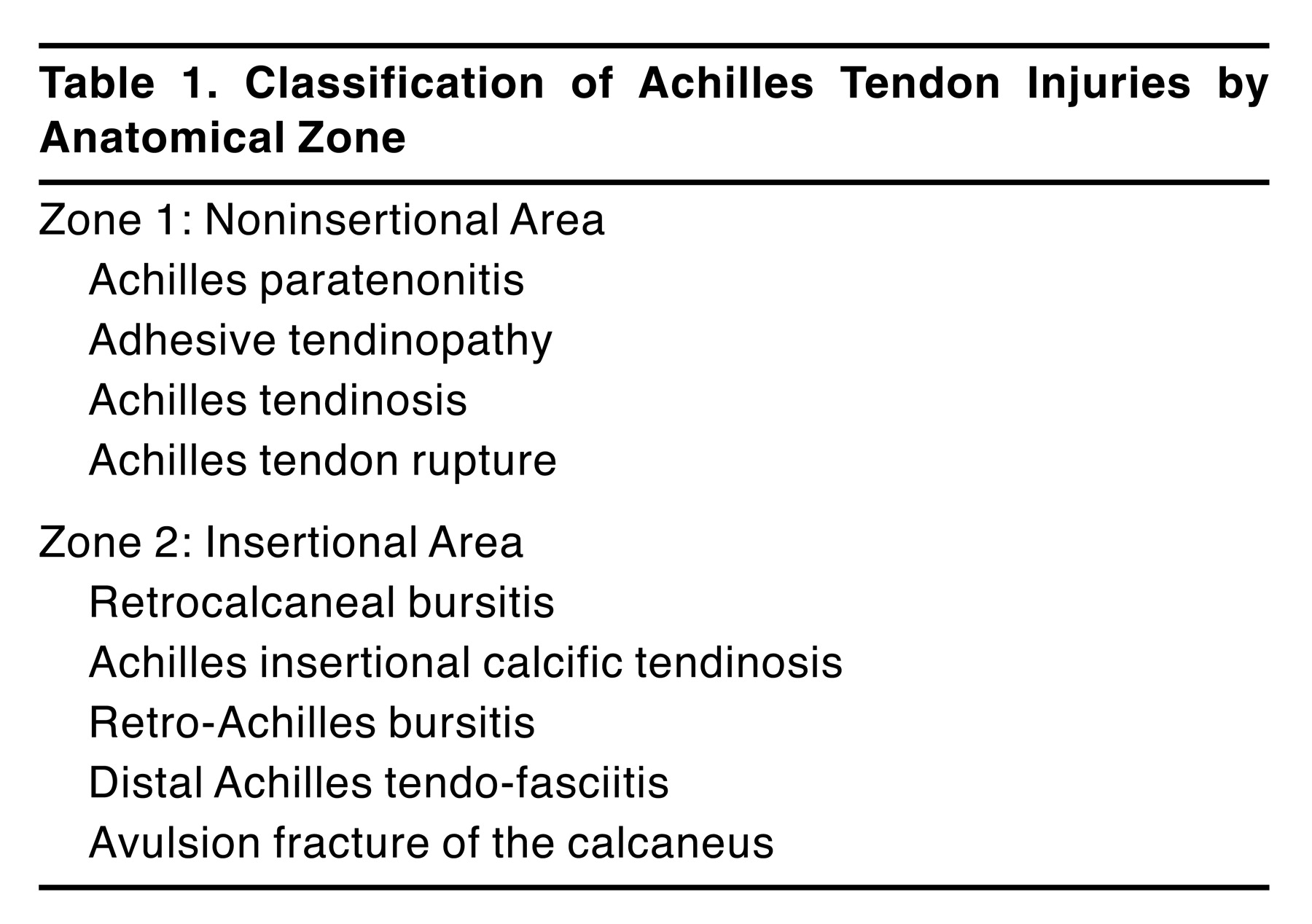
The Achilles tendon is the third most frequent site of foot and ankle injuries in the professional athlete; only ankle sprains and plantar fasciitis are more common.[13] Eighteen percent of all running injuries involve the Achilles tendon.[14] Injuries of the Achilles tendon are classified by anatomical location (Table 1),[15] occurring at either the noninsertional area (zone 1) or at the insertional area (zone 2). A third zone (zone 3) involves injuries to the proximal musculature of the Achilles tendon complex, but these occur much less frequently than zone 1 and 2 injuries and are not discussed in detail here. “Tennis leg” injuries (those involving strain of the medial head of the gastrocnemius muscle) and plantaris tendon ruptures occur in zone 3.

Table 1.
Classification of Achilles Tendon Injuries by Anatomical Zone
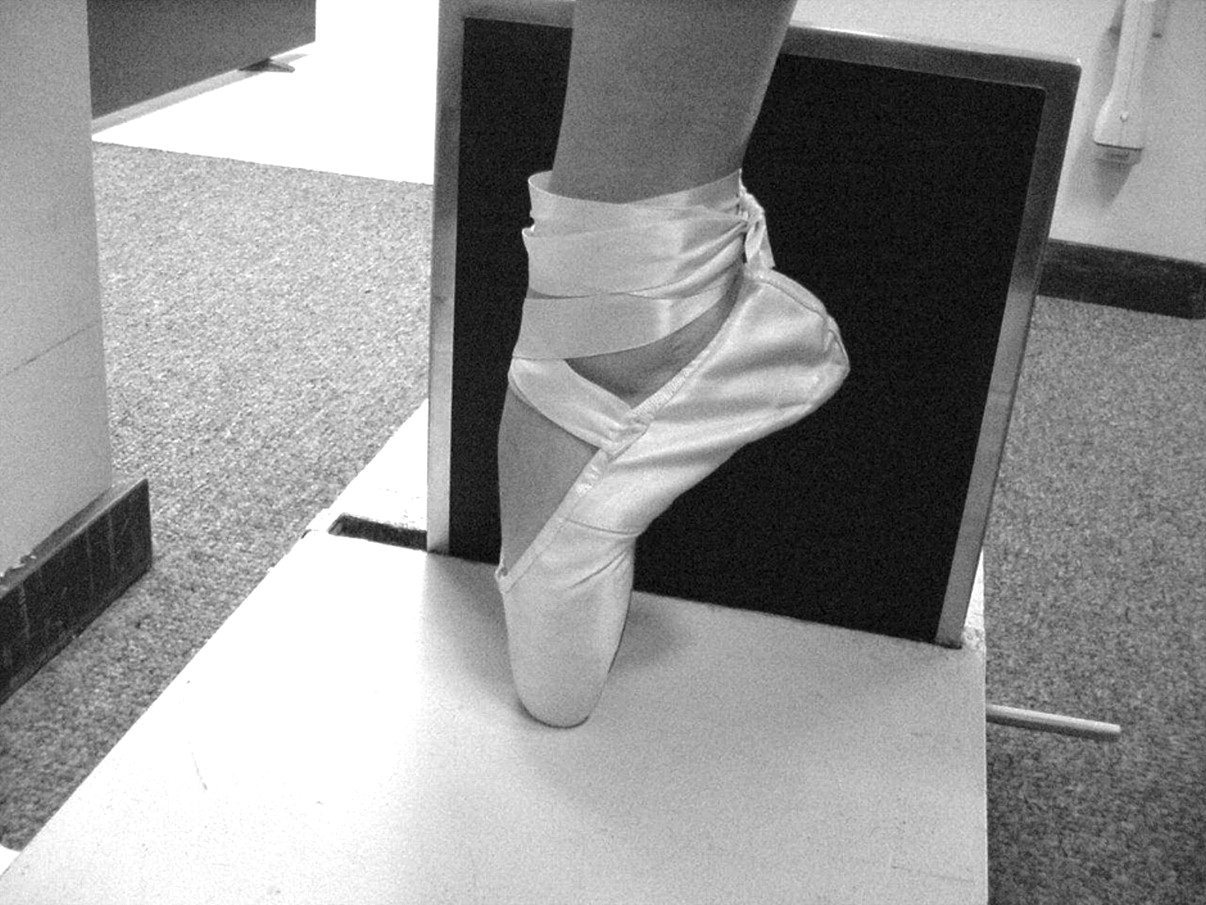
The type of sport, the athlete’s playing position, and the type of shoes should be assessed during the initial evaluation of an Achilles tendon injury. For example, soccer players wear “negative-heel” cleats on soft grass surfaces that place increased stress on the Achilles tendon. A player’s position in the sport may also be a contributing factor to the injury; midfielders and strikers may run up to a half-marathon every game, whereas goalkeepers spend the entire game moving laterally between the goal posts on the balls of their feet. Ballet dancers wear pointe shoes with a strap that may impinge on the Achilles tendon (Fig. 3).
Zone 1: Noninsertional Achilles Tendon Injuries
Achilles Paratenonitis. Pathology. Achilles paratenonitis is an inflammatory condition of the Achilles paratenon that results in effusion, warmth, swelling, and pain. The intratendinous substance of the Achilles tendon is normal; a biopsy of the tendon would reveal normal collagen fibers. The pathology of this condition involves only the sheath surrounding the tendon but not the tendon itself.
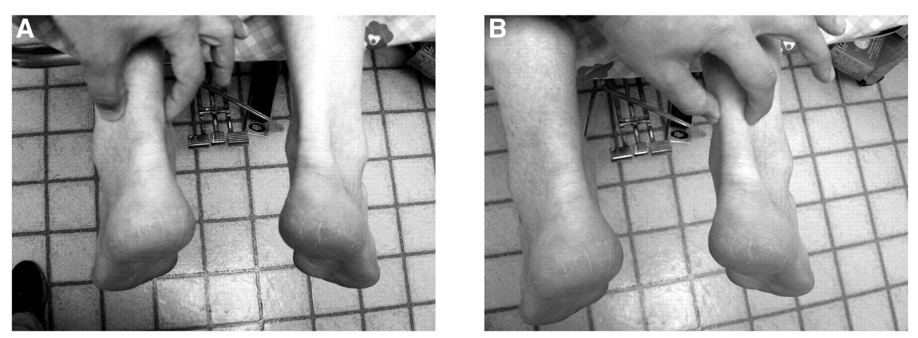
Diagnosis, Differentiating Paratenonitis from Tendinosis. Clinical examination confirms pain elicited with direct palpation, grasping of the painful area of the tendon between the thumb and forefinger. While the tendon is grasped, the pain does not move with plantarflexion and dorsiflexion motion of the Achilles tendon because the paratendinous sheath does not move while the Achilles tendon slides up and down in the sheath. With tendinosis, on direct palpation and grasping of the painful area of the tendon between the thumb and forefinger, the pain does move with motion of the Achilles tendon. As with all Achilles tendon injuries, the injured side should be compared with the contralateral side.
Figure 3.
It is important to assess shoes when evaluating Achilles tendon injuries in sports. The constrictive nature of a ballerina’s pointe shoe strap can contribute to Achilles tendon pathology.
Foot biomechanics may affect the location of symptoms. Excessive pronation tends to result in more pain at the medial border of the Achilles tendon, in part owing to the associated excessive calcaneal eversion (Fig. 4). Closed-kinetic-chain hypermobility increases the force of pull on the medial Achilles tendon fibers, whereas excessive supination tends to result in more pain laterally. A supinated foot positions the calcaneus in a more inverted position, making the lateral fibers of the Achilles tendon more taut.
Treatment. Acute treatment of this inflammatory condition should begin with PRICEN: Protection, Rest, Ice, Compression, Elevation, and Nonsteroidal anti-inflammatory drugs. Deep transverse friction soft-tissue techniques and ultrasound may stimulate collagen synthesis. Taping of the tendon in a plan-tarflexed position may allow return to play in mild cases. Functional orthoses and heel lifts that address a biomechanical fault (such as a hyperpronated rear-foot complex) may be useful in the rehabilitative and return-to-play phases of recovery. Also, poor flexibility (a tight gastrocnemius soleus complex) should be addressed with an appropriate stretching regimen.
Adhesive Tendinopathy. Pathology. Adhesive tendinopathy may result from chronic inflammation of the paratenon (untreated paratenonitis) leading to fibrosis, in which an adhesion forms between the paratenon and the Achilles tendon. Fibrillation, modulation, and “yellowing” of the edematous tendon may be present, and pathologic findings include myxomatous degeneration of collagenous tissue, fibrosis, round cell inflammatory infiltrate, and proliferation of fibrovascular connective tissue.[16]
Diagnosis. Typical findings are paratenonitis plus reduced motion and crepitus along the course of the tendon.
Treatment. Failure to reduce inflammation may lead to tendon degeneration from endogenous enzymes, a precursor to partial or complete rupture. Gould and Korson[16] reported on nine surgical patients treated for “stenosing tenosynovitis of the pseudosheath of the tendo Achilles” and found that eight of the nine patients returned to unrestricted activity within a few months. Surgery consisted of excision of the entire pseudosheath, allowing the tendon to assume a new, nonconstricting alignment.
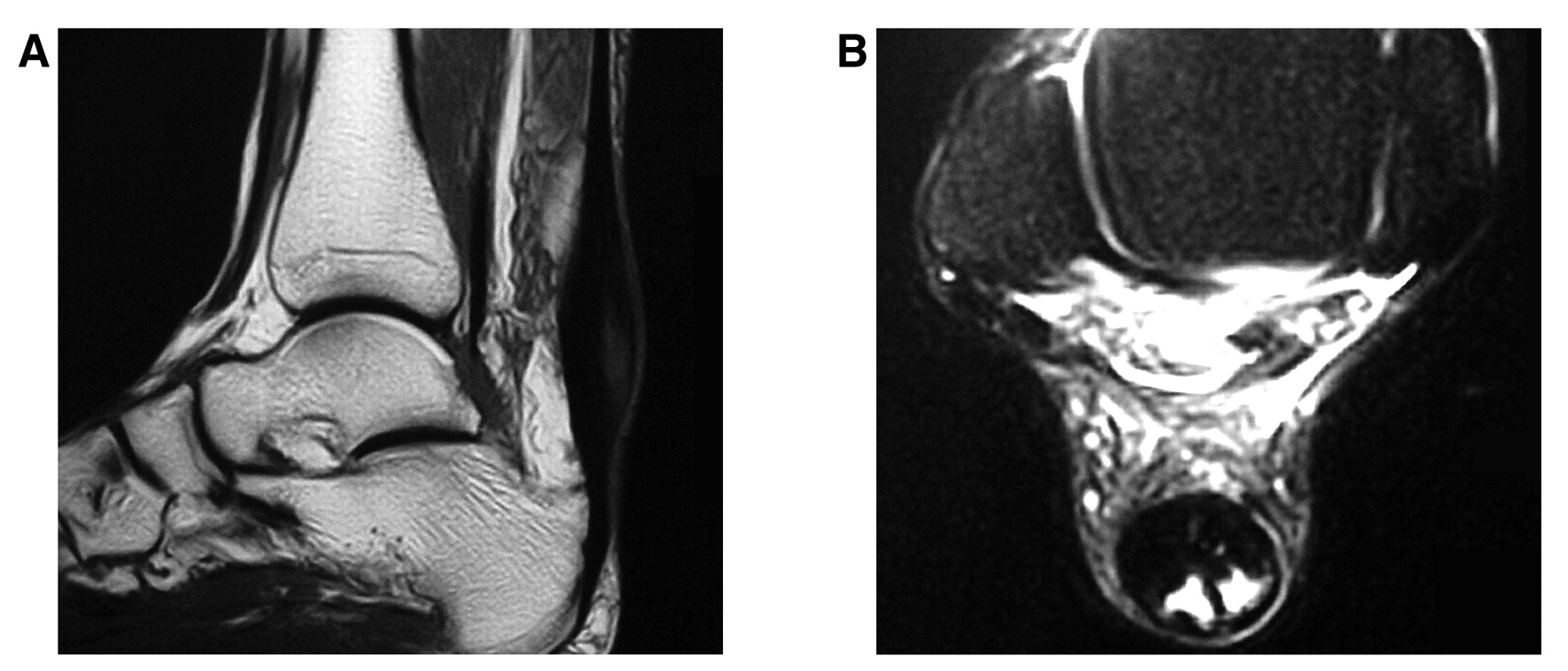
Achilles Tendinosis. Pathology. Achilles tendinosis is a more advanced chronic pathologic condition than paratenonitis, as tendinosis involves pathologic degeneration of the collagen fibers in the Achilles tendon substance (Fig. 5). A biopsy of the tendinosis will show pathologic degenerative changes, distinguishing this condition from paratenonitis, in which the tendon is normal on biopsy. Men aged 35 to 45 years are most commonly affected. The prognosis for this condition is worse than that for paratenonitis. Intratendinous changes may result from partial tears or microtears, incomplete rupture, longitudinal striations, or calcifications. Intratendinous mucoid degeneration is often present, including collagen degeneration (Fig. 6).
Diagnosis, Differentiating Tendinosis from Paratenonitis. Pain may or may not be present. Astute clinical examination can help distinguish between tendinosis and paratenonitis. With tendinosis, on direct palpation and grasping of the painful area of the tendon between the thumb and forefinger, the pain moves with motion of the Achilles tendon. Conversely, with paratenonitis, the pain does not move with motion of the Achilles tendon, which has classically been referred to as the “painful arc” sign. Clinically, a widened and thickened Achilles tendon will be much more prominent on the injured side compared with the contralateral side.
Figure 4.
A, A collegiate basketball player with excessively pronated subtalar joints bilaterally, causing Achilles paratenonitis along the medial aspect of the Achilles tendon complex. B, Functional orthotic devices are shown limiting the excessive pronation and alleviating the excessive pull along the medial Achilles tendon.
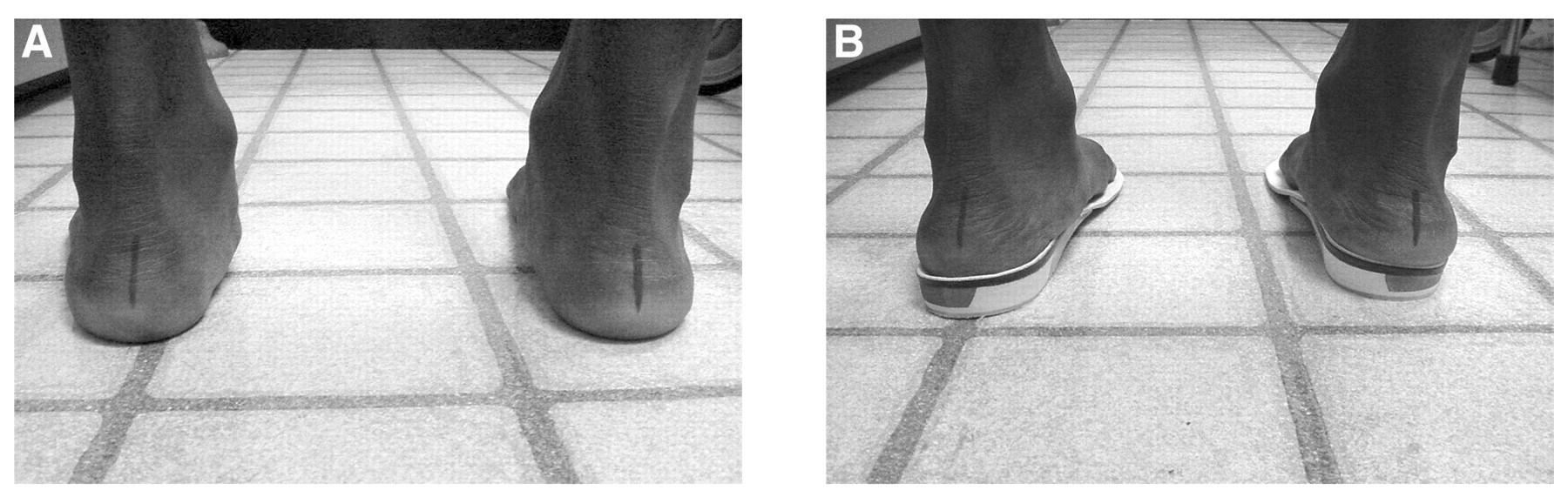
Figure 5.
Achilles tendinosis is shown on the patient’s left Achilles tendon (A) compared with a normal right Achilles tendon (B). Note the marked thickening and the lack of normal anatomical contour on the left compared with the normal right side.
Chronic tendinosis, which leads to calcification in the Achilles tendon, can be classified by radiographically observed lesions.[17] Type I radiopacities occur at the Achilles tendon insertion or the superior pole of the calcaneus. Type II intratendinous radiopacities are located 1 to 3 cm proximal to the distal Achilles tendon insertion. Type III radiographically observed lesions occur up to 12 cm proximal to the insertion zone and may be subclassified into IIIA (partial tendon calcification or ossification) or IIIB (total tendon involvement).
Treatment. Tendinosis may be seen in combination with a paratenonitis and, at least initially, may be treated in a similar manner, including deep transverse friction massage, ultrasound three times per week for 3 weeks, taping, stretching, strengthening, heel lifts, functional orthotic devices, and shoe modifications. Patient education should be a high priority when Achilles tendinosis is diagnosed. The patient must be made aware that he or she may be at increased risk for a complete rupture due to degeneration in the tendinous substance. Intratendinous calcification may also occur in chronic cases. The athlete should be advised to refrain from high-impact, explosive activities because tendinosis may be a precursor to rupture. “Playing through pain” with this injury should be discouraged.
Figure 6.
Sagittal (A) and axial (B) magnetic resonance images of Achilles tendinosis showing a “halo sign” confirming degenerative changes in the midsubstance of the tendon.
The use of nonsteroidal anti-inflammatory drugs for tendinosis has been controversial because this is not truly an inflammatory condition. Although nonsteroidal anti-inflammatory drugs may not be useful for their anti-inflammatory effects, they are still useful for their analgesic effects.
Eccentric training of the gastrocnemius soleus complex has been shown to improve clinical outcome in patients with chronic Achilles tendinosis. Shalabi et al[18] found that eccentric training resulted in decreased tendon volume and intratendinous magnetic resonance image signal, which was correlated with an improved clinical outcome. A recent Swedish randomized, prospective, multicenter study[19] compared treatment for chronic Achilles tendinosis with eccentric and concentric training. After 12 weeks of treatment, 18 of 22 patients in the eccentric loading group had returned to their pre-injury level of running, compared with only 8 of 22 who were treated with a concentric training regimen.
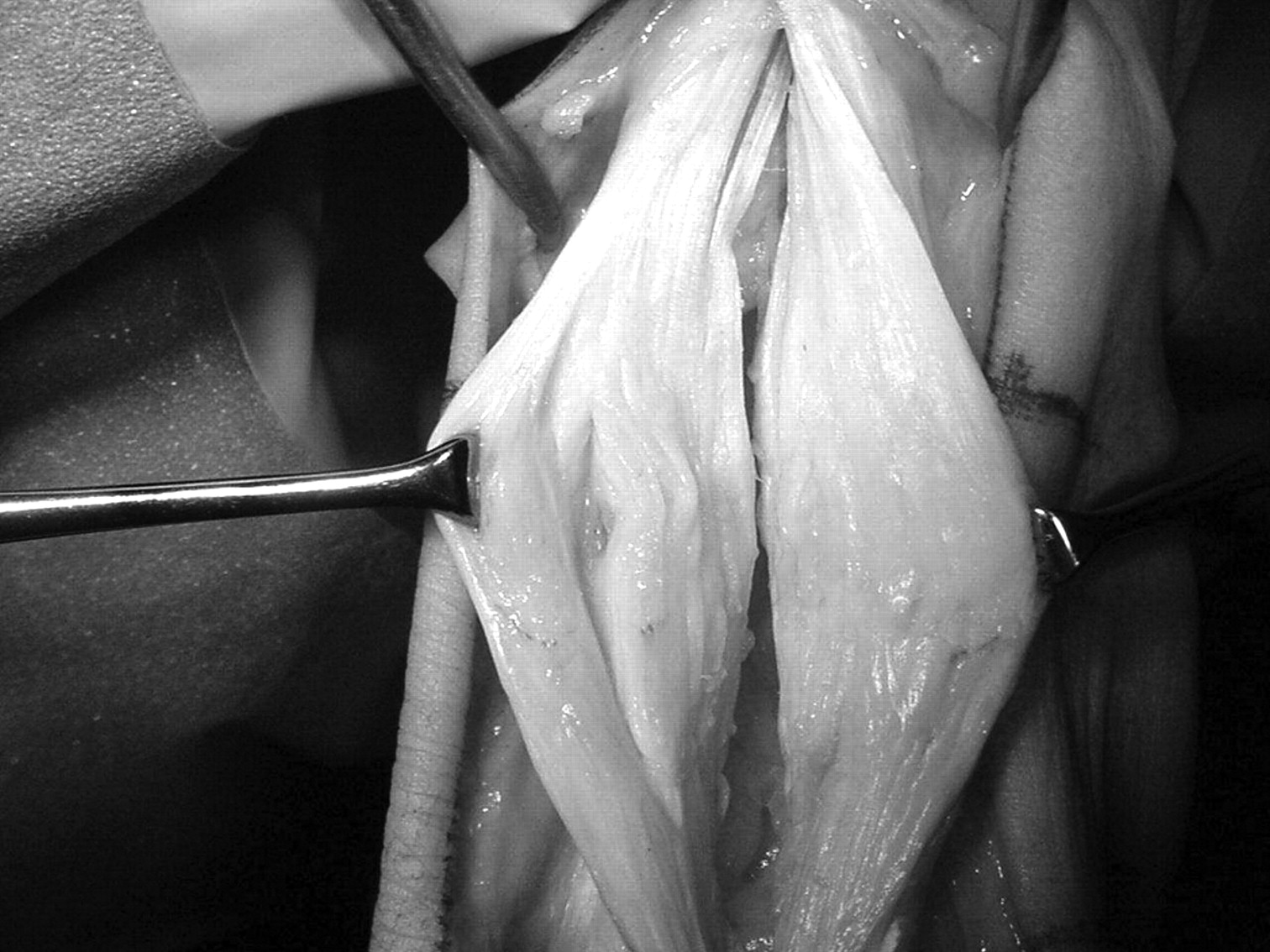
When symptoms are unremitting, surgical repair of Achilles tendinosis in the athlete has been well documented as a successful treatment, and many techniques are available (Fig. 7). Alfredson and coworkers[20,21] and Kvist[22] estimate that 25% of athletes with chronic Achilles tendinosis will not improve with conservative care and will require surgical intervention. Surgical procedures include paratendinous stripping, multiple longitudinal tenotomies, excision of a well-defined mass, and ultrasound-guided percutaneous tenotomy.[23–26]
Figure 7.
Intraoperative view of Achilles tendinosis repair using a longitudinal tendon-splitting approach. Note the central nidus on the left-sided tendon with pathologic degeneration of the central tendon, compared with the normal-appearing tendon proximally and distally.
Tallon et al[27] critically assessed the research methods of 26 studies that reported the surgical outcomes of patients with chronic Achilles tendinopathy and found a positive correlation between year of publication and overall methods score. Guidelines for improving study designs in the future for this area of clinical research are recommended.
Achilles Tendon Rupture. Pathology. Achilles tendon ruptures have an incidence of 1 in 10,000 per year and most commonly affect middle-aged athletic men (“weekend warriors”) (Fig. 8). Neglected, chronic, or partial ruptures also occur, but much less frequently.
Etiologic factors are of three types: mechanical, vascular, and related to poor tissue quality. Mechanical factors include sudden dorsiflexion of the ankle with the knee extended while an eccentric load is applied. Examples include a quarterback dropping back to pass and then suddenly stepping up into the pocket and a tennis player rushing the net and then quickly retreating for an overhead lob. Vascular factors include the watershed area 2 to 6 cm proximal to the insertion, an area of poor blood supply. Tissue quality of the ruptured area may involve a preexisting degenerative pathologic change in the tendon (tendinosis).
Use of fluoroquinolone antibiotics is associated with a higher risk of tendon rupture. Athletes who use these medications, especially those with known Achilles tendon pathology, must be warned of the risk.[28] An athlete with a urinary tract infection must be asked about current antibiotic drug therapy, as fluoroquinolone antibiotics are commonly used to treat urinary tract infections. Patients who take corticosteroids on a long-term basis may also be at risk for tendon rupture.[29]
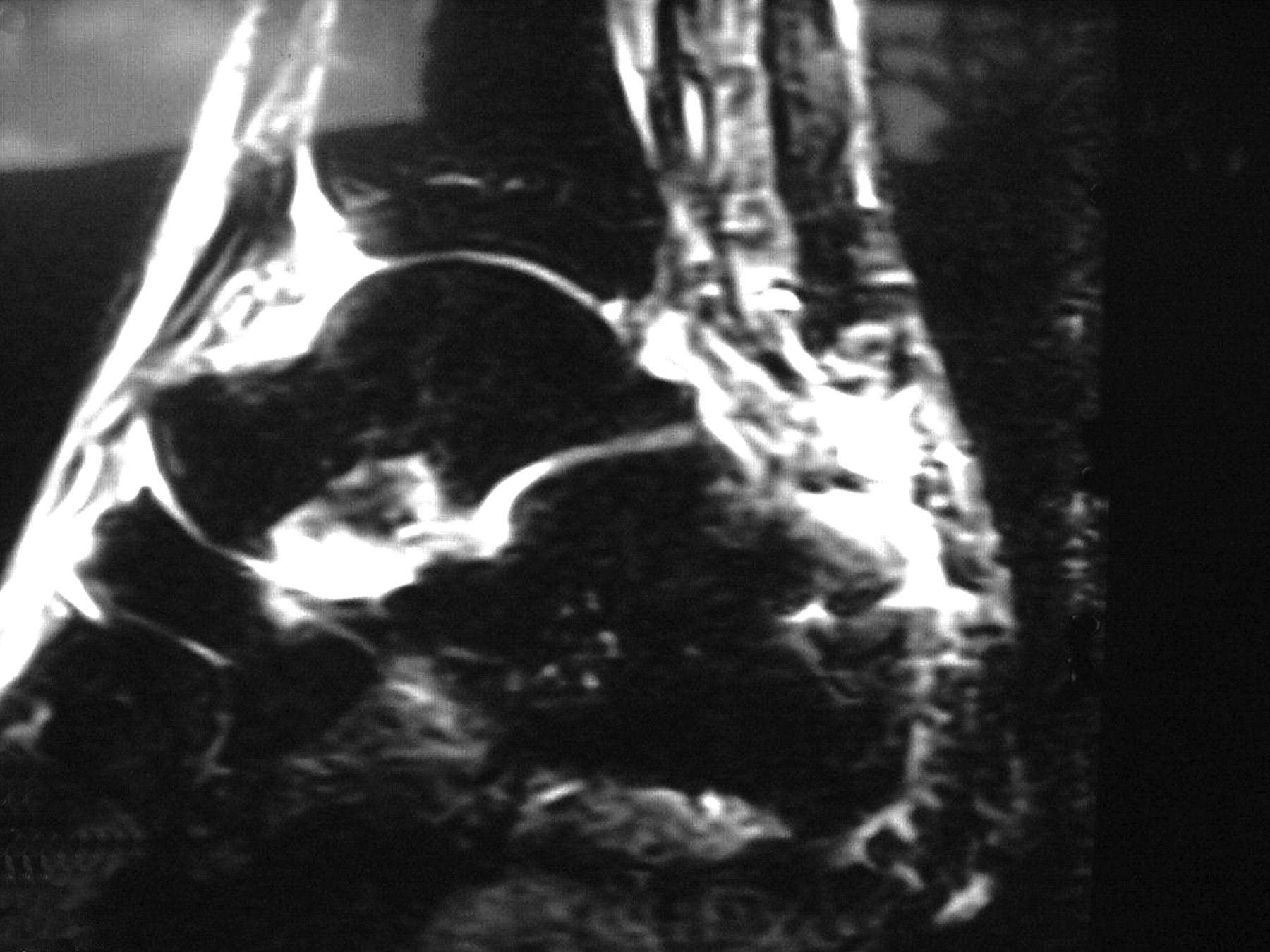
Figure 8.
Magnetic resonance image (short tau inversion recovery technique) of an Achilles tendon rupture.
A recent Scandinavian study[30] warns of an increased risk of Achilles tendon rupture in the contralateral limb after Achilles tendon rupture of the initial limb. These findings are analogous to those for anterior cruciate ligament injuries in that after an anterior cruciate ligament tear on one side, the other side is at higher risk for rupture, and bracing should therefore be applied to the unaffected knee. This study also found that nine of ten patients with bilateral Achilles tendon rupture had a blood type of B positive.
Diagnosis. Immediately after the rupture, patients often describe a feeling of being “hit with a bat,” only to look behind them and see nobody there. The result of Thompson’s test is positive when the calf musculature is squeezed and a corresponding plantar foot response is absent. A palpable defect may be present at the site of rupture. Diagnostic ultrasound can be helpful in assessing tendon pathology in real time, and magnetic resonance imaging is very useful in cases of questionable diagnosis.
Treatment. Some controversy still exists concerning surgical versus nonsurgical treatment of acute Achilles tendon ruptures.[31–36] Open primary repair of an acute Achilles tendon rupture is often the treatment of choice in the young, healthy athlete, although a recent study advocates conservative care.[37] Primary repair has been associated with a lower repeated rupture rate and a greater ability to perform toe-off and to return to sports activity, although it involves a greater risk of infection. Early weightbearing with the ankle plantigrade is not detrimental to the outcome of repair after acute rupture of the Achilles tendon and shortens rehabilitation time.[38]
Kauranen et al[39] found that the motor-performance function of the leg after Achilles tendon repair does not depend on whether the leg is in a plaster cast with the Achilles tendon in tension or in an active brace during the early postoperative period. Weber et al[40] found that the results of operative and nonoperative treatment for an acute Achilles tendon rupture were equivalent. Nonoperative care, however, may increase the risk of intratendinous calcification (Fig. 9). Closed percutaneous suturing techniques have been described more recently.[41]
Zone 2: Insertional Achilles Tendon Injuries
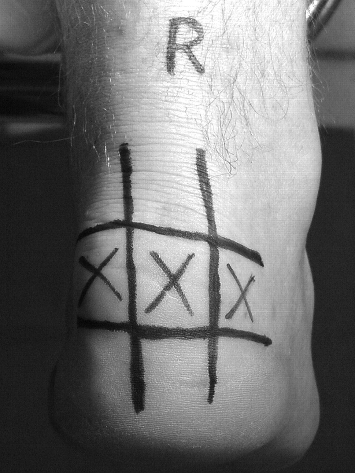
Zone 2 has been an area of considerable misunderstanding and confusion, leading to difficulty in making an accurate diagnosis of Achilles tendon pathology. This should not be the case if one has a good understanding of the anatomical structures involved and takes a thorough sports history. Topographic mapping of the athlete’s painful areas using a skin marker will help delineate a patient’s pathology, a method first described by Richard Bouché, DPM. “Bouché’s grid” is a tic-tac-toe–shaped grid drawn on the back of a patient’s heel and is helpful in pinpointing an athlete’s specific diagnosis (Fig. 10).[42] The landmarks for drawing the grid lines vertically are the Achilles tendon borders (medial and lateral), and the landmarks for drawing the grid lines horizontally should equally divide the calcaneus into thirds.
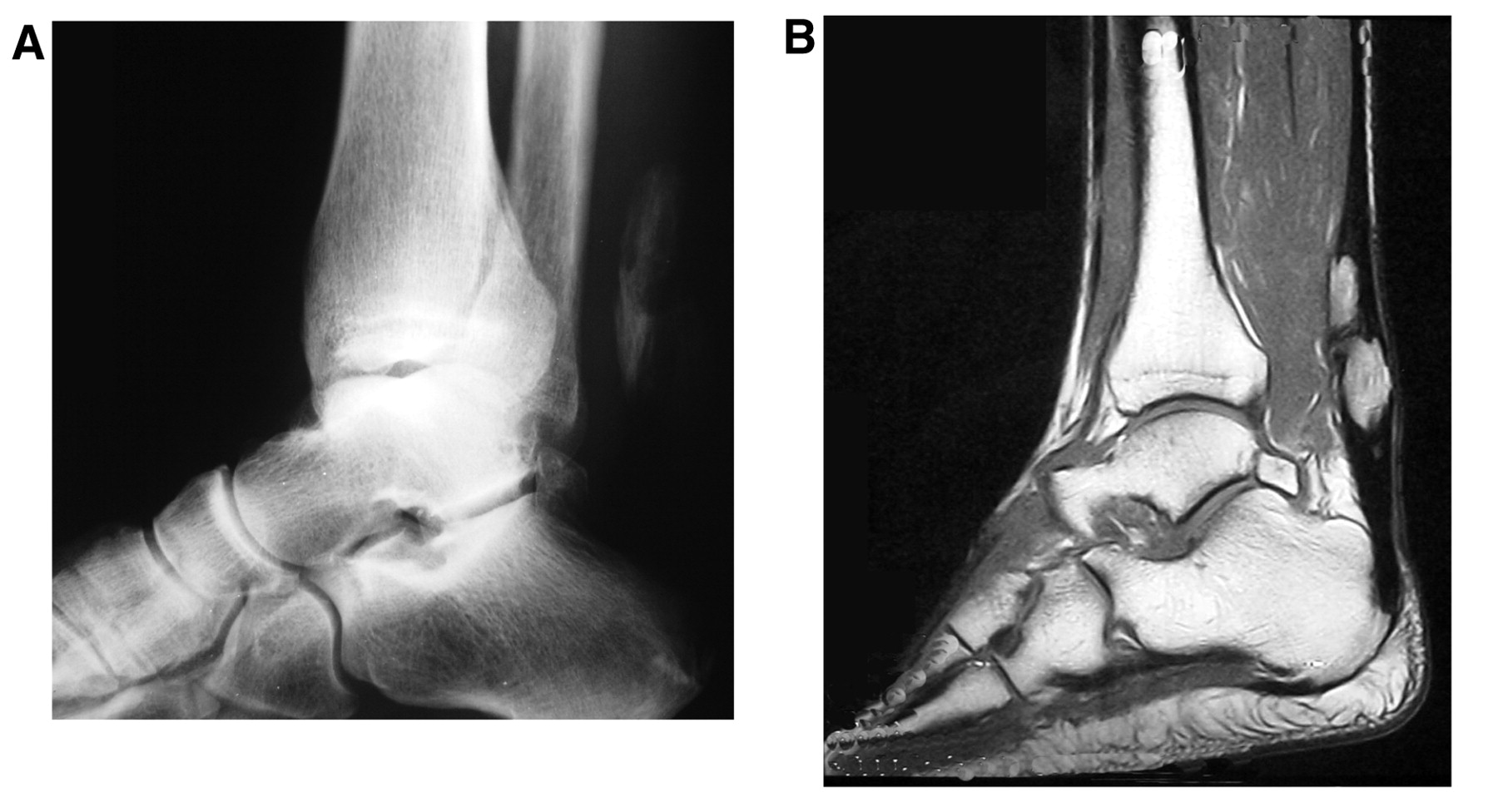
Figure 9.
Achilles tendon rupture in a tennis player who was treated for a rupture 15 years earlier with nonoperative care. A, Type IIIA radiopaque lesion of the tendo Achillis. B, Magnetic resonance imaging clearly documents the calcification that has occurred at the watershed area, the site of previous rupture. Note the similarity of the ossification in the Achilles tendon to the tibia, talus, and calcaneus.
Retrocalcaneal Bursitis. Pathology. Retrocalcaneal bursitis is an inflammatory condition of the bursal sac located between the posterosuperior border of the calcaneus and the anterior portion of the distal Achilles tendon.
Diagnosis. An enlarged posterosuperior margin of the calcaneus—Haglund’s deformity or “pump bump”—often is the source of pathology. Effusion and warmth may be present at the area surrounding the Achilles attachment.
Treatment. Treatment involves reducing inflammation and eliminating direct pressure. Heel lifts and cups and open-backed shoes are helpful, as are elastic compressive ankle sleeves. Unremitting cases may be improved with the use of a low-dose injection of a soluble corticosteroid, but extreme care must be taken not to inject into the tendinous substance. Also, postinjection immobilization should be used for several weeks. Arthroscopic debridement of the retrocalcaneal bursa may be helpful when all else fails. Pigmented villonodular synovitis may be present in the bursae.
Figure 10.
“Bouché’s grid” shown at zone 2 for Achilles insertional calcific tendinosis. The painful areas are marked by an X.
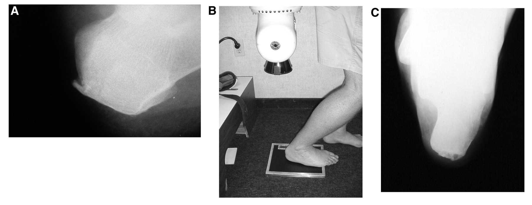
Achilles Insertional Calcific Tendinosis. Pathology. Achilles insertional calcific tendinosis is a pathologic, degenerative condition of the tendon at its insertion (Fig. 11). Calcification occurs in the tendinous substance after a cascade of cellular events. Microtears of the Achilles fibers at the insertion are followed by bleeding, which leads to calcification as seen radiographically.
Diagnosis, Differentiating Achilles Insertional Calcific Tendinosis from Haglund’s Deformity. Pain and symptoms from Achilles insertional calcific tendinosis usually occur with or without shoes, distinguishing Achilles insertional calcific tendinosis from Haglund’s deformity, which presents with pain exacerbated only by external shoe pressure. Also, Achilles insertional calcific tendinosis appears as a “fullness” of the entire posterior aspect of the heel, whereas Haglund’s deformity appears as a simple “bump” localized to the posterosuperior heel. On Bouché’s grid, Achilles insertional calcific tendinosis would manifest across the central row of boxes, whereas with Haglund’s pain would be located in the superolateral boxes.
Treatment. The prognosis for this condition is much worse than for retrocalcaneal bursitis, and chronic cases may require surgical intervention. Maffulli et al[43] studied 21 physically active patients with recalcitrant Achilles insertional tendinosis who were treated surgically. The Achilles tendon was disinserted from its calcaneal attachment, the calcific deposit was fully removed, and the Achilles tendon was then reinserted into the calcaneus using suture anchors. An average follow-up time of 48 months confirmed marked improvement in all of the patients; however, 5 of 21 patients were not able to return to their original level of physical activity.
Retro-Achilles Bursitis. Pathology. Retro-Achilles bursitis is an inflammatory condition of the bursal sac located between the posterior border of the distal Achilles tendon and the skin.
Diagnosis. Effusion and warmth may be present at the superficial area posterior to the Achilles attachment.
Treatment. Treatment involves reducing inflammation and eliminating direct pressure. Heel lifts and cups and open-backed shoes are helpful, as are elastic compressive ankle sleeves.
Distal Achilles Tendo-Fasciitis. Pathology. Achilles tendo-fasciitis involves the deep connective tissue that connects the Achilles tendon to the plantar fascia, which becomes inflamed.
Diagnosis. Pain is present on palpation of the posterior plantar portion of the calcaneus and is worsened by maximal ankle joint dorsiflexion and knee extension. Because this tissue is more prominent in the younger patient, it is most often seen in patients younger than 40 years. On Bouché’s grid, Achilles tendo-fasciitis would manifest across the lower boxes, whereas Achilles insertional calcific tendinosis pain would be located in the middle row of boxes.
Figure 11.
Achilles insertional calcific tendinosis. A, Lateral radiographic projection. B, Positioning for “90° axial calcaneal” radiographic projection. C, Radiographic view from part B shows medial-to-lateral involvement of the calcification at the Achilles tendon insertion.
Treatment. Flexibility exercises and a stretching regimen for all of the posterior leg muscles and plantar foot structures should be implemented.
Avulsion Fracture of the Calcaneus. Avulsion fracture of the calcaneus at the insertion of the Achilles tendon is rare in athletes. If it occurs, metabolic bone disease should be ruled out.
Rehabilitation Guidelines
Rehabilitation of most Achilles tendon injuries (other than ruptures) can be approached using the same sports medicine principles as for other sports-related injuries; advising an injured athlete to “rest” does not always mean completely stopping all athletic activity. Instead, “relative” rest and cross-training should be the goal for an athlete; an alternative activity must be found to allow the athlete to maintain cardiovascular fitness without inducing further injury or delaying healing. Initial treatment can often begin with a 25% decrease in the “FIT” (frequency, intensity, and time). Functional treatment involves a guided three-phase rehabilitation protocol focusing on protection and healing of the injured structures, with graduated strengthening aimed at remodeling injured tissue.[44]
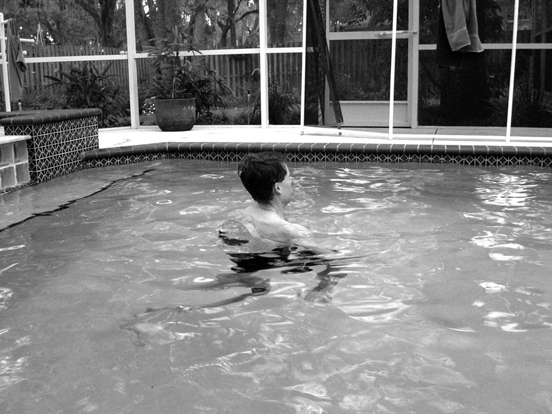
Rehabilitation with cross-training exercises will help speed recovery. Aqua-running is an excellent alternative activity that allows an athlete to maintain cardiovascular fitness while avoiding further injury to the Achilles tendon (Fig. 12). Athletes should be advised to run in the deep end of the pool (>8 feet deep). Other cross-training activities include swimming, cycling, elliptical training, and rowing. Prevention of further injury should also include improving flexibility, strength, and balance.
Return-to-play decisions should be based on an absence of pain, strength and range of motion equal to those of the contralateral limb, a gradual stepwise training protocol, and the ability of the athlete to perform the necessary skills of the sport without restriction.
Figure 12.
Aqua-running performed in the deep end of a pool is one of the best Achilles tendon rehabilitative exercises. It allows the athlete to maintain cardiovascular fitness while limiting the risk of repeated injury during the healing phase of recovery because there is no impact on the Achilles tendon.
New Therapies
A great deal of research has been published on improving Achilles tendon healing. Some newer treatments that had been experimental are now being used clinically. Several other treatments that may alleviate pain and shorten an athlete’s recovery time are still under investigation. Accelerating the biological healing process, strengthening the healed tendon, and reducing the pain associated with chronic tendon pathology are common goals of newer therapies.
A human-derived regenerative tissue matrix allograft (GraftJacket; Wright Medical Technology, Arlington, Tennessee) is now available and is being used to reinforce tenuous repairs of the Achilles tendon. A topical glyceryl trinitrate transdermal patch was more effective than placebo for reducing pain from chronic noninsertional Achilles tendinitis in the first 12 and 24 weeks of use.[45]
Soleus muscle transplantation into an Achilles tendon rupture repair has been successfully demonstrated in rabbits.[46] Studies using experimental Achilles rupture in rabbits in which fibrin glue was used showed an equivalence of primary functional treatment to a combination of operative and functional therapy.[47,48] Insulin-like growth factor may enhance Achilles tendon healing, repair, and regeneration.[49]
Marrow-derived stem cell–seeded collagen implants have been tested to enhance the structural and material properties of the Achilles tendon,[50] and growth differentiation factors 5 and 6 have been shown to increase tensile strength when used as collagen growth differentiation factor sponges in rats.[51] Injection of cartilage-derived morphogenetic protein-2 has recently been shown to improve tendon repair in rats.[52] Low-energy photostimulation causes release of growth factors from fibroblasts and, thus, increases collagen concentration.[53]
Although not yet approved by the US Food and Drug Administration for the treatment of Achilles tendon injuries, extracorporeal shockwave therapy is currently under investigation. Achilles insertional calcific tendinosis and Achilles tendinosis are specific conditions that may benefit from extracorporeal shockwave therapy, and it may alleviate the need for extensive surgical dissection and prolonged recovery. Until reports on long-term randomized prospective trials have been published, physicians must be cautious about the use of extracorporeal shockwave therapy for the Achilles tendon. Some are concerned that the treatment may actually precipitate tendon rupture.
Summary
Achilles tendon sports and fitness injuries are complex, diverse, and common. This review provides the sports medicine physician with a means of classifying specific Achilles tendon injuries and thus formulating an accurate diagnosis and an effective treatment plan. Thorough knowledge of Achilles tendon anatomy is essential in treating an Achilles tendon injury in an athlete. An understanding of the pathology, diagnosis, and treatment of the Achilles tendon should assist the sports medicine physician in returning an injured athlete to sports as quickly and safely as possible.
Financial Disclosure
None reported.
Conflict of Interest
References
- McCrory JL, Martin DF, Lowery RB, et al: Etiologic factors associated with Achilles tendinitis in runners. Med Sci Sports Exerc 31: 1374, 1999.
- Nelen G, Martens M, Bursens A: Surgical treatment of chronic Achilles tendinitis. Am J Sports Med 17: 754, 1989.
- Brunet M, Cook SD, Brinker M, et al: A survey of running injuries in 1505 competitive and recreational runners. J Sports Med Phys Fitness 30: 307, 1990.
- Clement D, Taunton J, Smart G: Achilles tendinitis and peritendinitis: etiology and treatment. Am J Sports Med 12: 179, 1984.
- Clement D, Taunton J, Smart G, et al: A survey of overuse running injuries. Phys Sports Med 9: 47, 1981.
- Krissoff W, Ferris W: Running injuries. Phys Sports Med 7: 55, 1979.
- Hootman JM, Macera CA, Ainsworth BE, et al: Epidemiology of musculoskeletal injuries among sedentary and physically active adults. Med Sci Sports Exerc 34: 838, 2002.
- Schepsis AA, Jones H, Haas AL: Achilles tendon disorders in athletes. Am J Sports Med 30: 287, 2002.
- Saxena A, Kim W: Ankle dorsiflexion in adolescent athletes. JAPMA 93: 312, 2003.
- Burdett RC: Forces predicted at the ankle during running. Med Sci Sports Exerc 14: 308, 1982.
- Scott SH, Winter DA: Internal forces of chronic running injury sites. Med Sci Sports Exerc 22: 357, 1990.
- Saxena D, Bareither D: Magnetic resonance and cadaveric findings of the “watershed band” of the Achilles tendon. J Foot Ankle Surg 40: 132, 2001.
- Mosely JB: “Survey of Professional ATC’s and Team Physicians,” in The Foot and Ankle in Sports, CV Mosby, Philadelphia, 1995.
- DeMaio M, Paine R, Drez DJ Jr: Achilles tendonitis. Orthopedics 18: 195, 1995.
- Puddu G, Ippolito E, Postachini F: A classification of Achilles tendon disease. Am J Sports Med 4: 145, 1976.
- Gould N, Korson R: Stenosing tenosynovitis of the pseudosheath of the tendo Achilles. Foot Ankle Int 23: 595, 2002.
- Morris KL, Giacopelli JA, Granoff D: Classifications of radiopaque lesions of the tendo Achillis. J Foot Surg 29: 533, 1990.
- Shalabi A, Kristoffersen-Wilberg M, Svensson L, et al: Eccentric training of the gastrocnemius. Am J Sports Med 32: 1286, 2004.
- Mafi N, Lorentzon R, Alfredson H: Superior short-term results with eccentric calf muscle training compared to concentric training in a randomized prospective multicenter study on patients with chronic Achilles tendinosis. Knee Surg Sports Traumatol Arthrosc 9: 42, 2001.
- Alfredson H, Pietila T, Jonsson P, et al: Heavy-load eccentric calf muscle training for the treatment of chronic Achilles tendinosis. Am J Sports Med 26: 360, 1998.
- Alfredson H, Lorentzon R: Chronic Achilles tendinosis. Sports Med 29: 135, 2000.
- Kvist M: Achilles tendon injuries in athletes. Sports Med 18: 173, 1994.
- Maffulli N, Binfield PM, Moore D, et al: Surgical decompression of chronic central core lesions of the Achilles tendon. Am J Sports Med 27: 747, 1999.
- Saxena A: Results of chronic Achilles tendinopathy surgery on elite and nonelite track athletes. Foot Ankle Int 24: 712, 2003.
- Saxena A: Sports medicine commentary. Foot Ankle Q 13: 58, 2000.
- Testa V, Capasso C, Benazzo F, et al: Management of Achilles tendinopathy by ultrasound-guided percutaneous tenotomy. Med Sci Sports Exerc 34: 573, 2002.
- Tallon C, Coleman B, Khan K, et al: Outcome of surgery for chronic Achilles tendinopathy. Am J Sports Med 29: 315, 2001.
- Vanek D, Saxena A, Boggs JM: Fluoroquinolone therapy and Achilles tendon rupture. JAPMA 93: 333, 2003.
- Hersh BL, Heath NS: Achilles tendon rupture as a result of oral steroid therapy. JAPMA 92: 355, 2002.
- Aroen A, Helgo D, Granlund O, et al: Contralateral tendon rupture risk is increased in individuals with a previous Achilles tendon rupture. Scand J Med Sci Sports 14: 30, 2004.
- Yinger K, Mandelbaum BR, Almekinders LC: Achilles rupture in the athlete: current science and treatment. Clin Podiatr Med Surg 19: 231, 2002.
- Khan RJ, Fick D, Brammar TJ, et al: Interventions for treating acute Achilles tendon ruptures. Cochrane Database Syst Rev 3: CD003674, 2004.
- Weber M, Niemann M, Lanz R, et al: Nonoperative treatment of acute rupture of the Achilles tendon: results of a new protocol and comparison with operative treatment. Am J Sports Med 31: 685, 2003.
- Cohen RS, Balcom TA: Current treatment options for ankle injuries: lateral ankle sprain, Achilles tendonitis, and Achilles tendon rupture. Curr Sports Med Rep 2: 251, 2003.
- Cetti R, Christensen SE, Ejsted R: Operative versus nonoperative treatment of Achilles tendon rupture: a prospective randomized study and review of the literature. Am J Sports Med 21: 791, 1993.
- Bhandari M, Guyatt G, Siddiqui F, et al: Treatment of acute Achilles tendon ruptures: a systematic overview and metaanalysis. Clin Orthop Relat Res 400: 190, 2002.
- Wallace RG, Traynor IE, Phil M, et al: Combined conservative and orthotic management of acute ruptures of the Achilles tendon. J Bone Joint Surg Am 86: 1198, 2004.
- Maffulli N, Tallon C, Wong J, et al: Early weight bearing and ankle mobilization after open repair of acute midsubstance tears of the Achilles tendon. Am J Sports Med 31: 692, 2003.
- Kauranen K, Kangas J, Leppilahti J: Recovering motor performance of the foot after Achilles rupture repair. Foot Ankle Int 23: 600, 2002.
- Weber M, Niemann M, Lanz R, et al: Nonoperative treatment of acute rupture of the Achilles tendon. Am J Sports Med 31: 685, 2003.
- Lim J, Dalal R, Waseem M: Percutaneous vs. open repair of the ruptured Achilles tendon: a prospective randomized controlled study. Foot Ankle Int 22: 559, 2001.
- Bouche R, McInnes B: “Posterior Heel Pain: Haglund’s Deformity, Pump-Bump Deformity, and Achilles Insertional Calcific Tendinosis (AICT),” in Master Techniques in Podiatric Surgery: The Foot and Ankle, Vol 1, ed by TJ Chang, p 266, Lippincott Williams & Wilkins, Philadelphia, 2005.
- Maffulli N, Testa V, Capasso G, et al: Calcific insertional Achilles tendinopathy. Am J Sports Med 32: 174, 2004.
- Sammarco VJ, Sammarco GJ: Principles and techniques in rehabilitation of the athlete’s foot: part 1. Tech Foot Ankle Surg 2: 51, 2003.
- Hunte G, Lloyd-Smith R: Topical glyceryl trinitrate for chronic Achilles tendinopathy. Clin J Sport Med 15: 116, 2005.
- Benazzo F, Gianluca S, Mosconi M, et al: Muscle transplant in the rabbit’s Achilles tendon. Med Sci Sports Exerc 33: 696, 2000.
- Thermann H, Frerichs O, Biewener A, et al: Healing of Achilles tendon: an experimental study: part 1. Foot Ankle Int 22: 478, 2001.
- Thermann H, Frerichs O, Holch M, et al: Healing of Achilles tendon, an experimental study: part 2. Foot Ankle Int 23: 606, 2002.
- Kurtz CA, Loebig TG, Anderson DD, et al: Insulin-like growth factor I accelerates functional recovery from Achilles tendon injury in a rat model. Am J Sports Med 27: 363, 1999.
- Young R, Butler D, Weber W, et al: Use of mesenchymal stem cells in a collagen matrix for Achilles tendon repair. J Orthop Res 16: 406, 1998.
- Aspanberg P, Forslund C: Enhanced tendon healing with GDF 5 and 6. Acta Orthop Scand 70: 51, 1999.
- Forslund C, Aspenberg P: Tendon healing stimulated by injected CDMP-2. Med Sci Sports Exerc 33: 685, 2000.
- Reddy G, Stehno-Bittel L, Enwemeka C: Laser photostimulation of collagen production in healing rabbit Achilles tendons. Lasers Surg Med 22: 281, 1998.
© 2007 American Podiatric Medical Association