


Abstract
Epidermal inclusion cysts are soft-tissue lesions that result from traumatic implantation of epidermal cells into the dermis. A case report is presented involving a large recurrent plantar inclusion cyst secondary to trauma sustained in the Vietnam War. The patient reported that the initiating trauma was the result of stepping on a punji stake in Vietnam. Punji stakes, sharpened spikes typically made of bamboo and dipped in poison or excrement, were used by the Vietnamese soldiers to wound enemy soldiers. Careful excision of the lesion is imperative to prevent recurrence. (J Am Podiatr Med Assoc 96(5): 445–447, 2006)
Epidermal inclusion cysts are common benign soft-tissue lesions that typically occur in young to middle-aged people on the trunk, neck, and face. [1] These lesions result from the implantation of epidermal cells into the dermal layers that continue to proliferate, slowly developing into a palpable mass. They may be solitary or multiple, and the etiology may be either congenital, resulting from the inclusion of ectodermal elements during embryogenesis, or acquired after mechanical implantation. Multiple lesions are often seen in patients with Gardner’s syndrome, which consists of multiple colon polyps, fibrous tissue tumors, osteomatosis, and dental abnormalities. [2] Most inclusion cysts, such as sebaceous cysts, are associated with hair follicles and rarely occur on the palms of the hands or the soles of the feet. When located on the palms or soles, these lesions are typically associated with traumatic induction of epidermal tissue. [3]
Several cases of inclusion cysts have been described in the podiatric medical literature. In 1971, Berlin [4] described three separate occurrences of inclusion cysts at the US army hospital in Fort Jackson, South Carolina. Berlin hypothesized that these lesions were the result of trauma, but the exact mechanism of trauma was not determined. In 1990, Marczak [2] described a unique case involving a sedentary elderly individual who developed an inclusion cyst in the absence of any known traumatic injury. In 1991, Sidoti and Emelianchik [5] described an inclusion cyst associated with a retained foreign body and infected granuloma. In this case, the inciting trauma and subsequent introduction of a foreign body resulted in the inclusion cyst.
Treatment of inclusion cysts is limited owing to their unique structure. Although areas of the cyst may contain fluid, typically these cysts are not fluid-filled; therefore, aspiration would probably be unsuccessful. [2] Definitive treatment for these lesions is surgical excision of the entire cyst and all aspects of the cyst wall. [6] Histologically, the walls of these lesions exhibit multiple layers of stratified squamous epithelium with several overlying layers of keratinous material. If any portion of the cyst wall remains after excision, the lesion will probably recur. [6]
Case Report
A 54-year-old man visited the Podiatry Clinic at the North Chicago Veterans Affairs Medical Center with the chief complaint of a painful mass on the plantar surface of his left foot. The patient stated that the mass had been present for nearly 10 years and had steadily increased in size during that time. The patient said that he had a similar mass in the same area removed approximately 10 years earlier. The patient reported that the mass occasionally fluctuated in size and that it became extremely painful after long periods of ambulation.
The original mass had developed 30 years earlier after the patient injured his foot on a punji stake during active duty in Vietnam. Punji stakes (sharpened spikes typically made of bamboo and dipped in poison or excrement) were used extensively in the Vietnam War by the Vietcong and North Vietnamese army. These “silent killers” could cause extensive damage when stepped on by a soldier. Often a single injured soldier would require the assistance of several fellow soldiers, slowing down the unit and putting the entire battalion at risk.
On physical examination, a circular mass measuring approximately 3 cm in diameter was noted deep to the plantar aspect of the first interspace at the level of the midshafts of the first and second metatarsals. The mass appeared solid and did not allow transillumination. A cicatrix was noted traversing proximally to distally over the plantar aspect of the mass. There was mild-to-moderate tenderness on palpation.
Radiographs of the lesion showed an increase in soft-tissue mass with no bony involvement. A magnetic resonance image obtained to determine the extent of the lesion showed a multilobulated semisolid mass with several fluid-filled cavities plantar to the musculature of the first interspace. After extensive discussion with the patient, it was decided to perform surgical excision.
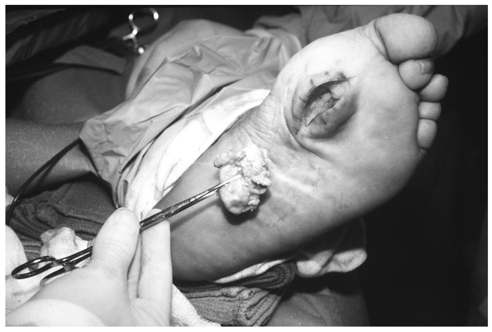
Through a linear plantar incision, the lesion was identified, and an attempt was made to excise the lesion. The cyst was punctured during dissection, revealing a thick, rust-colored exudate. After copious irrigation, the lesion was removed in toto (Fig. 1). The borders of the surgical wound were examined, and it was determined that no residual lesion remained. The wound edges were remodeled and coapted.
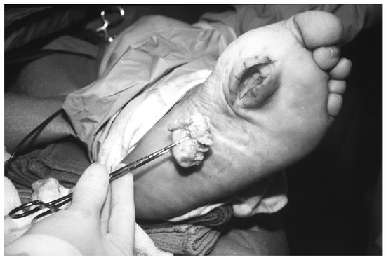
Figure 1.
Lesion, measuring 4.9 × 3.8 × 1.9 cm, after excision with exudate expressed from the mass.
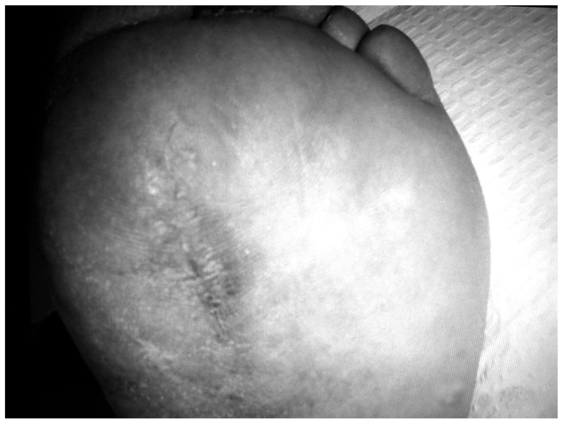
The patient was advised to remain nonweightbearing with crutches to reduce the chances of significant plantar scarring. The patient reported minimal postoperative pain, and the remainder of the postoperative period was unremarkable. The patient was returned to full weightbearing within 4 weeks and was released without limitations from the care of the podiatric medicine service after 4 months of follow-up (Fig. 2).
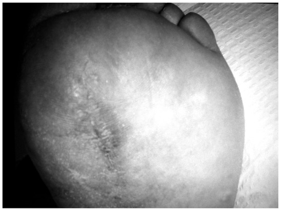
Figure 2.
Patient’s foot at the time of discharge from the podiatric medicine service, 4 months after surgery.
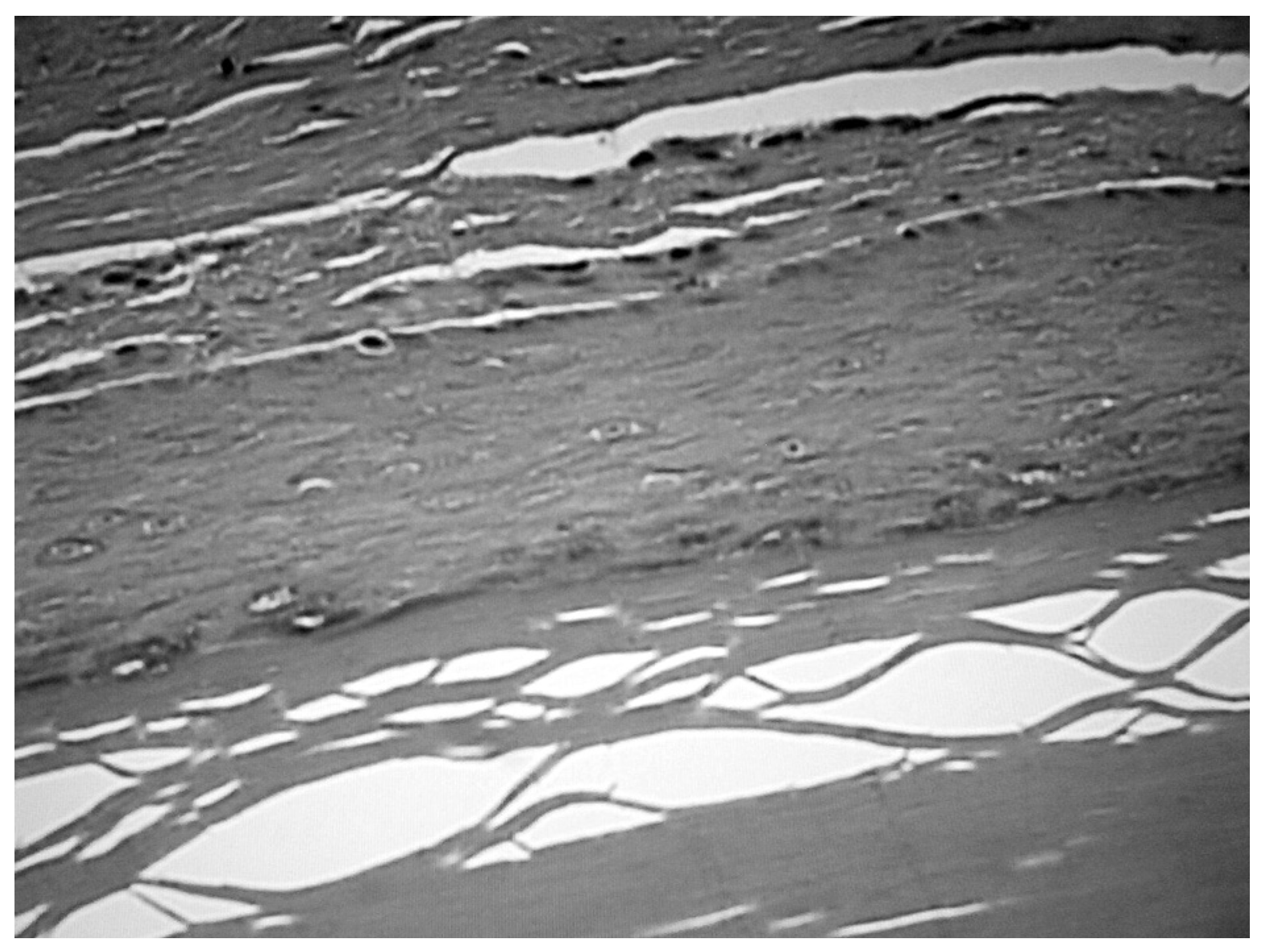
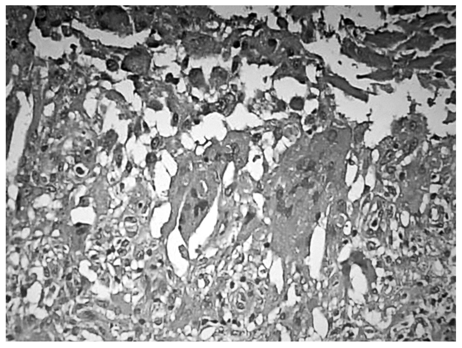
The mass measured 4.9 × 3.8 × 1.9 cm. The exterior color varied from yellow-tan to blue-gray, with attached segments of adipose tissue. Gross examination revealed a pouchlike structure 1.3 cm wide containing a small amount of colorless fluid. The lesion was bisected, revealing a fibrofatty composition with interior cartilage. Microscopic examination revealed a cystic mass lined by stratified squamous epithelium with laminated layers of keratinous material (Fig. 3). The expressed exudates consisted of multinucleated giant cells (Fig. 4). These findings were consistent with a diagnosis of epidermal inclusion cyst with associated chronic inflammation.
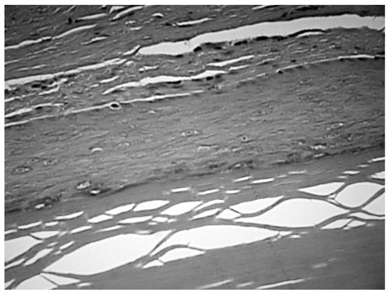
Figure 3.
Microscopic examination of the mass revealed stratified squamous epithelium with laminated layers of keratinous material; this is pathognomonic for inclusion cysts (H&E, ×40).
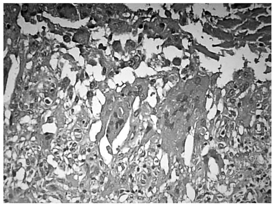
Figure 4.
Microscopic examination of exudate revealed a high concentration of multinucleated giant cells common to inclusion cysts (H&E, ×40).
Discussion
Whenever a patient presents with a painful mass in the foot, a thorough history and physical examination are imperative. This case is unique owing not only to the large size of the mass but also to the etiology. Although inclusion cysts have been reported as sequelae to trauma, rarely has a patient experienced such an obvious inciting event. Also significant is the fact that it was the second time his mass was excised. This illustrates the persistence of these lesions and the need to excise all remnants of the cyst.
Imaging also plays an important role in the treatment of these lesions. Plain film radiographs should always be obtained to rule out bony involvement. Computed tomography should be reserved for lesions with obvious osseous penetration. Magnetic resonance imaging, as obtained in this case, can be essential in assessing the size, location, density, and invasiveness into the surrounding soft-tissue structures of the lesion. The combination of a thorough history and physical examination and multiple imaging modalities can greatly assist the surgeon in planning the appropriate surgical course.
Conclusion
Epidermal inclusion cysts result from the implantation of epidermal cells into the dermal layers, which proliferate, causing a palpable mass. Although inclusion cysts are relatively rare on the plantar aspect of the foot, they should be included in the differential diagnosis of a patient with a history of trauma in the vicinity of the mass. These cysts, although often symptomatic, may cause significant pain when located under the weightbearing areas of the foot. In addition, it has been postulated that the repeated trauma of ambulation to the plantar aspect of the foot may cause the cyst to rupture into the adjacent soft tissue. [7] Ruptured cysts elicit considerable inflammation with abundant foreign-body giant cells. [8] Because of the recurrent nature of these cysts, excision should be considered the treatment of choice in most cases.
References
- Malay D: “Tumors, Part 1,” in McGlamry’s Comprehensive Textbook of Foot and Ankle Surgery, 3rd Ed, Vol 2, ed by AS Banks, MS Downey, DE Martin, et al, p 1317, Lippincott, Williams & Wilkins, Philadelphia. ,2001. .
- Marczak L: Epidermal inclusion cyst of the heel. .JAPMA80::660. ,1990. .
- Lemont H: Traumatic inclusion cyst versus pilar cyst of the foot. .JAPMA82::218. ,1992. .
- Berlin S: Epidermal inclusion cyst of the foot: a report of three cases. .JAPA61::55. ,1971. .
- Sidoti SP, Emelianchik AJ: Infected granuloma and inclusion cyst secondary to a retained foreign body. .J Foot Surg30::369. ,1991. .
- Sheff J, Bregman P, Curran T: Bilaterally symmetrical epidermal inclusion cysts with foreign-body giant-cell reaction. .JAPMA88::410. ,1998. .
- Misner S, Mariash S, Alvarez G: Ruptured plantar epidermal inclusion cyst with foreign body giant cell reaction. .J Foot Surg30::38. ,1991. .
- Robbins SL: “The Skin,” in Pathologic Basis of Disease, p 1263, WB Saunders, Philadelphia. ,1984. .
© 2006 American Podiatric Medical Association