


Abstract
Schwannomas of the foot are rare. We describe three cases that mimicked compressive neuropathy, two resembling tarsal tunnel syndrome and a third resembling Morton’s neuroma. All three patients had complete resolution of their pain after resection of the tumor.
Schwannomas are benign tumors that arise from peripheral nerves with a Schwann cell sheath. Schwannomas of the foot have rarely been reported, with approximately 30 cases described in the literature.[1,2,3] They are usually located on the flexion surface of the limbs[4] and tend to be solitary lesions, although multiple lesions are occasionally reported.[5,6] Schwannomas may occur in the subcutaneous, intramuscular, intermuscular, or intraosseous planes. The symptoms are a matter of debate. Some authors report that the schwannoma is usually asymptomatic and that clinically it presents as a slow-growing soft-tissue tumor with occasional neurologic symptoms, such as altered sensation or motor weakness,[1,3,5,7,8] or that pain arises only when the mass exceeds 5 cm.[9] Other researchers, however, report that most schwannomas prompt local or radiating pain (or both) and a positive Tinel sign.[4] The pain sometimes resembles that associated with tarsal tunnel syndrome[10,11] or Morton’s neuroma.[3,12]
There is no sex predilection for schwannomas, and although they may occur at any age, they are commonly found during the fourth decade of life.[13] The lesions grow slowly at the periphery of the nerve sheath, without invading the interior of the nerve. The treatment of choice for these lesions is excision; because malignant transformation is rare, wide excision is not indicated. Tumor removal does not jeopardize the integrity of the nerve and can result in relief of symptoms and maintenance of nerve function.[7,12,14] There are, however, reports of neurologic complications after excision of a schwannoma, including pain and muscle weakness.[15]
We describe three cases that mimicked compressive neuropathy, two resembling tarsal tunnel syndrome and a third resembling Morton’s neuroma. All three patients had complete resolution of their pain after resection of the tumor.
Case Reports
Case 1
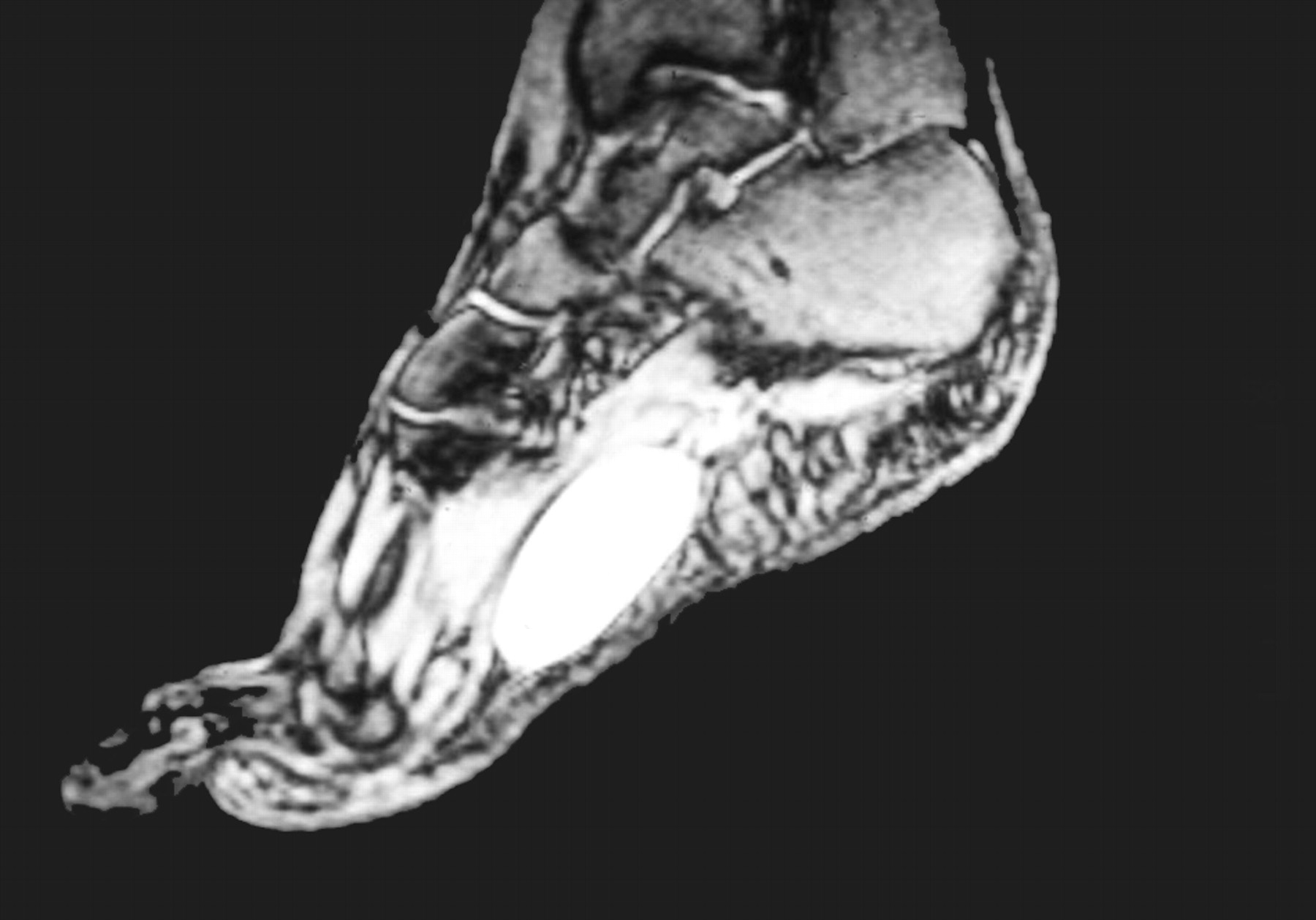
A 48-year-old woman presented for evaluation of a mass on the plantar aspect of her left foot. She first became aware of the mass 2 years earlier but had felt no pain until 1 month before consultation, when weightbearing elicited pain that radiated toward the toes. Examination showed a 2.5 × 1.5-cm firm mass on the medial plantar aspect of the left foot. It was moderately tender on deep palpation, and there was evidence of partial numbness on the plantar aspect of the first and second toes. Results of radiographic examinations of the left foot were negative. Magnetic resonance imaging of the same foot showed a 4 × 2-cm clearly defined mass embedded in the plantar musculature (Figure 1).
Figure 1
.
Sagittal gradient-echo T2-weighted image of the left foot showing a clearly defined mass with high signal intensity located in the plantar musculature.
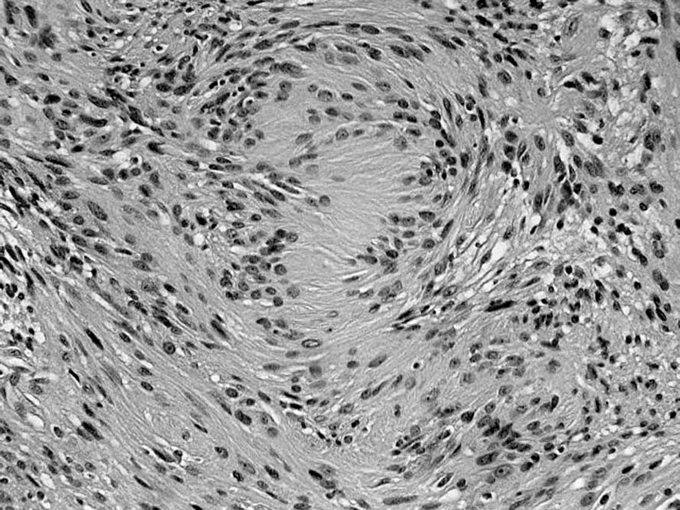
Surgery was performed under regional anesthesia and a thigh tourniquet was used. A well-encapsulated mass adhering to the medial plantar nerve was dissected from the nerve and excised. On histologic examination, the mass was identified as a benign schwannoma. Hematoxylin and eosin staining exhibited numerous nuclei in a palisade arrangement (Antoni A pattern) (Figure 2). Three years later, the patient had no sign of a mass and no painful foot symptoms.
Figure 2
.
Hematoxylin and eosin staining (×200) exhibiting numerous nuclei in a palisade arrangement (Antoni A pattern).
Case 2
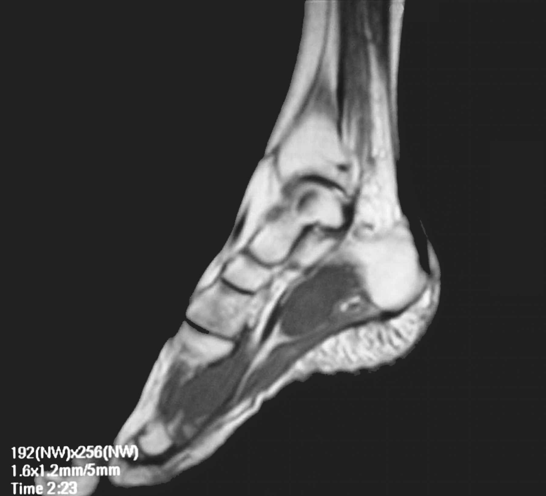
A 46-year-old woman presented for evaluation of a mass on the medial aspect of the ankle and sole of her left foot. The mass was first noticed 2 months earlier, coinciding with pain that radiated toward the medial aspect of the foot and first toe. The pain was intense, obliging the patient to assume an antalgic gait, and was present even at rest, particularly in bed at night. Examination of the foot and ankle showed a 5 × 3-cm mass on the medial aspect of the ankle and sole. The mass was tender on deep palpation, and Tinel’s sign was present; there was also evidence of numbness on the plantar aspect and the first and second toes. Magnetic resonance imaging of the foot disclosed a clearly defined mass embedded in the plantar musculature close to the calcaneus (Figure 3).
Figure 3
.
Sagittal T1-weighted image of the left foot showing a mass isointense to muscle involving the plantar musculature near the calcaneus.
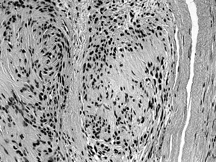
As in Case 1, surgery was performed under regional anesthesia and a thigh tourniquet was placed. A well-encapsulated lobular mass adhering to the posterior tibial nerve was dissected from the nerve and excised. On histologic examination, the mass was identified as a benign schwannoma. Hematoxylin and eosin staining exhibited numerous nuclei in a palisade arrangement (Antoni A pattern) (Figure 4). The pain subsided during the postoperative period, and 18 months later the patient displayed no sign of a mass or painful symptoms.
Figure 4
.
Hematoxylin and eosin staining (×200) exhibiting numerous nuclei in a palisade arrangement (Antoni A pattern).
Case 3
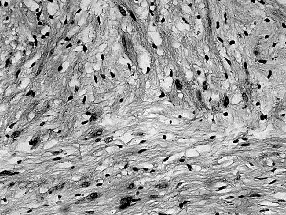
A 57-year-old man presented with persistent pain in the ball of his right foot that radiated into the second and third toes. Ultrasonographic examination disclosed a hypoechoic mass under the second and third metatarsal heads (Figure 5). Magnetic resonance imaging of the foot showed a rounded, clearly defined mass under the second and third metatarsal heads, leading to an initial diagnosis of Morton’s neuroma (Figure 6). During surgery, under regional anesthesia and using a thigh tourniquet, a slightly lobular mass measuring approximately 2 cm was dissected from the interdigital nerve to which it adhered. At histologic examination, the mass was identified as a benign schwannoma. Hematoxylin and eosin staining exhibited numerous nuclei in a palisade arrangement (Antoni A pattern) (Figure 7).
Figure 5
.
Ultrasonographic scan showing a hypoechoic mass measuring approximately 2 cm.
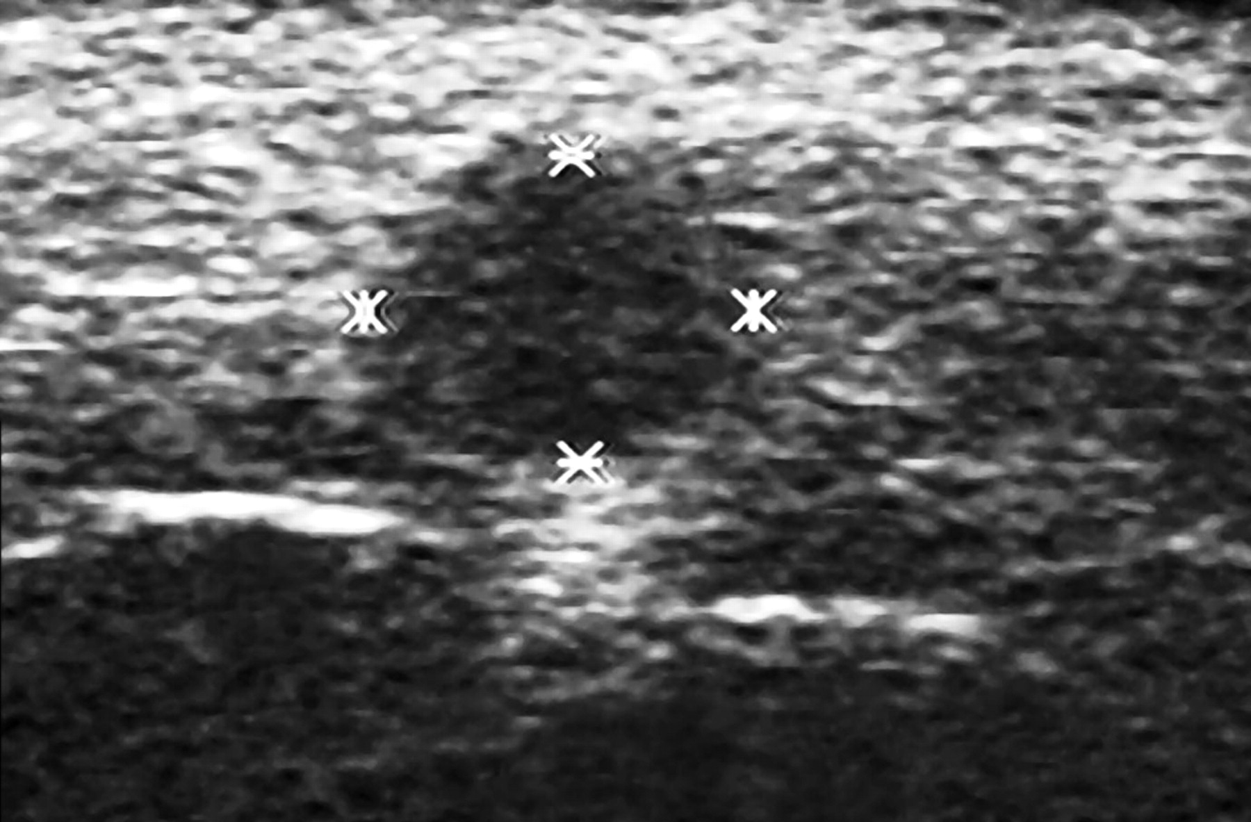
Figure 6
.
Coronal spin-echo T1-weighted image of the right foot showing a small mass under the second and third metatarsal heads.
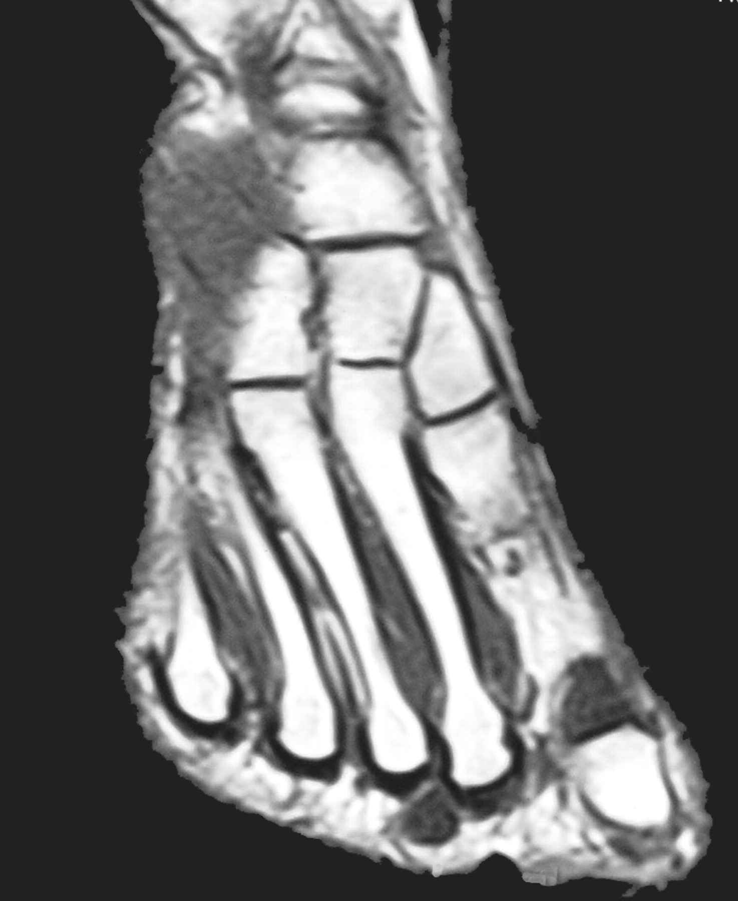
Figure 7
.
Photomicrograph of a specimen from Case 3 (H&E, ×200). These findings are similar to those of the other cases (Antoni A pattern).
Discussion
In the three cases of schwannoma reported here, the main symptom was pain, although in two patients the tumor measured less than 5 cm; this may be because the location of the mass on the plantar aspect of the foot more readily prompted irritation due to weightbearing. Two patients displayed symptoms resembling those of tarsal tunnel syndrome, whereas in the third patient clinical signs were more reminiscent of Morton’s neuroma. In all three patients, the tumor was located on a flexor surface, as in most of the cases reported in the literature[4]; the mass was a solitary finding, as is also usually the case, although multiple lesions are occasionally reported.[5,6] The age of our patients also matched that of other cases reported in the literature,[13] with the lesion appearing in the fourth decade of life in two of our patients. Although schwannomas may occur in the subcutaneous, intramuscular, intermuscular, or intraosseous planes, the location was intramuscular in the three cases reported here.
Differential diagnosis should include other potentially painful tumors,[11] radiculopathy,[9] and, in the foot, tarsal tunnel syndrome and Morton’s neuroma; as the published literature illustrates, many schwannomas in the lower limb prompt symptoms that resemble those reported in these nerve-entrapment syndromes.[11,12] The initial diagnosis in the cases reported here was compression neuropathy. In all three patients, simple removal of the tumor led to the disappearance of all symptoms, with no tumor recurrence. In conclusion, as Liebau et al[13] suggested, although schwannomas are rare, they should be considered in all cases of pain and paresthesia of the foot when other possible sites of injury have been ruled out.
References
- Torossian JM, Augey F, Salle M, et al: Giant foot schwannoma. Br J Plast Surg, 54, p. 742001.
- Mott RC, Dellon AL: Multiple schwannomas of the foot: case report and strategy for treatment. JAPMA, 93, p. 512003.
- Pace JF, Spinosa FA: Benign schwannoma of the foot. JAPMA, 79, p. 2931989.
- Ahmed AR, Watanabe H, Aoki J, et al: Schwannoma of the extremities: the role of PET in preoperative planning. Eur J Nucl Med, 28, p. 15412001.
- Kwon BC, Baek GH, Chung MS, et al: Intramuscular neurilemoma. J Bone Joint Surg Br, 85, p. 7232003.
- Joyce M, Laing AJ, Mullet H, et al: Multiple schwannomas of the posterior tibial nerve. J Foot Ankle Surg, 8, p. 1012002.
- Zuckerman JD, Powers B, Miller JW, et al: Benign solitary schwannoma of the foot: a case report and review of the literature. Clin Orthop Relat Res, 228, p. 2781988.
- Thomas J, Jaffe KA: Foot and ankle tumors. Curr Opin Orthop, 11, p. 1172000.
- Ghaly RF: A posterior tibial nerve neurilemoma unrecognized for 10 years: case report. Neurosurgery, 48, p. 6682001.
- Finkbeiner GS, Marks RM: Nerve problems of the foot and ankle: evaluation, diagnosis, and treatment. Curr Opin Orthop, 13, p. 932002.
- Grossman MR, Mandracchia VJ, Urbas WM, et al: Neurilemoma of the posterior tibial nerve with an uncommon case presentation. J Foot Surg, 31, p. 2191992.
- Laurencin CT, Bains M, Yue JJ, et al: Schwannoma of the superficial peroneal nerve presenting as web space pain. J Foot Ankle Surg, 34, p. 5321995.
- Liebau C, Baltzer AW, Schneppenheim S, et al: Isolated peripheral neurilemoma attached to the tendon of the flexor digitorum longus muscle. Arch Orthop Trauma Surg, 123, p. 982003.
- Graviet S, Sinclair G, Kajani N: Ancient schwannoma of the foot. J Foot Surg, 34, p. 461995.
- Kang HJ, Shin SJ, Kang HJ: Schwannomas of the upper extremity. J Hand Surg Br, 25, p. 6042000.
© 2006 American Podiatric Medical Association