


Abstract
This investigation evaluates the effects of diabetes on the mechanical properties of human bone, specifically, the tibia. Seven diabetic and seven nondiabetic human (male) cadaveric distal tibiae were used in this study. The average age of the diabetic cadaveric samples was 51 years (range, 46–61 years), and the average age of the nondiabetic cadaveric samples was 75 years (range, 67–85 years). Three-point bending tests for strength and stiffness were performed on a small sample of each distal tibia. Each specimen was loaded at a constant rate until failure. From the recorded curve of load versus displacement, the ultimate and yield strength of bone and the bending modulus of bone were calculated. The diabetic samples were generally weaker than the older, nondiabetic samples, but no statistically significant differences were found in the elastic modulus (P = .29), yield strength (P = .90), ultimate strength (P = .46), and fracture toughness (P = .78), leading to speculation that diabetes has an effect similar to that of aging on the musculoskeletal system.
Despite the extensive research concerning diabetes and its overall systemic effects, little experimental evidence exists to help identify the effects of diabetes on human bone, cartilage, and the musculoskeletal system in general. Recent findings[1–6] have demonstrated that diabetes has a demineralizing effect on bone, with a concomitant decrease in mass and strength. Bone loss secondary to diabetes has been attributed to metabolic abnormalities, abnormal calcium concentration within cells, and high blood glucose levels.[3,7–9] The alterations of bone and mineral metabolism created by diabetes may lead to weakening of bone, thereby rendering the diabetic patient more susceptible to fracture or delayed fracture healing.[10–15] Some of these alterations in bone and cartilage are similar to those seen with advancing age in nondiabetic specimens.[6,10–12,16–21]
Although several studies have evaluated the mechanical properties of diabetes on animal bones, few can be found that tested the effects of diabetes on the mechanical properties of human bones of the lower extremity. Therefore, the purpose of this study was to evaluate the effects of diabetes on the mechanical properties of human bone, specifically, the tibia, and to determine whether these effects are similar to the aging process of bone.
Materials and Methods
Institutional review board approval was obtained from The University of Texas Health Science Center at San Antonio for the use of recently amputated lower-extremity specimens of diabetic and nondiabetic patients. All of the diabetic specimens were obtained from lower-extremity amputations performed at The University of Texas Health Science Center at San Antonio. A medical record review was performed to evaluate the factors that led to each amputation. These amputations were elective surgical procedures or were required because of trauma, infection, or tumor in the lower extremity (the tibial bone was not affected). Patients who were nonambulatory for an extended period before their lower-extremity amputation were excluded. Any bone that demonstrated signs of possible osteomyelitis (noted on imaging before amputation or by clinical suspicion) was also excluded from the study, including any bone exposed through a wound or ulcer or that demonstrated visual signs of cortical erosion or altered structural integrity. The specimens were frozen at −80°C immediately after amputation and evaluation of the specimens by the pathology department. The control specimens (along with information regarding the age, sex, and medical history of the donors) were obtained from the Musculoskeletal Transplant Foundation (Edison, New Jersey).
To prepare the specimens for testing, the amputated feet were allowed to thaw for 12 hours at room temperature. Each tibia was dissected, and all surrounding soft tissue was removed. The bones were then wrapped in saline-moistened gauze, sealed in plastic bags, labeled, and refrozen at −80°C until needed for mechanical testing. When ready for testing, the tibia samples were thawed in phosphate buffered saline (pH 7.4) at room temperature for 12 hours. Two specimens of cortical bone (30 × 4 × 2 mm) were machined from the anterior aspect of each distal tibia, just proximal to the plafond. The longitudinal axis of the specimens was parallel to the longitudinal axis of the tibia. The bone specimens were gripped in a vise attached to a precision bench-top milling machine, and all of the dimensions were machined for each specimen. The variation of the dimensions was controlled within 1%. One of these two specimens was used to measure the transverse fracture toughness of bone, and the other was used to determine the ultimate strength and stiffness of bone.
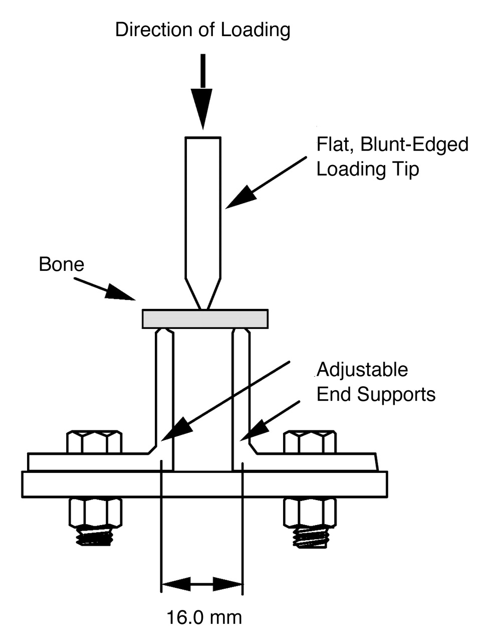
Fracture toughness testing was performed in a three-point bending apparatus according to the appropriate standard recommended by the American Society for Testing and Materials (Fig. 1).[22] A pre-crack was introduced in the middle of the bone specimen using a circular saw and a sharp razor blade. The calculations take into account the variation of the pre-crack size, and, therefore, its effect on measurements is not an issue because the crack size was measured accurately. In this study, the crack size was measured under the microscope, and the error of measurement was within 0.5%. Such an error has little effect on the final results. In addition, all pre-cracks were prepared in a bench-top milling machine in a consistent manner to avoid any error induced by the shape of the pre-cracks.
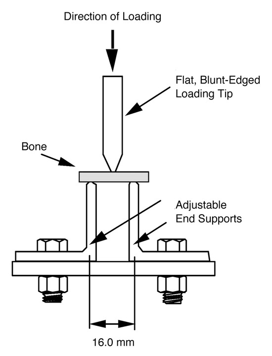
Figure 1.
Three-point bending apparatus.
Test specimens were supported in a custom-designed fixture with a support span of 16 mm. A compression load was applied in the middle of the specimens at a loading rate of 5 mm/min in a material testing machine (model 1011; Instron Corp, Canton, Massachusetts) until failure. The curve of load versus displacement was recorded using a computer-controlled data-acquisition system, and fracture toughness values were calculated according to the standard.[22] Because the pre-crack propagates in a direction perpendicular to the longitudinal axis of the diaphysis, the property actually measured in this test was the transverse fracture toughness of bone.
The three-point bending tests for strength and stiffness were performed according to the experimental procedures and the following calculation formulae recommended in American Society for Testing and Materials standard D790-86[23]:

where σy and σs are yield strength and ultimate strength, respectively; Fy, the force at which yielding occurs; Fs, the maximum applied force; E, Young’s modulus; c, the distance from the center of mass of the cross-section of the specimen; δ, the displacement at the loading point; L, the span of the support; and I, the cross-sectional moment of inertia.

where Kc is fracture toughness (critical stress intensity factor); Pc, the force at failure; B, the thickness of the specimen; W, the height of the specimen; a, the crack length; and f(a/W), a function of the ratio of a/W.
Each specimen was loaded at a constant rate of 10 mm/min until failure. From the recorded curve of load versus displacement, the ultimate and yield strength and bending modulus of bone were calculated. Because of the limitation of bone stock, specimen sizes in the fracture toughness and three-point bending tests were smaller than those recommended in the standards. However, a relative comparison between these specimens should be valid because all specimens in each test had the same shape and size and were tested under the same conditions.
The data were analyzed using unpaired t-tests to detect differences in biomechanical properties between diabetic and nondiabetic bone samples. Significant differences were considered only when P < .05.
Results
Seven diabetic and seven nondiabetic human cadaveric distal tibiae were used in this study. All of the bone samples were collected from male donors. The average age of the diabetic bone samples was 51 years (range, 46–61 years), and the average age of the nondiabetic bone samples was 75 years (range, 67–85 years). The mean ± SD values of the intrinsic biomechanical properties of bone samples from the diabetic and nondiabetic distal tibiae are given in Table 1. Overall, the values for nondiabetic bone samples were greater than those for diabetic samples. However, the results of the statistical analysis demonstrate that no significant differences were found in the elastic modulus, yield strength, ultimate strength, and fracture toughness between diabetic and nondiabetic bone samples tested in this study.

Table 1.
Intrinsic Biomechanical Properties of Bone Samples from Diabetic and Nondiabetic Distal Tibiae
Discussion
Although physicians generally believe that diabetic bone is inferior to bone in nondiabetic patients, alterations in the material and structural properties of bone in the diabetic patient have not been fully detailed. Previous studies[6,24] support this notion in human metatarsal bones, and the present study further suggests this correlation in the tibia. The present investigation demonstrates that diabetic tibial bones have strength and stiffness similar to nondiabetic tibial bones from a much older patient population (24 years older). We may then speculate that diabetes affects bone quality in much the same way as the process of aging.[6,16] It has been well documented in the scientific literature that aging adversely affects bone quality.[6] Most of the research in this area has focused on femoral neck and hip fractures in the elderly, but human tibiae have also been shown to decrease ultimate strain as a function of age.[25,26] Most recently, Courtney et al[24] demonstrated that the bending strength and bone density of human metatarsal specimens decrease with age. The precautions taken in treating fractures in the elderly population may, therefore, also be prudent in diabetic patients.
Although the increased morbidity in orthopedic patients with diabetes is well documented,[1,10–12,14–16] the literature regarding the specific effects of diabetes on bone quality is not as strong.[6,14,15,24] Some studies[10,17] have demonstrated that diabetic bones are weaker, whereas other studies[11,12,18,27] have demonstrated that the fracture-repair process, in the diabetic state, seems to be defective. Experimental studies using streptozotocin-induced diabetes in rats, a well-established model of diabetes, have supported the clinical observation of diabetes-associated osteopenia[4,28–31] and have documented decreased protein and collagen metabolism in the presence of diabetes.[32–39] Metabolic and structural changes observed in experimental diabetes models of young adult rats were found to be similar to changes found in nondiabetic animals of advanced age.[31,40] In a human model, Fleischli et al[6] demonstrated that diabetic metatarsals behave in a similar manner as those of a much older nondiabetic population. These studies seem to suggest, as does the present investigation, that diabetes may accelerate the normal aging process with respect to bone quality.
Further studies may provide more insight into how these diabetes-related bone changes affect fracture healing in diabetic individuals or even the pathogenesis of Charcot’s arthropathy. The present study merely suggests that diabetes adversely affects bone quality. The exact nature of these changes, although still unknown, will have far-reaching implications in the care of diabetic patients with orthopedic conditions.
The limitations of this study are important to note. Our results would have been more valuable had our two study groups been matched for age. The random collection of amputation specimens, and the need to use bone bank specimens, made it impossible to obtain age-matched groups. Therefore, the control group was significantly older than the diabetic group. In addition, the factors that led to amputation must also be considered. Patients may have experienced medical conditions that adversely affected their ambulatory status before their lower-extremity amputation. Inactivity or confinement to a bed or wheelchair may have created a disuse osteopenia, and, thus, patients who were known to be nonambulatory for an extended period before their lower-extremity amputation were excluded. It may be safe to assume, however, that most patients with a severe infection or nonhealing wound that requires a below-the knee or above-the-knee amputation are not fully ambulatory before the amputation. However, the older control group might also have been quite sedentary before their death, and, thus, this may be of no consequence.
Conclusion
This investigation found that diabetic tibial bone specimens demonstrate no significant differences in area of elastic modulus, yield strength, ultimate strength, and fracture toughness compared with much older, nondiabetic tibial bone specimens.

References
- Levin ME, Boisseau VC, Avioli LV: Effects of diabetes mellitus on bone mass in juvenile and adult-onset diabetes. N Engl J Med 294: 241, 1976.
- Matsumoto M, Goriya Y, Sekimoto H: Osteopenic changes in high sugar diet-induced diabetic rabbits (HSDD-R). Diabetes Res 18: 115, 1991.
- Santiago JV, McAlister WH, Ratzan SK, et al: Decreased cortical thickness and osteopenia in children with diabetes mellitus. J Clin Endocrinol Metab 45: 845, 1977.
- Goodman WH, Hori MT: Diminished bone formation in experimental diabetes: relationship to osteoid maturation and mineralization. Diabetes 33: 825, 1984.
- Locatto ME, Abranzon H, Caferra D, et al: Growth and development of bone mass in untreated alloxan diabetic rats: effects of collagen glycosylation and parathyroid activity on the bone turnover. Bone Miner 23: 129, 1993.
- Fleischli JG, Laughlin TJ, Lavery LA, et al: The effects of diabetes mellitus on the material properties of human metatarsal bones. J Foot Ankle Surg 37: 195, 1998.
- McNair P, Madsbad S: Bone mineral loss in insulin-treated diabetes mellitus: studies on pathogenesis. Acta Endocrinol 90: 463, 1979.
- Levy J, Reid I, Halstad L, et al: Abnormal cell calcium concentrations in cultured bone cells obtained from femurs of obese and noninsulin-dependent diabetic rats. Calcif Tissue Int 44: 131, 1989.
- Heinze E, Vetter U: Skeletal growth of fetuses from streptozocin diabetic rat mothers: in vivo and in vitro studies. Diabetologia 30: 100, 1987.
- Wray JB, Stunkle E: The effects of experimental diabetes upon the breaking strength of the healing fracture in the rat. J Surg Res 5: 479, 1965.
- Herbsman H, Powers JC, Hirschman A, et al: Retardation of fracture healing in experimental diabetes. J Surg Res 8: 424, 1968.
- Dixit PK, Ekstrom RA: Retardation of bone fracture healing in experimental diabetes. Indian J Med Res 85: 426, 1987.
- Levin SL, Nunley JA: The management of soft-tissue problems associated with calcaneal fractures. Clin Orthop Relat Res 290: 115, 1993.
- Seeley DG, Kelsey J, Jergas M, et al: Predictors of ankle and foot fractures in older women: the Study of Osteoporotic Fractures Research Group. J Bone Miner Res 11: 1347, 1996.
- Ivers RQ, Cumming RG, Mitchell P, et al: Diabetics and risk of fracture: the Blue Mountain Eye Study. Diabetes Care 24: 1198, 2001.
- Athanasiou KA, Fleischli JG, Bosma J, et al: Effects of diabetes mellitus on human ankle cartilage biomechanics. Clin Orthop Relat Res 268: 182, 1999.
- Dixit PK, Ekstrom RA: Decreased breaking strength of diabetic rat bone and its improvement by insulin treatment. Calcif Tissue Int 32: 195, 1980.
- Macey LR, Kana SM, Jingushi S, et al: Defects of early fracture-healing in experimental diabetes. J Bone Joint Surg Am 71: 722, 1989.
- Schnider SL, Kohn RR: Effects of age and diabetes mellitus on the solubility and nonenzymatic glucosylation of human skin collagen. J Clin Invest 67: 1630, 1981.
- Schnider SL, Kohn RR: Glucosylation of human collagen in aging and diabetes mellitus. J Clin Invest 66: 1179, 1980.
- Hamlin CR, Kohn RR, Luschin JH: Apparent accelerated aging of human collagen in diabetes mellitus. Diabetes 24: 902, 1975.
- American Society for Testing and Materials: Standard E399-90: Standard Test Method for Plane Strain Fracture Toughness of Metallic Materials, American Society for Testing and Materials, Philadelphia, 1993.
- American Society for Testing and Materials: Standard Test Methods for Flexural Properties of Unreinforced and Reinforced Plastics and Electrical Insulating Materials, American Society for Testing and Materials, Philadelphia, 1986.
- Courtney AC, Davis BL, Manning T, et al: Effects of age, density, and geometry on the bending strength of human metatarsals. Foot Ankle Int 18: 216, 1997.
- Burstein AH, Reilly DT, Martens M: Aging of bone tissue: mechanical properties. J Bone Joint Surg Am 58: 82, 1976.
- Courtney AC, Wachtel EF, Myers ER, et al: Age-related reductions in the strength of the femur tested in a fall-loading configuration. J Bone Joint Surg Am 77: 387, 1995.
- Topping RE, Bolander ME, Balian G: Type X collagen in fracture callus and the effects of experimental diabetes. Clin Orthop Relat Res 308: 220, 1994.
- Anacker SL, Di Fabio RP: Influence of sensory inputs on standing balance in community-dwelling elders with recent history of falling. Phys Ther 72: 575, 1992.
- Lassila V, Virtanen P: Impact of streptozotocin-induced diabetes on rat blood and alveolar bone affected by occlusal stress. Scand J Dent Res 97: 216, 1989.
- Verhaeghe J, Herck EV, Visser WJ, et al: Bone and mineral metabolism in BB rats with long-term diabetes: decreased bone turnover and osteoporosis. Diabetes 39: 447, 1990.
- Ziv I, Moskowitz RW, Kraise I, et al: Physiochemical properties of the aging and diabetic sand rat intervertebral disc. J Orthop Res 10: 202, 1992.
- Black CT, Hennessey PJ, Ford EG, et al: Protein glycosylation and collagen metabolism in normal and diabetic rats. J Surg Res 47: 200, 1989.
- Chang K, Uitto J, Rowold EA, et al: Increased collagen cross-linkages in experimental diabetes. Diabetes 29: 778, 1980.
- Schneir M, Ramamurthy N, Golub L: Skin collagen metabolism in the streptozotocin-induced diabetic rat: enhanced catabolism of collagen formed both before and during the diabetic state. Diabetes 31: 426, 1982.
- Spanheimer RG: Direct inhibition of collagen production in vitro by diabetic rat serum. Diabetes 37: 371, 1988.
- Spanheimer RG, Umpierrez GE, Stumpf V: Decreased collagen production in diabetic rats. Diabetes 37: 371, 1988.
- Spanheimer RG: Collagen production in bone and cartilage after short-term exposure to streptozotocin. Matrix 9: 172, 1989.
- Umpierrez GE, Goldstein S, Phillips LS, et al: Nutritional and hormonal regulation of articular collagen production in diabetic animals. Diabetes 38: 758, 1989.
- Weiss RE, Gorn AH, Nimmi ME: Abnormalities in the biosynthesis of cartilage and bone proteoglycans in experimental diabetics. Metabolism 31: 848, 1981.
- Monnier VM, Kohn RR, Cerami A: Accelerated age-related browning of human collagen in diabetes mellitus. Proc Natl Acad Sci U S A 81: 583, 1984.
© 2006 American Podiatric Medical Association