


Abstract
A 19-year-old woman presented with pain at the lateral side of the fifth toe of her left foot, which was separated from the adjacent toe. Initial examination suggested dislocation of the fifth metatarsophalangeal joint due to a past fracture. Radiographs showed a mass arising from the proximal phalanx of the little toe, with no medullary and cortical continuity. Excisional biopsy of the mass was performed, and a histologic diagnosis of bizarre parosteal osteochondromatous proliferation of bone (Nora’s lesion) was made.
Bizarre parosteal osteochondromatous proliferation (BPOP) is a rare tumor usually seen in small bones (hands less often than feet) of young adults. This lesion grows rapidly and has aggressive features on imaging studies as well as confusing findings on histopathologic analysis, leading to many errors in diagnosis and inappropriate treatments.
This lesion, which arises from the periosteum, is composed of a cartilaginous cap and bone tissue. Histologically, it exhibits hypercellular cartilage with calcification and ossification, a blue tinctorial quality in osteocartilaginous interfaces, spindle cell stroma with cytologic atypia, and binucleated or bizarre enlarged chondrocytes.[1–4] The classic radiologic feature of BPOP is a well-marginated mass with a lack of medullary continuity with the bone from which it originates.[5-9]
Hemmadi and Patel[10] presented the case of a 3 × 2-cm BPOP lesion in the great toe of a 60-year-old man. The patient experienced three relapses after his first complaint, which occurred when he was only 8 years old. Bandiera et al[8] described a 47-year-old man with a lesion on the proximal phalanx of the great toe. There had been no recurrence 19 months after surgery. A 22-year-old man with a tumor on the fifth metatarsal of his right foot was described by Horiguchi et al.[4] Their findings supported the concept that BPOP is a reparative process similar to processes that occur in enchondral ossification in the growth plate.[4]
Case Report
A 19-year-old woman with pain at the lateral side of the fifth toe of her left foot was seen at an outpatient clinic. She explained that the toe had been progressively separating from the adjacent toe in the past year following a trauma, causing pain when she wore a shoe.
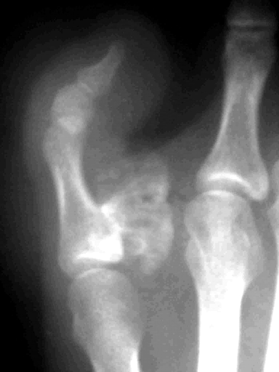
Physical examination revealed lateralization of the fifth toe with a palpable mass at the fourth web space and tenderness at the dorsolateral aspect of the fifth toe (Figs. 1 and 2). These findings were consistent with a previous displaced fracture of the phalanx or dislocation of the fifth metatarsophalangeal joint. However, radiographs showed a mass located at the medial side of the proximal phalanx of the fifth toe (Figs. 3 and 4). The mass was pedunculated, resembling an osteochondroma; however, there was no cortical and medullary continuity of the lesion. There was no vascular and neurologic deficiency due to compression of the mass.
Figure 1.
Preoperative appearance of the patient’s foot.
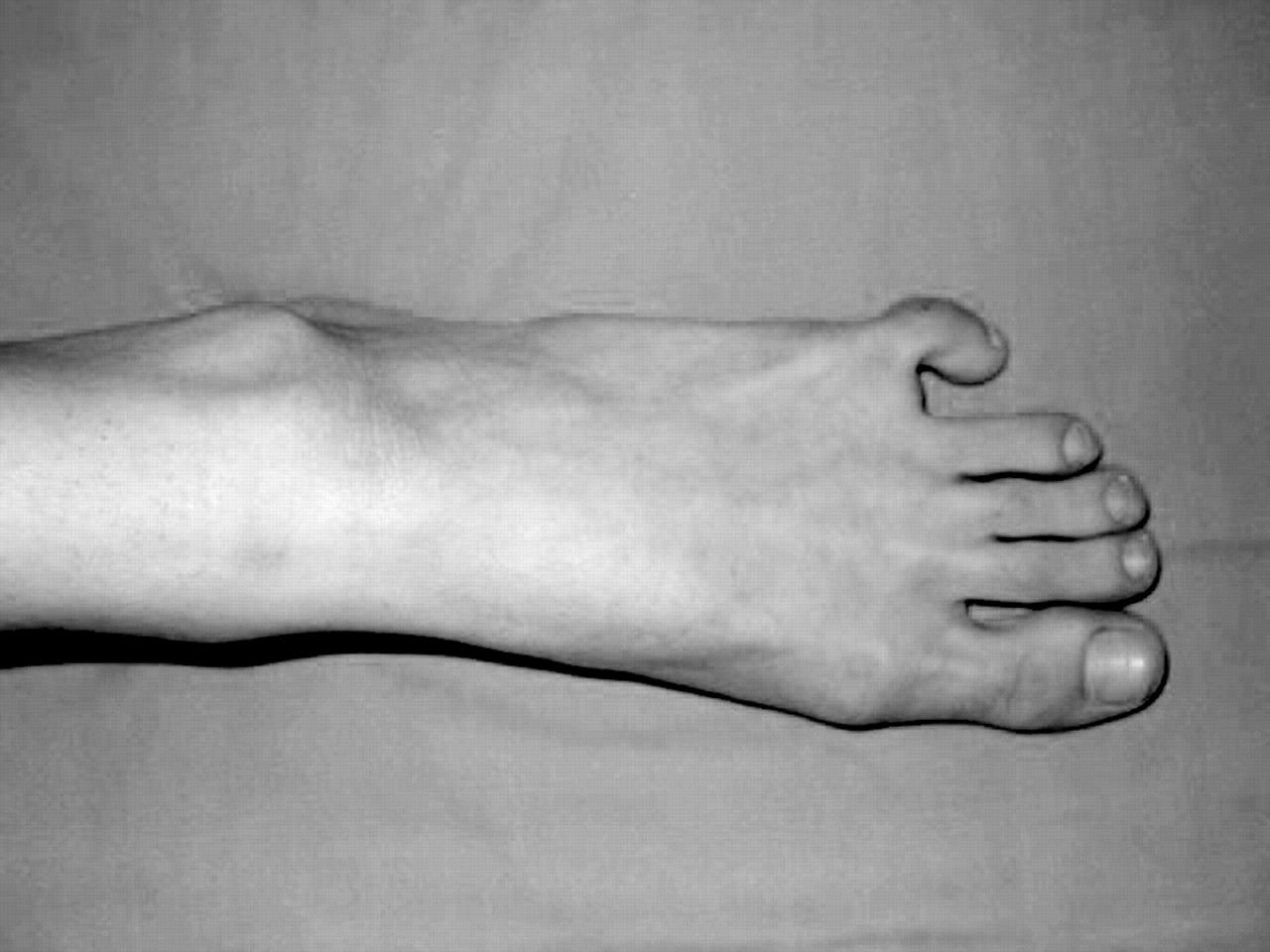
Figure 2.
Preoperative appearance of the affected toe.
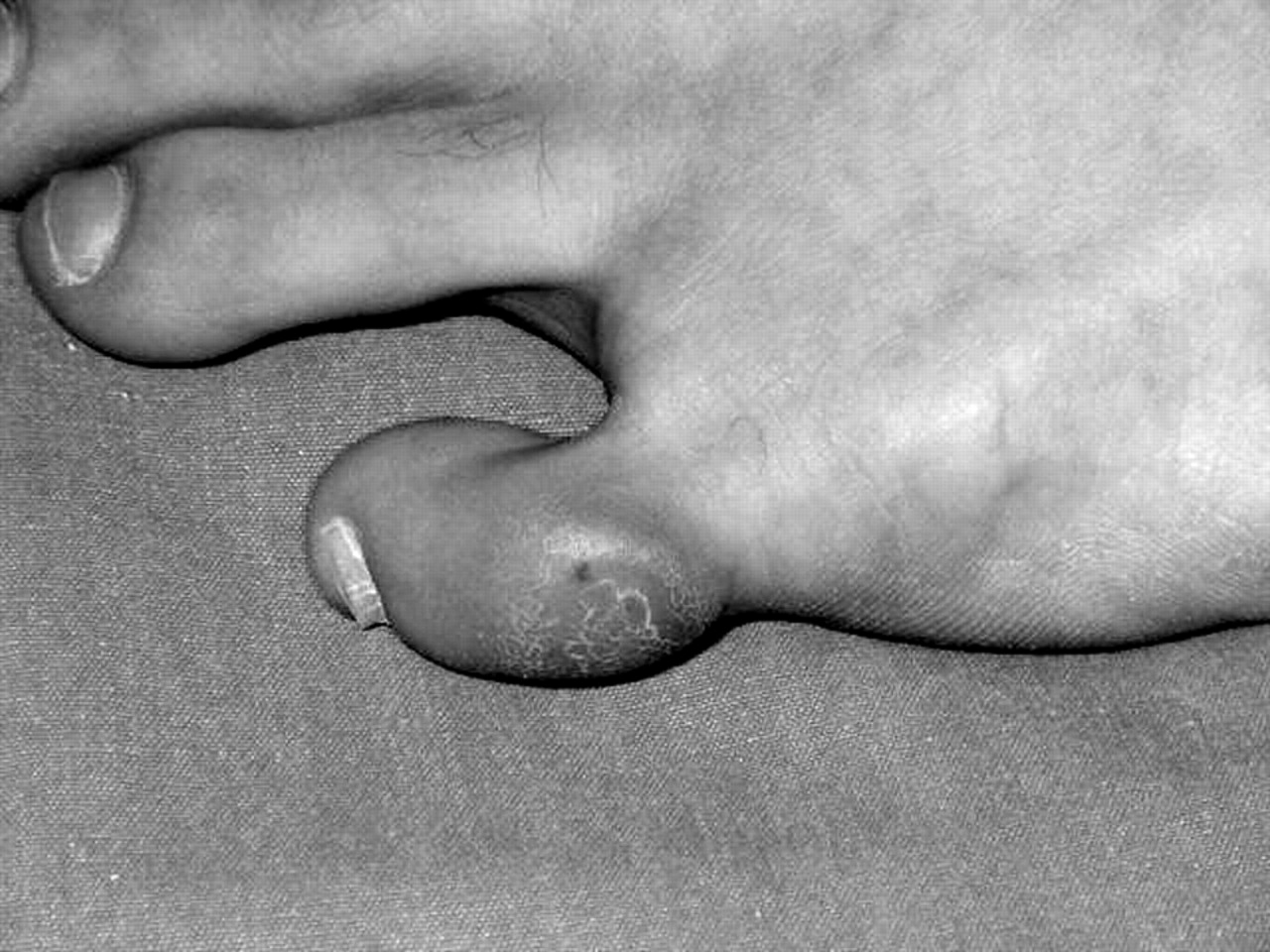
Figure 3.
Preoperative radiograph of the patient’s foot.
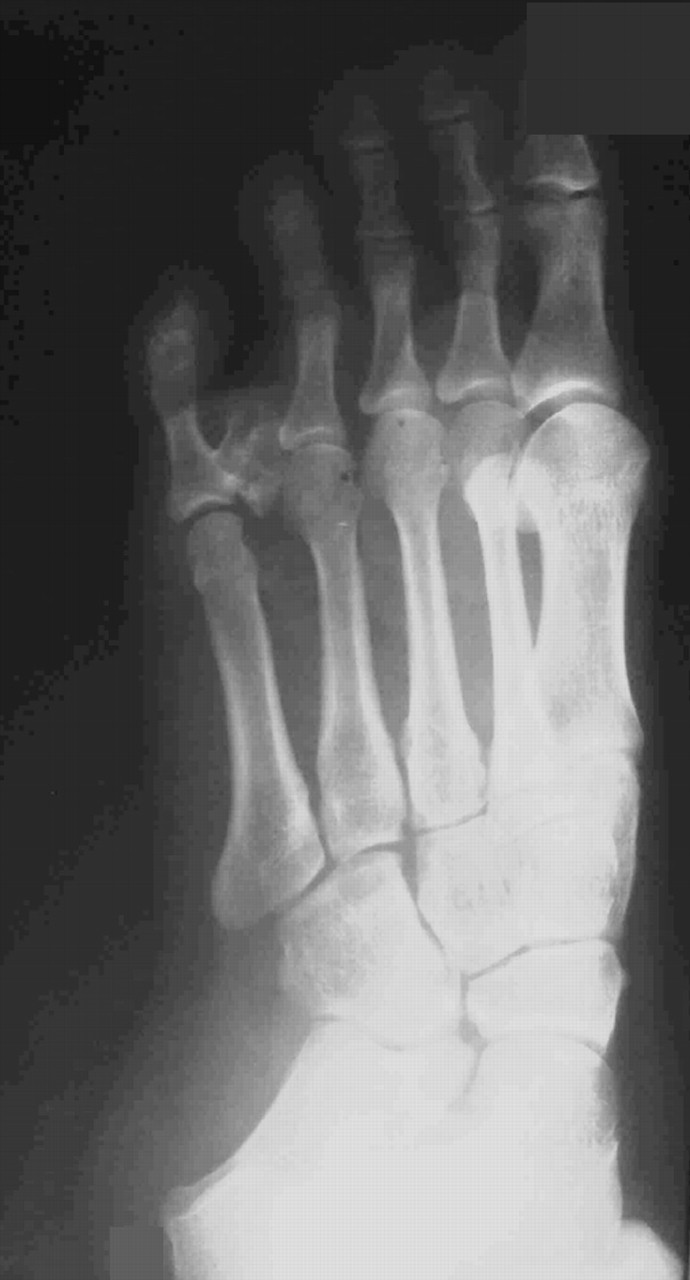
Figure 4.
Preoperative radiograph of the lesion of the affected toe.
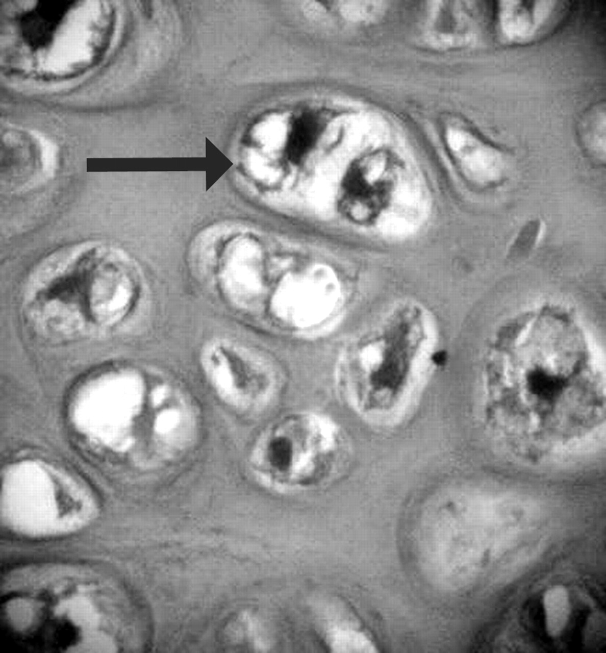
The lesion was excised. The mass measured 1.8 × 1.5 × 1.2 cm macroscopically (Fig. 5). It was pedunculated and capped by a hyaline cartilage layer microscopically, and this hyaline cartilage showed irregular growth into the trabecular bone responsible for its characteristic blue tinctorial quality in the osteochondroid interface zone (Fig. 6). The cartilaginous component of the lesion contained disordered, atypical, bizarre, and sometimes binucleated chondrocytes (Fig. 7). Calcification and ossification of cartilage with benign osteoblasts covering irregular osseous trabeculae and spindle cell stroma were present. In general, the spindle cell stroma in the intertrabecular space of the osteochondroid interface zone is an important characteristic that cannot be seen in osteochondromas (Fig. 6). Therefore, the histopathologic diagnosis was BPOP. No recurrence was seen clinically or radiologically at follow-up 30 months after surgery.
Figure 5.
The surgically excised bone mass.
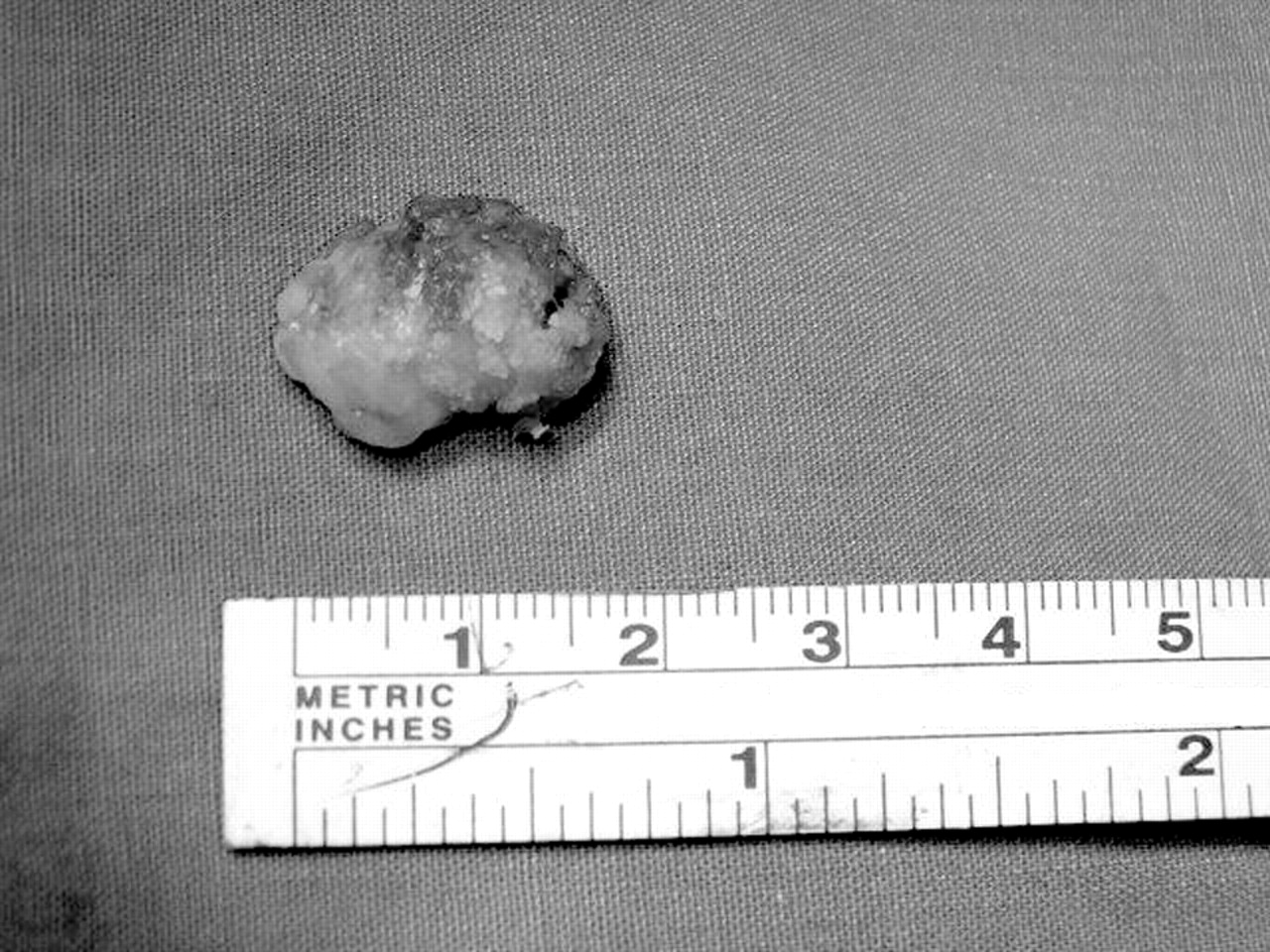
Figure 6.
The lesion is covered with a cartilage cap, closely resembling an osteochondroma, but disordered growth into the trabecular bone (giving rise to its irregular osteochondroid interface zone of blue tinctorial quality) is another diagnostic histologic sign of BPOP (H&E, ×40).
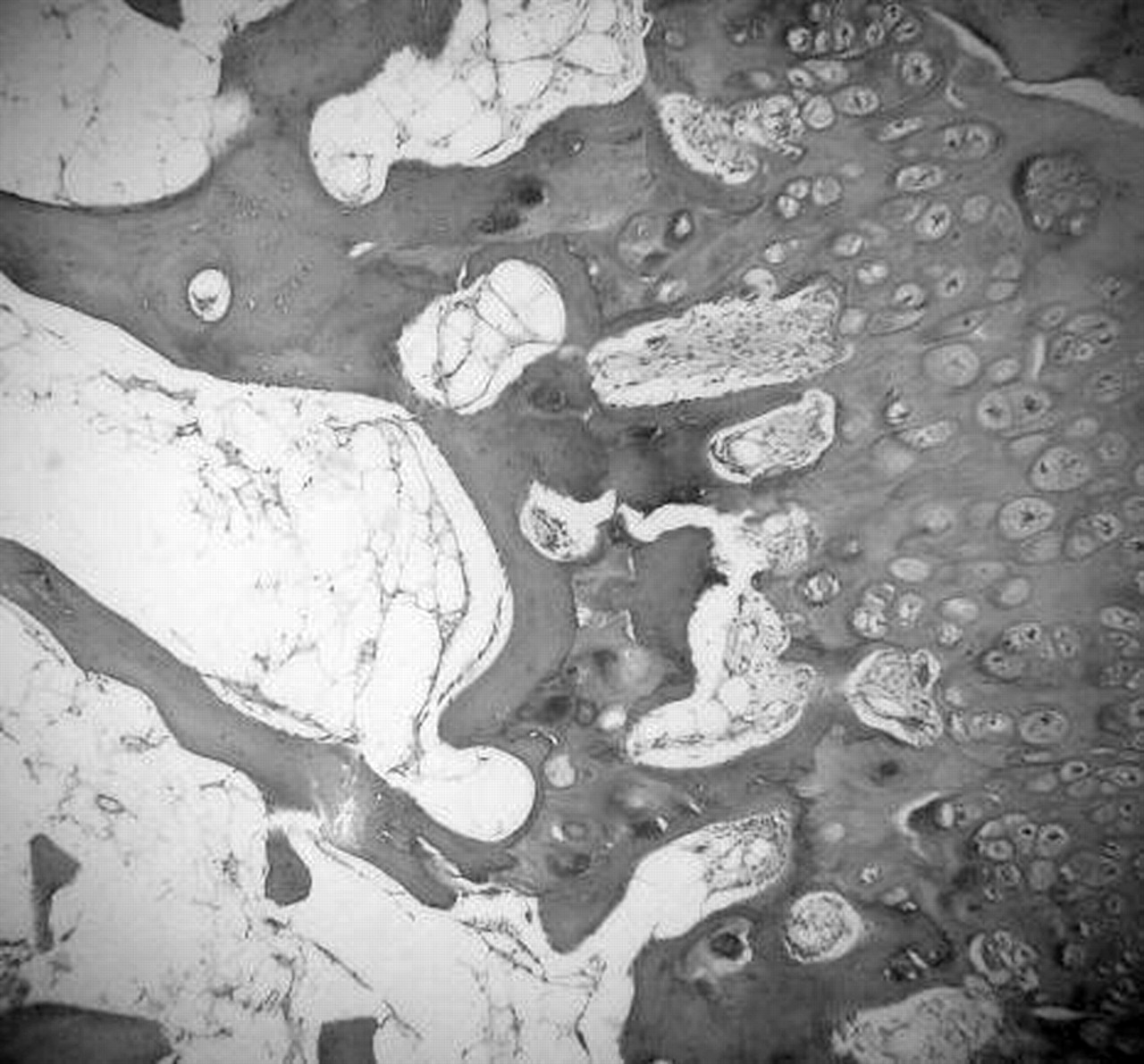
Figure 7.
The presence of atypical, bizarre, and even binucleated chondrocytes (arrow) in the cartilaginous component is an important characteristic of the lesion (H&E, ×100).
Discussion
Bizarre parosteal osteochondromatous proliferation (Nora’s lesion) was first described by Nora et al in 1983.[1] It is a rare tumor and is usually seen in adults in the second and third decades of life. The male-female ratio is equal. The most frequent locations of BPOP are the proximal and middle phalanges, metacarpals, and metatarsals. It is rarely located at the long tubular bones.
Nora’s lesions differ from other lesions on radiologic and histologic examinations. The base of the lesion is wide, without continuity between the cortex and the medullary cavity of the bone on radiologic examination.[5-9] It may be pedunculated or may arise directly from the surface of the cortex. Macroscopically, the lesion has a nodular surface covered with glistening cartilage and a grossly distinct blue tinctorial bone. Histologically, this lesion contains a disordered cartilage layer with atypical, bizarre, and sometimes binucleated chondrocytes. The chondrocytes are enlarged but lack atypia and hypochromasia. The lesion is hypercellular, with fibroblast proliferation and disorganized growth into the bone, with spindle-shaped fibroblasts in the intertrabecular spaces.[5,6] The disorganized and irregular cartilage has patchy ossification areas, unlike normal columniation of the cartilage in osteochondromas. Also, in BPOP, osteoblasts are uniformly scattered on bony trabeculae and do not show cellular atypia.
Similar to the present case, Bandiera et al[8] and Harty et al[11] described cases with masses arising from the plantar aspect of the great toe and the tibial sesamoid of the first metatarsophalangeal joint, respectively. The radiologic evaluations, although showing no continuity of the medullary canal and the cortex, suggested osteochondroma for the case described by Bandiera et al and parosteal osteosarcoma for the case described by Harty et al. However, the histologic diagnosis of the excised masses was BPOP for both.
The differential diagnosis of BPOP includes osteochondroma, turret exostosis, myositis ossificans, chondroma, chondrosarcoma, and osteosarcoma. Radiologically, osteochondroma shows continuity with the medullary cavity. Histologically, there is no cellular atypia, and chondrocytes are arranged in parallel rows.[1] Turret exostosis is a dome-shaped osseous proliferation generally seen on the dorsal aspect of the phalanx. Myositis ossificans, which characteristically presents with radiologic ossification, lacks a cartilaginous cap. As in the previous two lesions, florid reactive periostitis is related to trauma. It shows a laminar or mature periosteal reaction.[2,5,8] Periosteal chondroma, rarely seen in the feet, appears as an erosive lesion on the cortex. Chondrosarcoma presents with an infiltrative growth pattern and greater cytologic atypia than chondroma and with a popcorn-calcified lesion.[2] Osteosarcoma, which is rare in the feet, can present with a cartilaginous cap and atypical stroma. Radiologically, it appears as a heavily mineralized and sclerotic lesion.[2,5]
Bizarre parosteal osteochondromatous proliferation is a benign reactive lesion with characteristic radiologic and histologic findings. Local recurrence is common. The first-time recurrence rate is 50%, and the secondary recurrence rate is 25%. The recurrence usually occurs 2 months to 2 years after surgery.[2,5,8] The treatment of choice for a BPOP lesion is marginal excision.[7] Careful clinical, radiologic, and histologic evaluation is important in making the diagnosis of BPOP. Awareness of Nora’s lesion is necessary for accurate diagnosis and the prevention of inappropriate and destructive therapy.
References
- Nora F, Dahlin D, Beabut J: Bizarre parosteal osteochondromatous proliferation of the hands and feet. Am J Surg Pathol 7: 245, 1983.
- Campanacci M: “Pseudotumoral Soft Tissue and Periosteal Ossifications,” in Bone and Soft Tissue Tumors, p 1265, Springer-Verlag, New York. ,1999.
- Kissel CG, McQuaid M, Lucas DR, et al: Bizarre parosteal osteochondromatous proliferation: an unusual bone tumor. JAPMA 85: 301, 1995.
- Horiguchi H, Sakane M, Matsui M, et al: Bizarre parosteal osteochondromatous proliferation (Nora’s lesion) of the foot. Pathol Int 51: 816, 2001.
- Meneses MF, Unni KK, Swee RG: Bizarre parosteal osteochondromatous proliferation of the bone (Nora’s lesion). Am J Surg Pathol 17: 691, 1993.
- Smith N, Ellis A, McCarthy S, et al: Bizarre parosteal osteochondromatous proliferation: a review of seven cases. Aust N Z J Surg 66: 694, 1996.
- Davies CWT: Bizarre parosteal osteochondromatous proliferation in the hand: a case report. J Bone Joint Surg Am 67: 648, 1985.
- Bandiera S, Bacchini P, Bertoni F: Bizarre parosteal osteochondromatous proliferation of bone. Skeletal Radiol 27: 154, 1998.
- De Smet L, Lambert I, Sciot R: Bizarre parosteal osteochondromatous proliferation in the hand: report of two cases. Chir Main 20: 247, 2001.
- Hemmadi SS, Patel BR: Bizarre parosteal osteochondroma in the foot. J Postgrad Med 38: 45, 1992.
- Harty JA, Kelly P, Niall D, et al: Bizarre parosteal osteochondromatous proliferation (Nora’s lesion) of the sesamoid: a case report. Foot Ankle Int 21: 408, 2000.
© 2006 American Podiatric Medical Association