


Abstract
Measurement of large-fiber peripheral nerve function is critical to the assessment of patients with nerve injury, chronic nerve compression, and neuropathy. We evaluated the Semmes-Weinstein nylon monofilament (SWM), vibrometry, and the Pressure-Specified Sensory Device (PSSD) (Sensory Management Services LLC, Baltimore, Maryland) prospectively on the plantar surface of the hallux, bilaterally, in 35 patients with peripheral nerve problems related to nerve compression and neuropathy. Five patients had carpal tunnel syndrome and, therefore, had normal hallux measurements. Normative data for the SWM were obtained for 59 age-stratified people. A moderately strong Pearson product moment correlation was found for large-fiber nerve function between the PSSD and the SWM and between the PSSD and vibrometry. However, when these functions were compared with normative values for each neurosensory testing technique, sensitivity for detecting the presence of a peripheral nerve problem was 100% for the PSSD, 63% for the SWM, and 30% for vibrometry. False-positive test results were obtained for the hallux in 0% of normal feet when the PSSD was used, in 20% when vibrometry was used, and in 30% when the SWM was used as the test instrument. The PSSD was the most sensitive in identifying the presence of a large-fiber peripheral nerve problem in patients with pain or paresthesia in the foot related to the posterior tibial nerve.
Evaluation of the patient with chronic nerve compression or peripheral neuropathy requires the ability to measure the population of large myelinated nerve fibers that transmit neural impulses for the perception of touch. The sensation of touch is the perception of moving and static stimuli, commonly referred to as movement, vibration, touch, and pressure.[1] The devices available today to quantify these perceptions include the Semmes-Weinstein nylon monofilament (SWM), vibrometry, and the Pressure-Specified Sensory Device (PSSD) (Sensory Management Services LLC, Baltimore, Maryland).[2] Chronic nerve compression and peripheral neuropathy may or may not be associated with pain. Evaluation of the small myelinated or unmyelinated nerve fibers can be done by measuring thermal detection thresholds for hot or cold. This, however, is not usually necessary in clinical practice except for the neurologic evaluation of two rare problems: a spinal cord cyst that compresses the anterolateral spinal thalamic tract (syrinx), and neuropathy of unknown etiology.[3,4] Use of a visual analog scale appropriately quantifies the degree of pain associated with neuropathy. If a patient primarily has painful neuropathy, it is essential to evaluate the large myelinated nerve fiber population, because if the large fibers are functioning normally, the patient has a rare small-fiber neuropathy.[5] This form of neuropathy can be documented by cutaneous biopsy to evaluate intraepithelial nerve fibers.[6,7] Such a patient would not be a candidate for surgical decompression of peripheral nerves.
The patient with chronic nerve compression or peripheral neuropathy may become a candidate for surgical decompression of nerves if nonoperative treatment fails. It is, therefore, essential to be able to measure the function of the large myelinated nerve fibers before surgery to document the problem and to stage the degree of peripheral nerve dysfunction.[8] Although vibratory stimulation with a tuning fork[9] or vibrometer[10] has been demonstrated to be useful clinically in the identification of a patient with a peripheral nerve injury or nerve compression, this method of evaluating large myelinated nerve fiber function has a significant limitation. Vibration, being a waveform stimulus, will stimulate nerve fibers to the dorsal (radial sensory nerve) and volar (median nerve) surfaces of the thumb in the upper extremity and to the dorsal (peroneal nerve) and plantar (tibial nerve) surfaces of the big toe in the lower extremity. It is self-evident that a surgeon cannot direct a surgical procedure to a unique nerve if a waveform stimulus is used for the measurement. Although vibratory testing has been demonstrated to identify populations of patients with diabetic polyneuropathy and, at a threshold of 25 V, a population of patients with diabetes mellitus at risk for ulceration,[3,11–13] no study has compared the vibratory threshold of a skin surface with the cutaneous pressure threshold of the same skin surface in a series of patients with a wide spectrum of peripheral nerve problems.
This study compared the measurements obtained using a vibratory stimulus with those obtained using two different measures of touch and pressure stimuli—the SWM and the PSSD—in a series of patients with peripheral nerve problems that might benefit from surgical decompression. The null hypothesis to be tested was that measurement of the cutaneous pressure threshold for distinguishing one-point from two-point static touch stimuli will not identify the peripheral nerve problem at an earlier stage than will estimation of the cutaneous pressure threshold for one-point static touch or vibration at 128 Hz.
Materials and Methods
A consecutive series of 35 patients being evaluated for peripheral nerve symptoms in the hands, feet, or both were tested for 10 weeks during the summer of 2004. Each patient underwent bilateral testing and had the same skin surfaces tested: the index (median nerve) and little (ulnar nerve) finger pulp, the pulp of the hallux (tibial nerve), and the dorsal web space of the foot between the first and second toes (deep peroneal nerve). A single examiner (E.A.) used the PSSD to obtain measurements. Another examiner (H.R.) who was blinded to the measurements obtained using the PSSD used the SWM and the vibrometer to obtain measurements. Data analysis was performed by a third person (G.D.R.). Only data for the hallux are reported in this article. Five patients had carpal tunnel syndrome and no symptoms in their feet, and these patients (ten toes) are considered in the “hallux normal” group for statistical analysis.
The PSSD testing was performed with the patient in a relaxed position in a chair in a quiet recovery room, separate from the room in which the other sensibility testing was performed. The patient had 15 min of test-free time between the PSSD testing and the subsequent testing. The SWM testing was performed using a new set of 20 monofilaments (Touch-Test Sensory Evaluators; North Coast Medical, Morgan Hill, California). The vibrometer was the Bio-Thesiometer (Bio-Medical Instrument Co, Newbury, Ohio), a single-frequency (128-Hz), variable-intensity instrument. The SWM and vibrometer testing was performed with the patient in a comfortable, reclining chair. The SWM testing was performed before the vibrometer testing to minimize any effect of residual “buzzing” in the skin of the test site on subsequent testing.
The inclusion criterion was referral to the senior author (A.L.D.) for evaluation of a peripheral nerve problem. Therefore, we included patients with upper (5 patients with carpal tunnel syndrome) and lower (4 patients with tarsal tunnel syndrome) nerve compression problems and those with peripheral neuropathy (20 patients with diabetic neuropathy and 6 with neuropathy of unknown etiology). The population included patients who were early in the course of their clinical problem as well as late in the course, thereby presenting the full spectrum of abnormal large myelinated nerve fiber function for evaluation. Criteria for exclusion were central nervous system disease, cognitive impairment related to neuropathic pain medications or narcotics, and post-traumatic or iatrogenic painful neuromas.
Normative data for the PSSD were taken from published age-adjusted values for the upper[14] and lower[15] extremities. Normative data for the Bio-Thesiometer were taken from data supplied by the manufacturer. Values for grading the results of the SWM testing were taken from the hand-rehabilitation literature[16] and from a recent study of data for the foot.[17] Because the latter study included only college-aged subjects, and patients with neuropathy are an older population, new normative data were obtained. Fifty-nine people aged 18 to 86 years were evaluated (11 were aged 18–29 years, 7 were aged 30–39 years, 11 were aged 40–49 years, 8 were aged 50–59 years, 11 were aged 60–69 years, and 11 were aged ≥70 years). None of the participants had a known history of peripheral nerve injury, nerve compression, peripheral neuropathy, or systemic disease associated with neural dysfunction (diabetes mellitus, thyroid disease, lupus, or renal disease). Study participants were patients from the clinical practice of one author (J.S.). These patients each had a unilateral foot problem related to a forefoot problem, with a noninvolved contralateral foot. The measurements were made on the contralateral foot. The population consisted of 14 men and 45 women with a mean ± SD age of 51 ± 17.6 years. Twenty-two left feet and 37 right feet were tested.
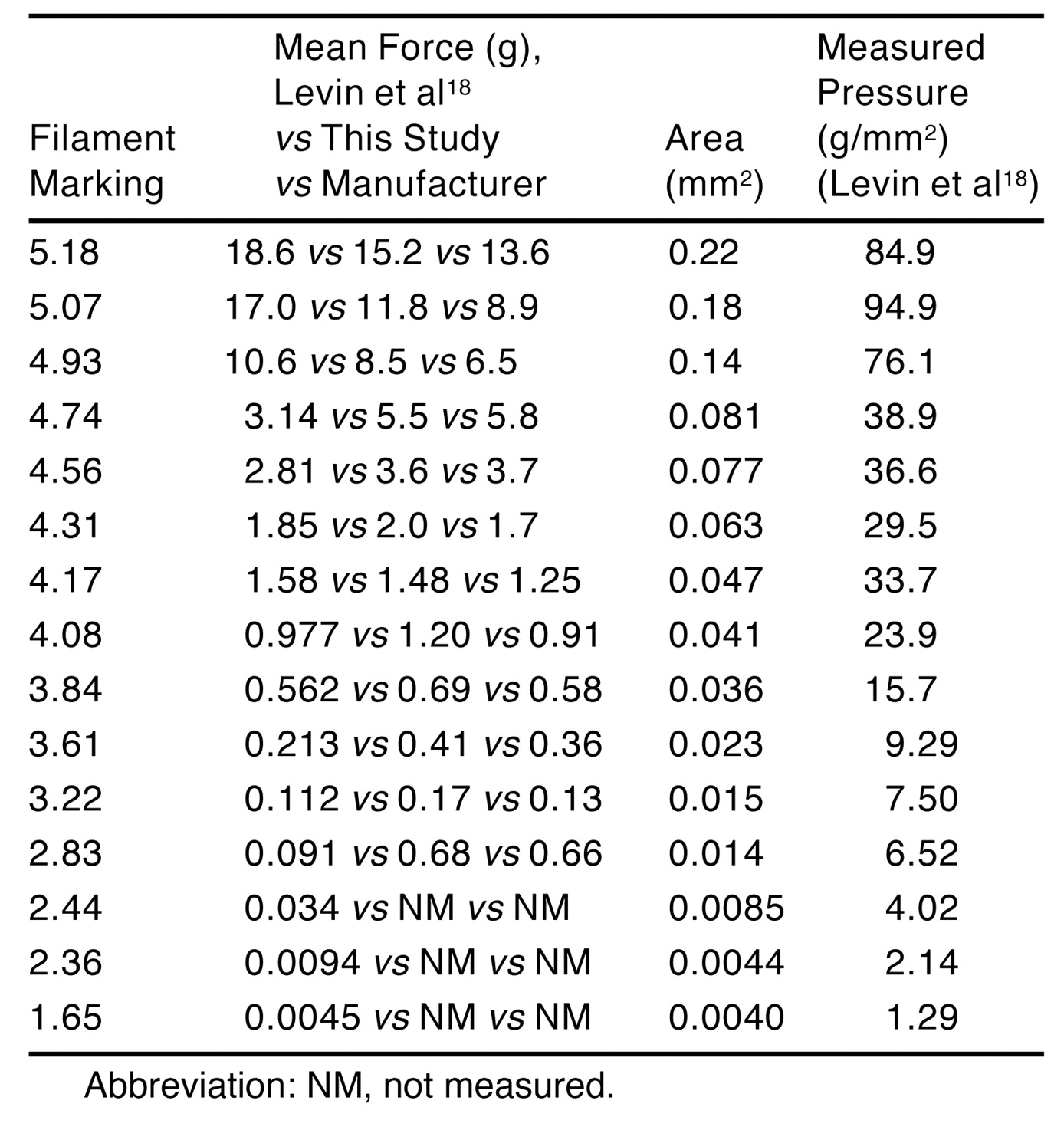
A 20-piece SWM set was used to evaluate five sites on the foot (the pulp of the big toe and the little toe, the first metatarsal head, the medial heel, and the dorsal first/second web space) for each patient. Testing was conducted in a quiet, distraction-free room with the leg resting comfortably on a padded chair. The filaments were applied perpendicular to the skin surface until buckling occurred, were held in place for approximately 1.5 sec, and were then removed. The filaments were applied in order of decreasing stiffness, beginning with the 5.07 marking. Per the manufacturer’s instructions, monofilaments were applied at the same location up to three times to elicit a positive response. Care was taken to avoid areas of excessive callus formation. The last filament size (the lowest SWM marking) that the subject could identify and locate correctly was recorded as the cutaneous (force) threshold for that site. The filaments used in this study were independently calibrated by measuring their buckling force directly using a Mettler electronic scale (accurate to the 0.001-mg level) in the pharmacy department of Union Memorial Hospital, Baltimore, Maryland by obtaining the average of three force recordings. This mean force is given for each filament in the set in Table 1, which includes the force analysis of a set of filaments by Levin et al.[18] For statistical analysis, the SWM markings were not converted to pressure (grams per square millimeter).
Data analysis was performed by comparing the three different test results for each skin area tested. For each skin surface tested, it was determined whether the data from the SWM, the vibrometer, or the PSSD were abnormal as a method of determining which of the three test results became abnormal first. Data analysis was also performed by comparing, for a given skin surface, the degree of correlation between the measurement from the SWM versus the PSSD and the vibrometer versus the PSSD. The coefficient of correlation was calculated.

Table 1.
Force Measurements for Semmes-Weinstein Monofilaments.
Results
The age-related normative data for the five sites of the foot tested using the SWM are given in Table 2: the big toe pulp (medial plantar nerve), the heel (medial calcaneal nerve), the dorsal first/second web space (deep peroneal nerve), the first metatarsal head (medial plantar nerve), and the fifth toe pulp (lateral plantar nerve). These data demonstrate that for each site tested there is a wide range of filament markings within each age group but that, in general, the numeric value of the markings increases with age. Each skin site can be characterized statistically by the mode (the filament marking to which the greatest number of patients responded). Using this criterion, the big toe pulp and the first dorsal web space were the most sensitive, with a marking of 3.22. The least sensitive skin surface by this criterion would be the heel, with a marking of 4.17. The other two sites were intermediary, with markings for the mode of 3.61. Defining normal, for example, for the big toe pulp for all ages, by giving the 90th percentile (not confidence limit) as the upper limit would mean that anyone who could perceive the 4.56 filament marking would be considered normal. For the age range 40 to 69 years, the age range of the patients in the present study, the 90th percentile filament marking would still be 4.56. Defining normal, for example, for the dorsal first web space for all ages, by giving the 90th percentile (not confidence limit) as the upper limit would mean that anyone who could perceive the 4.08 filament marking would be considered normal. For the age range 40 to 69 years, the age range of the patients in the present study, the 90th percentile filament marking would still be 4.08.
Table 2.
Semmes-Weinstein Monofilament Normal Data.
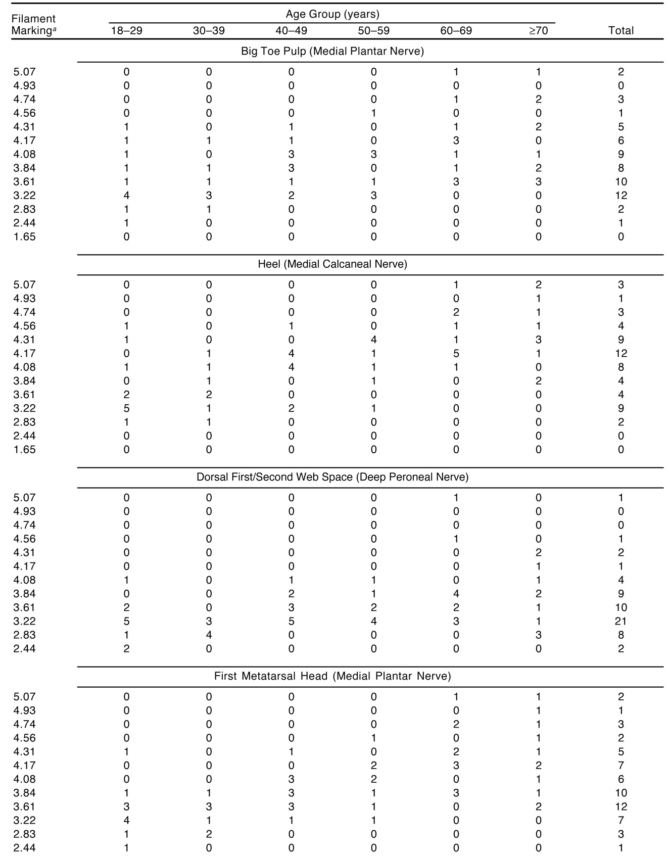
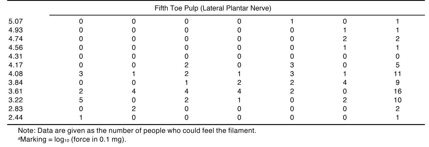
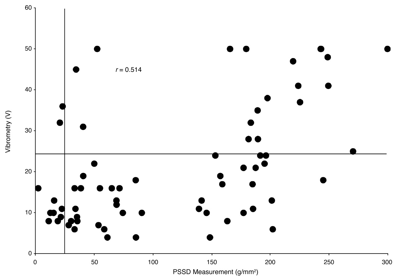
Figure 1 presents the data for the PSSD measurements versus those obtained using the SWM. Figure 2 presents the data for the PSSD measurements versus those obtained using the vibrometer. With the PSSD measurements recorded on the x-axis, the vertical line represents the 99% upper limit of normal for an individual 45 years of age. In Figure 1, with the SWM measurements recorded on the y-axis, the horizontal line represents the 90th percentile for the SWM. In Figure 2, with the vibrometry measurements recorded on the y-axis, the horizontal line represents the upper limit of normal for the vibrometer. Analysis of the number of points above the horizontal line in Figure 1 and Figure 2 demonstrates the number of patients found to have abnormal measurements as obtained with the SWM and vibrometry, respectively. Analysis of the number of points to the right of the vertical line in Figure 1 and Figure 2 demonstrates the number of patients found to have abnormal measurements using the PSSD.
Statistical analysis for sensitivity and specificity using the data in Figure 1 and Figure 2 demonstrates a sensitivity for the PSSD of 100%, for the SWM of 63%, and for vibrometry of 30%. The specificity for the PSSD is 100%, for the SWM is 70%, and for vibrometry is 80%. Using χ2 analysis, the PSSD was found to be significantly more sensitive than the SWM and vibrometry (P < .001 for both) in identifying patients with abnormal large-fiber function in the posterior tibial nerve. The SWM is more significantly able to identify abnormal large-fiber function than vibrometry in the posterior tibial nerve (P < .001).
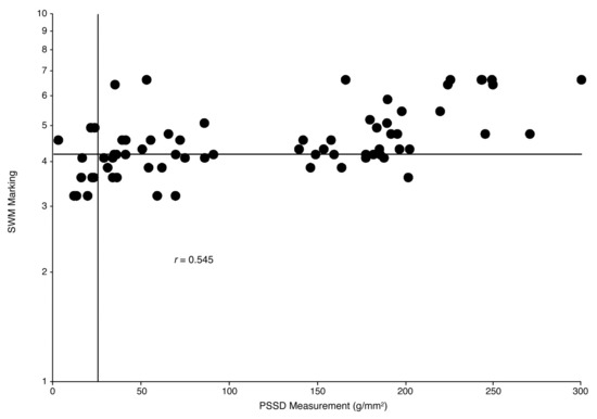
Figure 1.
Relationship between Pressure-Specified Sensory Device (PSSD) measurements and Semmes-Weinstein monofilament (SWM) estimates for the hallux. The SWM values below the horizontal line are considered normal, and the PSSD values to the left of the vertical line are considered normal. The ten dots to the left of the vertical line represent hallux values for normal feet, so that those dots in the bottom left quadrant of the graph represent measurements for those without disease and correctly identified as such by the PSSD and the SWM. Those dots in the upper right quadrant of the graph represent measurements that are abnormal by the PSSD and the SWM for patients with a peripheral nerve problem. Those dots in the lower right quadrant of the graph represent patients for whom the PSSD identified abnormal sensibility but the SWM did not. In the upper left are patients found to have a normal measurement using the PSSD who were normal but for whom the SWM test demonstrated an abnormality.
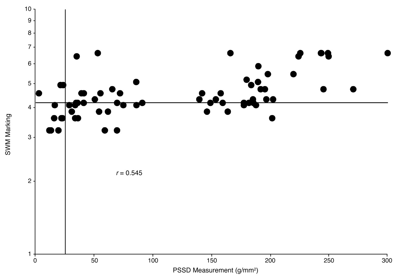
Figure 2.
Relationship between Pressure-Specified Sensory Device (PSSD) measurements and vibrometry findings for the hallux. Vibrometry values below the horizontal line are considered normal, and PSSD values to the left of the vertical line are considered normal. The ten dots to the left of the vertical line represent hallux values for normal feet, so that those dots in the bottom left quadrant of the graph represent measurements for those without disease and correctly identified as such by the PSSD and vibrometry. Those dots in the upper right quadrant of the graph represent measurements that are abnormal by the PSSD and vibrometry for patients with a peripheral nerve problem. Those dots in the lower right quadrant of the graph represent patients for whom the PSSD identified abnormal sensibility but vibrometry did not. In the upper left are patients found to have a normal measurement using the PSSD who were normal but for whom vibrometry demonstrated an abnormality.
Discussion
The results of this study demonstrate that the first change in large myelinated nerve fiber function that can be demonstrated for the posterior tibial nerve is a change in the pressure required to discriminate one-point from two-point static touch stimuli, as measured using the PSSD. The cutaneous vibratory threshold obtained using the vibrometer and the estimate of the cutaneous pressure threshold for one-point static touch obtained using the SWM identified significantly fewer patients as having abnormal measurements (P < .001), demonstrating that these testing modalities are less sensitive than the PSSD in identifying the presence of a peripheral nerve problem in a patient with symptoms related to the tibial nerve.
The results of this study are not surprising regarding the inability of the SWM to identify early changes in nerve compression or neuropathy. The filaments have been repeatedly criticized with regard to 1) the problem with their being interpreted as measuring a pressure threshold, when they actually measure a force threshold; 2) the statistical problem of the markings not being treated as logarithms; 3) the inability to know the true surface area in contact with the skin at the moment the filament buckles; 4) the variance in manufacturing that gives a 10% error in the initial applied force of new filaments; 5) the fatigue of the nylon with more than 100 uses, which causes a 10% change in the applied force; and 6) the fact that the SWM does not make a true measurement but, rather, gives an estimate of a range of the given skin surface tested.[18–21] These problems are compounded by the fact that it is not statistically possible to state a mean or a 99% confidence interval for a testing device that does not make a measurement, so that normative data for the SWM for the foot do not exist in the scientific literature.
The results of this study show a neurophysiologic correlation between the PSSD and the SWM for large-fiber function, with a Pearson product moment correlation coefficient of 0.545, which is significant at P < .001. However, when these data are plotted against normative data to obtain a relationship that is clinically relevant, the interpretation of these data changes. The outcome of this study—that the results of using SWM to evaluate a piece of skin do not correlate clinically with the results of using the PSSD to evaluate that same piece of skin—confirms the results of the only previous study[22] to compare these two instruments. A psychophysical explanation for this lack of clinical correlation is that the varying cross-sectional area of the nylon filaments means that each time a patient is tested, the patient is really being asked two questions: can you detect a difference in cross-sectional area between the two stimuli, and can you detect a difference in force between the two stimuli? In contrast, the cross-sectional area of the hemispheric metal prong of the PSSD is constant, and only the force of application changes. Another reason to expect these two instruments not to clinically correlate is that a regression analysis of the first measurable PSSD variable to change in patients with chronic nerve compression is the threshold for two-point static touch.[14] Therefore, even if the same information were to be given by the SWM and the one-point static touch mode of testing for the PSSD, the one-point static touch variable would not be expected to identify the earliest changes.
The normative data reported in this study for the SWM deserve comment. Two previous studies[22,23] used 20 nondiabetic patients older than 60 years as a control group in their evaluation of patients with diabetic neuropathy at risk for ulceration. One study used the SWM to test the feet of 40 college-aged volunteers.[17] The present group of patients was aged 18 to 86 years. In the present study, the data were interpreted after plotting individual patient SWM estimates for a threshold and calculating the 90% level for a healthy person to respond to the filament. This approach recognizes that the SWM gives an estimate of a range and cannot, therefore, have the logarithmic markings used to give either a mean value or a 99% upper confidence limit as a normative value.
Of the three test techniques evaluated in the present study, the poorest performer in determining whether a patient has a specific peripheral nerve problem was vibrometry, probably owing to the inherent problem with using a waveform to stimulate tissue. The wave travels through the hallux and stimulates the peroneal and tibial nerve innervations of this tissue. Furthermore, the wave travels to the lateral side of the foot and stimulates the lateral plantar and medial plantar nerves. The results of this study suggest that although vibratory testing is appropriate for studying cohorts of patients with a problem compared with other cohorts of patients (eg, patients with diabetes with and without neuropathy), vibrometry is not the best test to use to attempt to distinguish a peroneal nerve problem from a tibial nerve problem in the hallux.
Conclusion
The PSSD is significantly more sensitive than the SWM or vibrometry in identifying peripheral nerve problems in the hallux pulp.
Acknowledgment
James J. Zulty, PhD, RPH, Medstar Hospital Pharmacy at Union Memorial Hospital in Baltimore, Maryland, for his help with the calibration of the nylon monofilaments; Sam Spadone, DPM, Temple University School of Podiatric Medicine, Philadelphia, Pennsylvania, for statistical consultation regarding normative data calculations for the Semmes-Weinstein nylon monofilaments.
References
- Dellon AL: Evaluation of Sensibility and Re-education of Sensation in the Hand, Williams & Wilkins, Baltimore. ,1981. .
- Dellon AL: “Instrumentation,” in Somatosensory Testing and Rehabilitation, p 98, American Occupational Therapy Association, Bethesda, MD. ,1997. .
- Valk GD, Grootenhuis PA, van Eijk JT, et al: Methods for assessing diabetic polyneuropathy: validity and reproducibility of the measurement of sensory symptom severity and nerve function tests. .Diabetes Res Clin Pract47::87. ,2000. .
- Mendell JR, Sahenk Z: Painful sensory neuropathy. .N Engl J Med348::1243. ,2003. .
- Magda P, Latov N, Renard MV, et al: Quantitative sensory testing: high sensitivity in small fiber neuropathy with normal NCS/EMG. .J Peripher Nerv Syst7::225. ,2002. .
- Hermann DN, Griffin JW, Hauer P, et al: Intraepidermal nerve fiber density and sural nerve morphometry in peripheral neuropathies. .Neurology53::1634. ,1999. .
- Periquet MW, Novak V, Collins MP, et al: Painful sensory neuropathy: prospective evaluation using skin biopsy. .Neurology53::1641. ,1999. .
- Dellon AL: Clinical grading of peripheral nerve problems. .Neurosurg Clin N Am12::229. ,2001. .
- Dellon AL: Clinical use of vibratory stimuli to evaluate peripheral nerve injury and compression neuropathy. .Plast Reconstr Surg65::466. ,1980. .
- Dellon AL: The vibrometer. .Plast Reconstr Surg71::427. ,1983. .
- Maser RE, Viggo KN, Bass EB, et al: Measuring diabetic neuropathy: assessment and comparison of clinical examination and quantitative sensory testing. .Diabetes Care12::270. ,1989. .
- Young MJ, Breddy JL, Veves A, et al: The prediction of diabetic neuropathy foot ulceration using vibration perception thresholds: a prospective study. .Diabetes Care17::557. ,1994. .
- Coppini DV, Young PJ, Weng C, et al: Outcome on diabetic foot complications in relation to clinical examination and quantitative sensory testing: a case-control study. .Diabet Med15::765. ,1998. .
- Dellon AL, Keller K: Computer-assisted quantitative sensorimotor testing in patients with carpal and cubital tunnel syndrome. .Ann Plast Surg38::493. ,1997. .
- Tassler PL, Dellon AL: Pressure perception in the normal lower extremity and in the tarsal tunnel syndrome. .Muscle Nerve19::285. ,1996. .
- Bell JA: “Light Touch–Deep Pressure Testing Using Semmes-Weinstein Monofilaments,” in Rehabilitation of the Hand, ed by JM Hunter, LH Schneider, EJ Macklin, et al, p 399, CV Mosby, Philadelphia. ,1984. .
- Jeng C, Michelson J, Mizel M: Sensory thresholds of normal human feet. .Foot Ankle Int21::501. ,2000. .
- Levin S, Pearsall G, Ruderman RJ: von Frey’s method of measuring pressure sensibility in the hand: an engineering analysis of the Weinstein-Semmes pressure aesthesiometer. .J Hand Surg [Am]3::211. ,1978. .
- McGill M, Molyneaux L, Spencer R, et al: Possible sources of discrepancies in the use of the Semmes-Weinstein monofilament: impact on prevalence of insensate foot and workload requirements. .Diabetes Care22::598. ,1999. .
- Booth J, Young MJ: Differences in the performance of commercially available 10-g monofilaments. .Diabetes Care23::984. ,2000. .
- Dellon AL, Mackinnon SE, Brandt KE: The markings of the Semmes-Weinstein nylon monofilaments. .J Hand Surg [Am]18::756. ,1993. .
- Holewski JJ, Stess RM, Graf PM, et al: Aesthesiometry: quantification of cutaneous pressure sensation in diabetic peripheral neuropathy. .J Rehabil Res Dev25::1. ,1988. .
- Rith-Najarian SJ, Stolusky T, Gohdes DM: Identifying diabetic patients at high risk for lower-extremity amputation in a primary health care setting: a prospective evaluation of simple screening criteria. .Diabetes Care15::1386. ,1992. .
© 2005 American Podiatric Medical Association