


Abstract
Onychomycosis, most commonly caused by two species of dermatophyte fungi—Trichophyton rubrum and Trichophyton mentagrophytes—is primarily treated with regimens of topical and systemic antifungal medications. This study was undertaken to evaluate in vitro the efficacy of low-voltage direct current as an antifungal agent for treating onychomycosis. Agar plate cultures of T rubrum and T mentagrophytes were subjected to low-voltage direct current electrostimulation, and antifungal effects were observed as zones in the agar around the electrodes lacking fungal growth. Zones devoid of fungal growth were observed for T rubrum and T mentagrophytes around anodes and cathodes in a dose-dependent manner in the current range of 500 μA to 3 mA. Low-voltage direct current electrostimulation has great clinical potential for the treatment of onychomycosis and perhaps other superficial maladies of fungal etiology.
Onychomycosis is one of the most common problems encountered by podiatric physicians. Onychomycosis is a fungal infection of one or more elements of the nail system, which consists of the nail matrix, the nail bed, and the nail plate.[1] Several studies[1,2,3,4] suggest that onychomycosis affects 2% to 18% of the world’s population. In North America, onychomycosis accounts for approximately 50% of all nail disease, is several times more common in the toenail than in the fingernail, and is most commonly found in older individuals (some studies suggest that nearly 50% of people older than 70 years may be affected).[4] The incidence of onychomycosis in the United States and other developed countries has been increasing in recent years. This increase is thought to have several contributing factors, including the general aging of the population, the increasing incidence of diabetes mellitus, the greater use of immunosuppressive drugs and antibiotics, the increased exposure of the general population to the etiologic fungi, and the spread of the human immunodeficiency virus.[4,5]
Onychomycosis can be caused by three different groups of fungi: dermatophytes, yeasts, and nondermatophytic molds.[6,7] Dermatophytes are the most common etiology, accounting for 85% to 90% of all cases.[8] Only two dermatophyte species—Trichophyton rubrum and Trichophyton mentagrophytes—are believed to be responsible for nearly 80% of all cases of onychomycosis. Several different yeast species can also cause onychomycosis. These species together are responsible for 5% to 10% of all cases. In approximately 70% of these cases, the etiologic agent is the yeast Candida albicans. Finally, several different species of the nondermatophyte molds can cause onychomycosis. As a group, these molds are responsible for approximately 3% to 5% of all cases.
Although onychomycosis is not a fatal infection or a very debilitating condition in most afflicted individuals, it can have serious emotional and physical consequences.[9] The condition can be associated with significant pain and discomfort, and in severe cases it may lead to disfigurement and various degrees of functional loss. In addition to physical impairment, the psychological and social consequences of onychomycosis can be significant.[10] Thus onychomycosis represents more than a mere cosmetic problem for many afflicted individuals, and treatment from health-care providers is often sought.
The treatment of onychomycosis has historically proved difficult.[1] The three traditional approaches to treatment are debridement of the nail unit, application of topical medication, and systemic antifungal agents.[11,12] The most successful of these approaches has been the use of systemic antifungal drugs. During the past 40 years, oral systemic antifungal agents have been the mainstay of onychomycosis therapy.[1,4,5] However, because of several negative factors,[1] including drug toxicity, potential adverse interactions of antifungal agents with other drugs, and the prolonged course of treatment required with many antifungal therapeutic regimens, the search for new, alternative treatments that are efficacious and have minimal side effects is still an important research goal.
During the past 10 years, new treatment protocols that use electrostimulation have begun to be used in the treatment of certain types of wounds.[13] The clinical efficacy of such treatment is now well documented.[13,14] One of the ways in which electrostimulation probably enhances wound healing is by its antimicrobial effect. Several in vitro studies have conclusively demonstrated that application of electric current is antibacterial to most of the pathogenic bacteria commonly found in wounds.[15,16] In the course of our own clinical use of low-voltage direct current (LVDC) electrostimulation in wound treatment during the past decade, we noticed that in addition to its enhancement of the wound-healing process, it also seemed to significantly ameliorate many cases of onychomycosis of the toenail (Michael S. Brogan, MS, DPT, personal communication, 1995). Subsequently, without yet knowing the reasons for its efficacy, we began to use this modality in the treatment of onychomycosis, with very good clinical results (Michael S. Brogan, MS, DPT, personal communication, 1995).
This study was conducted to begin determining the factors responsible for the observed efficacy of this modality in the treatment of onychomycosis. Principally, we performed in vitro experiments to evaluate the antifungal effects of LVDC electrostimulation on the two primary etiologies of onychomycosis: T rubrum and T mentagrophytes. In this article, we describe the methods used and the results obtained, and we discuss the potential for the clinical use of LVDC electrostimulation in the treatment of onychomycosis in conjunction with or as an alternative to current antifungal drug regimens.
Materials and Methods
The medium used to culture all fungi was Sabouraud dextrose agar in 100-mL Petri plates (Becton Dickinson Microbiology Systems, Cockeysville, Maryland). Pure cultures of the dermatophyte fungi T rubrum and T mentagrophytes were obtained from Presque Isle Cultures (Presque Isle, Pennsylvania). Permanent stock cultures of T rubrum were established by inoculation of the organism onto a solid growth medium consisting of Sabouraud dextrose agar in Petri plates and subsequent incubation of these plates at 25°C for 7 days. Each plate was then covered by numerous colonies of T rubrum that had coalesced into a homogeneous, fluffy, fungal “lawn” across the entire surface of the agar. Permanent stock cultures of T mentagrophytes were established in the same manner. The stock cultures of both fungi were maintained at 4°C throughout the experiments and were monitored weekly for viability.
All of the LVDC electrostimulation experiments described in this article were performed in a NuAire Class II, Type A/B3 biological safety cabinet (NuAire, Inc, Plymouth, Minnesota) using standard aseptic techniques. All LVDC electrostimulation was performed using a Rich-Mar VI LIDC Stimulator (Inola, Oklahoma). Independent current measurements were made using a BK Precision ampmeter (Dynascan Corp, Chicago, Illinois).
For each electrostimulation experiment, a 1.0-cm2 section of agar containing either T rubrum or T mentagrophytes growing on the surface was removed aseptically from the respective stock culture plates using a sterile dissecting needle. This piece of agar was then transferred to 5 mL of sterile 0.9% saline. The sterile saline tube was mixed by vortexing for 10 sec to dislodge the fungal hyphae, conidia, and spores from the surface of the agar. Using a sterile micropipette, 250 mL of the fungal-saline solution was transferred to a new, sterile Sabouraud dextrose agar Petri plate. If multiple plates were to be inoculated in a given experiment, this procedure was performed multiple times from the same fungal-saline solution. The fungal-saline solution was evenly distributed over the entire surface of each of the inoculated plates using a sterile, glass spreading rod. The plates were then incubated at 25°C for 24 hours. At the end of this period, a barely visible film of fungal growth covered the entire growth medium surface. Noninoculated control plates were included in each experimental group to ensure the sterility of the medium.
After 24 hours of incubation, electric current was applied to each experimental plate containing either T rubrum or T mentagrophytes using a Rich-Mar VI LIDC Stimulator. The electrodes used to accomplish this consisted of two pieces of stainless steel (1 mm in diameter and 2.5 cm long) permanently inserted through the top portion of a sterile Petri plate 1.9 cm apart and secured with epoxy cement on the outer surface. The electrodes were disinfected and stored in 95% ethanol before and between experiments. Just before each experiment, the top portion containing the electrodes was removed from the 95% ethanol storage unit, the ethanol was allowed to evaporate, and the electrodes were inserted into the agar in the bottom portion of a plate containing a 24-hour growth of either fungus by closing the top portion over the lower portion of a Petri plate. Then, either the electrodes remained in the agar for 30 min at room temperature (22° to 24 °C) without current application (0 A) or electric current was applied using the electro-stimulation apparatus described previously. A current of 500 μA, 1 mA, 2 mA, or 3 mA of LVDC was applied for 30 min at room temperature (22° to 24 °C). All amperages were confirmed by the use of an independent ampmeter connected in series. In each experiment, to confirm fungal viability and media soundness, a control plate containing the particular fungus used in the experiment but not subjected to either electrostimulation or electrode intrusion was also included. This plate was inoculated at the same time, in the same manner, and from the same saline tube as the experimental plates.
After LVDC electrostimulation, each Petri plate was incubated at 25°C for 24 hours to allow for additional fungal growth, which could then be easily visualized. After this 24-hour incubation, the diameter of any zone lacking fungal growth at the positive and negative electrodes was measured to the nearest 0.1 mm using a millimeter ruler and a dissecting microscope. The plates were then incubated for an additional 3 to 5 days, with additional observations and measurements made daily.
To determine whether the electrode material itself was toxic to either of the two fungi, or if any ethanol (used in disinfection) remained on the electrodes during the electrostimulation application (which might kill the fungus or inhibit its growth), Sabouraud dextrose agar plates were inoculated with either T rubrum or T mentagrophytes and were incubated for 24 hours. Following incubation, the electrodes were inserted into the plate containing either of the fungi and allowed to remain for 30 min, but no electric current was applied (0 A). The plates were then incubated for 7 days and were checked for the presence of any zones lacking fungal growth during each of these days.
To determine whether any toxic or inhibitory antifungal metabolites were generated in the Sabouraud dextrose agar growth medium as a result of the application of electric current, LVDC of either 3 or 8 mA was applied to plates for 30 min before inoculation of either T rubrum or T mentagrophytes. Immediately after this procedure, 250 mL of fungal-saline solution of either T rubrum or T mentagrophytes was evenly distributed over the surface. The plates were then incubated at 25°C for 7 days, and the presence of any zones lacking fungal growth near the site of the electrode contacts (or elsewhere) was observed and measured each day.
To determine whether LVDC electrostimulation acts primarily in a fungistatic manner (inhibits fungal growth but does not kill fungal cells) or a fungicidal manner (kills fungal cells), the following procedure was performed during each experiment with either T rubrum or T mentagrophytes. Low-voltage direct current electrostimulation was applied, and after 24 hours of incubation at 25°C, samples were carefully taken in the areas lacking fungal growth around each electrode using a sterile swab to determine whether viable fungal cells were present in these zones. This swab was then used to inoculate fresh, sterile Sabouraud dextrose agar plates. These plates were incubated for 7 days at 25°C and were observed daily for the presence of any fungal growth that would result from the transfer of any viable fungal hyphae or spores from an experimental plate to the new plate. A control plate inoculated with fungi taken from the same experimental plate but from a region away from the electrodes and containing fungal growth was also included with each of these experiments.
Results
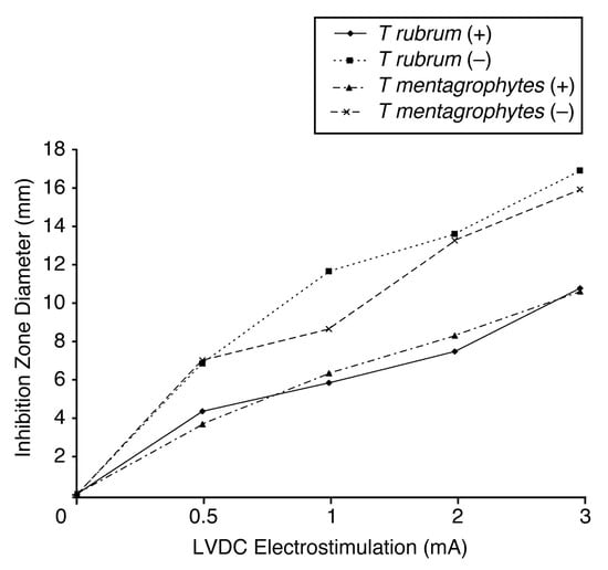
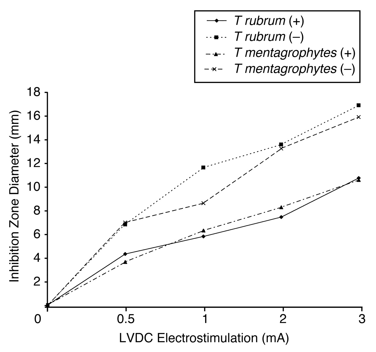
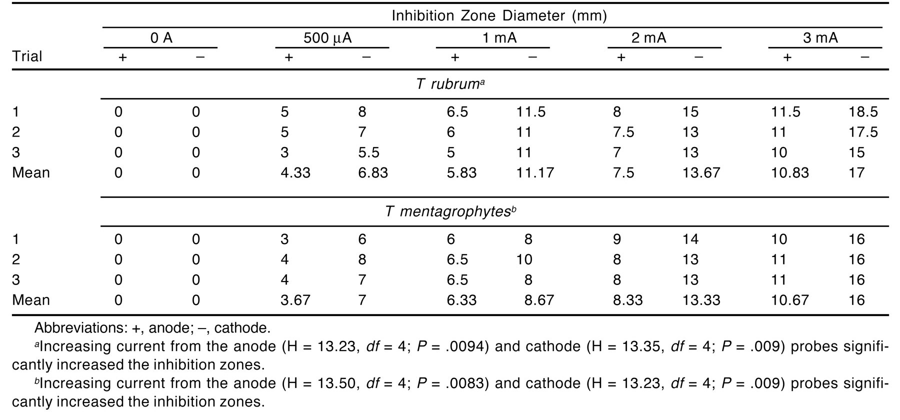
For each fungus, each experiment was replicated three times at the specified current and time settings. The sizes of the zones around each electrode that lacked fungal growth for T rubrum or T mentagrophytes owing to LVDC electrostimulation at the various amperages used are displayed in Table 1 and Figure 1. For all amperages except 0 A, circular or roughly circular zones devoid of fungal growth were observed around the positive (anode) and negative (cathode) electrodes. In addition, we observed increases in zone diameters with increasing amperages for both fungi (Fig. 1). Because we inserted 1-mm electrodes into the semisolid agar surface during each experiment to generate a current, a depression was produced in the agar surface at the location of these electrodes on all plates. This depression was probably caused by compression or liquefaction of the agar. Consequently, the absence of growth observed directly under each electrode in this study, as noted in a previous study,[16] was not considered to be indicative of fungicidal or fungistatic activity.
Table 1.
Inhibition Zone Diameters Around the Electrodes for Trichophyton rubrum and Trichophyton mentagrophytes
Figure 1.
Antifungal activity of low-voltage direct current (LVDC) electrostimulation on Trichophyton rubrum and Trichophyton mentagrophytes. Plot of the mean inhibition zone diameter of fungicidal activity of three trials versus the dose of LVDC electrostimulation applied. +, anode;−, cathode.
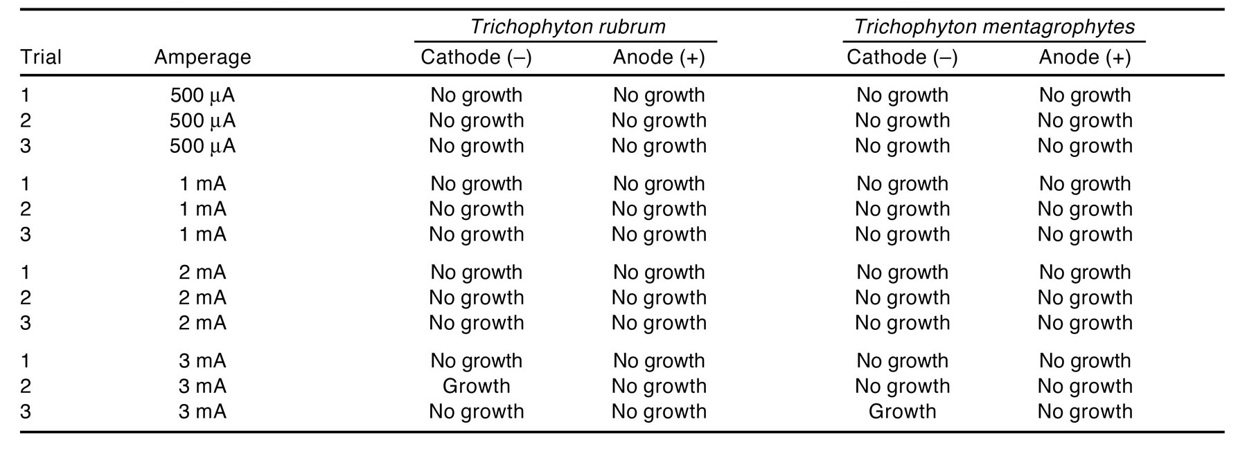
To confirm that the antifungal effects on T rubrum and T mentagrophytes observed in this study were due to LVDC electrostimulation rather than to other possible causes, the following controls were undertaken. To rule out the possibility that the observed antifungal effects were due to the electrode material itself, each experiment included a plate with electrodes inserted but no current applied. On these plates, no areas devoid of fungal growth around either electrode were ever observed with T rubrum or T mentagrophytes (Table 1). This same control was also used to determine whether the lack of fungal growth around the two electrodes was due to any remaining ethanol on the electrodes. If this were the case, some area devoid of fungal growth would have been observed around the region where the electrodes were inserted into the agar plates, even without the application of LVDC. As Table 1 demonstrates, no such region was observed.
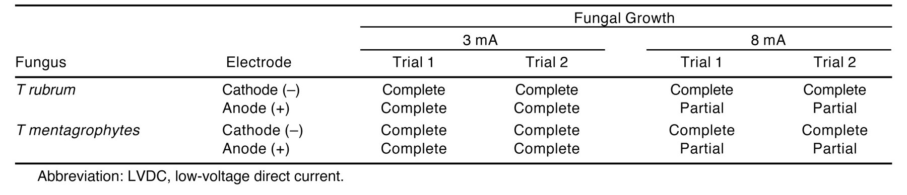
Another possible reason for the antifungal effects observed in this investigation was that changes produced in the fungal growth medium as a result of LVDC electrostimulation made the medium no longer supportive of fungal growth. To investigate this possibility, we carried out the following experiments. Before the inoculation of either fungus onto the Sabouraud dextrose agar medium, LVDC of either 3 mA (the highest clinical amperage used in this study) or 8 mA (beyond the clinical range used) was applied to eight plates for 30 min each (four plates at 3 mA and four plates at 8 mA). Trichophyton rubrum was then inoculated onto two of the 3-mA and two of the 8-mA plates and incubated for 7 days; T mentagrophytes was inoculated similarly. If the medium was indeed changed by LVDC electrostimulation in such a way as to prevent or inhibit fungal growth, this would be observed as a lack of growth in the agar in the region around the electrodes (or possibly some other region of the agar). The results of these experiments are given in Table 2. No such region lacking fungal growth was observed for T rubrum or T mentagrophytes in the areas surrounding either of the electrodes (or any other area) when we inoculated the fungi onto a Sabouraud dextrose agar plate after 3 mA of LVDC application. At 8 mA, no region lacking fungal growth was observed for either fungus at the cathode. However, at this amperage, some discoloration, liquefaction, and subsequent depression of the agar were observed in the area in which the anode was placed. Neither fungus grew very well over this discolored, depressed anode region at 8 mA. Otherwise, growth occurred throughout the remainder of the plate.
Table 2.
Growth Measurement of Trichophyton rubrum and Trichophyton mentagrophytes Inoculated onto Sabouraud Dextrose Agar Plates After LVDC Electrostimulation
To determine whether the range of LVDC electrostimulation used in these experiments was acting primarily as a fungicidal agent or a fungistatic agent, the areas around each electrode that lacked fungal growth were sampled using a sterile swab 24 hours after electrostimulation for the presence of viable fungal cells or spores. A total of 24 samples for T rubrum and 24 for T mentagrophytes were taken. A sample was taken from the area around each electrode on all plates, which received a dose of 500 μA, 1 mA, 2 mA, or 3 mA. These results are given in Table 3. In 2 of the 24 samples, growth was observed on the fresh Sabouraud dextrose agar plates inoculated with a swab after sampling an experimental plate. In the remaining 22 samples, no growth was observed.
Table 3.
Viability Sampling Results from the Zones Around the Electrodes Devoid of Fungal Growth
Finally, as has previously been reported,[16] we also observed the production of gas bubbles at the cathode during all experiments and, occasionally, at the anode. In addition, a blue discoloration was observed around the cathode, possibly owing to a pH change.
The effects of LVDC electrostimulation amperages were tested using Kruskal-Wallis nonparametric analysis of variance tests owing to the narrow range of the zone-of-inhibition measurements. The results of the statistical analysis are presented in the footnotes to Table 1. All analyses were conducted using StatView for Windows, version 5.0.1 (SAS Institute, Inc, Cary, North Carolina).
Discussion
Our investigation was undertaken to determine whether the clinically observed efficacy of LVDC electrostimulation in the treatment of onychomycosis is due, at least in part, to an antifungal effect of the applied electric current. Antibacterial effects against many of the common bacterial wound pathogens have been demonstrated in vitro for LVDC and high-voltage pulsed current in several studies.[15,16,17] However, such in vitro documentation is negligible with respect to the effect of electric current on the common fungal pathogens, the yeast C albicans being one of the few exceptions.[18] The results of our investigation clearly demonstrate that clinically relevant amperages of LVDC electrostimulation are antifungal in vitro to the two primary causes of onychomycosis—the fungi T rubrum and T mentagrophytes. The antifungal effects occur in a dose-dependent manner in the clinically relevant ranges (500 μA to 3 mA) that we chose to use in our in vitro experiments.
A second and related determination we wanted to make was whether LVDC electrostimulation in the amperage range we used acts primarily as a fungistatic agent or as a fungicidal agent. We carefully sampled the areas devoid of fungal growth around the electrodes in each of our experiments 24 hours after electrostimulation using a sterile swab to assay for any viable fungal cells or spores that could give rise to fungal colonies on new Sabouraud dextrose agar plates. Similar methods have been used to determine whether electrostimulation is bactericidal or bacteriostatic.[15] In 46 of the 48 samples, we observed no fungal growth on the newly inoculated plates. The two plates that did show some growth were from the cathode region of the 3-mA dose plate. These zones had the largest diameter, and the sampling swab may have inadvertently touched some viable fungal cells at the periphery of the zone. If so, they would represent an artifact of these experiments rather than lack of fungicidal activity. We strongly believe that this is the case. Consequently, the data strongly suggest that LVDC electrostimulation acts fungicidally in the amperage range used in this study.
Cellular death can be brought about by a variety of factors, including damage or denaturation of key cellular enzymes, damage to DNA, damage or disruption of the cell membrane, and damage or destruction of key cellular transport systems.[19] Electricity is believed to kill cells by affecting the molecular structure of the cell membrane, leading to fatal changes in cell membrane permeability.[18,20] Such cell membrane damage could explain the antifungal effects that we observed in vivo and in vitro. However, other factors might also play a role. Application of electric current to the agar medium can result in changes in the pH of the medium, increases in temperature, and the generation of toxic metabolites. All of these factors (and perhaps others) could act antimicrobially to one degree or another. Such factors have been considered in other studies[15,16] examining the antibacterial effects of electricity. We did not examine these factors individually in this study because we were mainly concerned with determining whether LVDC electrostimulation itself was antifungal. If the application of electricity to a fungus growing on a toenail or an agar surface results in the death of all or some of those fungal cells, then whether that fungicidal activity was due to fatal changes in the cell membrane of fungal cells and also to changes in pH or increases in cellular temperature is secondary to our present interest. Such aspects should be addressed in subsequent investigations.
In our study, we wanted to demonstrate that the electric stimulation itself, rather than the experimental methods, produced the observed antifungal effects. As we did not observe any fungal inhibition around either electrode during any trial when the electrodes were inserted but current was not applied, we are confident that the stainless steel electrode material itself is not antifungal. Another concern we had was that some of the ethanol, which we used to disinfect the electrodes before and between experiments, might remain on the electrodes on insertion into the Sabouraud dextrose agar medium. Because ethanol is a disinfectant, it can kill or inhibit fungal and bacterial cells.[21] Thus we might see areas around each electrode lacking fungal growth because of the disinfection action of ethanol rather than the antifungal effects of LVDC electrostimulation. To minimize this possibility, after we removed the electrodes from their ethanol storage unit (stored in our biological hood), we allowed them to dry for a minimum of 30 sec before they were inserted into any Sabouraud dextrose agar experimental plates. Because we never observed any areas lacking fungal growth around either electrode at 0 A, we are confident that little or no ethanol remained on the electrodes on insertion into any plate. It is clear that the ethanol had evaporated and was not a cause of the observed zones lacking fungal growth.
Another possible reason for any observed areas lacking fungal growth around either electrode was that LVDC electrostimulation in the current range we used changed the Sabouraud dextrose agar medium in some way so that it could no longer support the growth of either T rubrum or T mentagrophytes. As the data show, no lack of growth was observed at 3 mA around the cathode or the anode or anywhere else on the agar plates that initially received LVDC electrostimulation. Growth occurred throughout each plate. At 8 mA, more than twice the highest amperage used in our study, growth occurred at the cathode but not at the anode, where liquefaction and depression were observed. It is probable that at this amperage, lack of growth was due to some physical changes produced in the medium (ie, liquefaction), the generation of toxic metabolites, the denaturation of vital nutrients, or a combination of several factors.[15] However, because 3 mA was the highest dose of LVDC electrostimulation used in our antifungal studies, we are confident that at this and lower amperages the medium was not altered in a substantial way so as to negatively affect the growth of either fungus. To further support this assertion, we noted that in our experiments involving 500 μA, 1 mA, 2 mA, and 3 mA, the areas of the agar lacking fungal growth owing to the LVDC application were still capable of supporting the growth of both fungi. This is evidenced by the fact that if these agar plates were incubated at 25°C for 7 to 10 days, we would eventually clearly see viable fungi from outside the diameter of the fungicidal zone begin to recolonize that zone until a solid fungal lawn was again formed over the entire area. Together, these results strongly support the assertion that any possible changes produced in the Sabouraud dextrose agar medium were not responsible for the observed zones lacking fungal growth around the electrodes. The results of the two experiments described here are given in Table 2 and Table 3.
Conclusion
The data presented here strongly support the conclusion that the application of electric current in the form of LVDC electrostimulation to fungal cells is the only factor responsible for the observed in vitro fungicidal activity reported in this study. These data are consistent with our clinical observations of the efficacy of LVDC electrostimulation when used to treat onychomycosis. Consequently, we believe that LVDC electrostimulation (and possibly other types of electrostimulation), a nonpharmaceutical, noninvasive, cost-effective treatment, can play an important role in the amelioration and cure of onychomycosis caused by T rubrum or T mentagrophytes (and possibly other fungi) when used as an alternative therapy or in conjunction with traditional antifungal medications. The results of this in vitro study will serve as a catalyst for future investigation involving human trials in which we hope to determine the optimal parameters of treatment with this modality.
Dr. Kalinowski, Dr. Edsberg, and Mr. Brogan have proprietary interests in the patents covering the low-voltage direct current application and techniques described in this article. In addition, the same three authors are partners in LMD Technologies, LLC, Buffalo, NY, which is in the process of developing, manufacturing, and marketing devices based on this technology. |
References
- Scher RK: Onychomycosis: therapeutic update. .J Am Acad Dermatol40::S21. ,1999. .
- Elewski BE, Hay RJ: Novel treatment strategies for superficial mycoses: introduction. .J Am Acad Dermatol40::S1. ,1999. .
- Elewski BE, Charif MA: Prevalence of onychomycosis in patients attending a dermatology clinic in northeastern Ohio for other conditions. .Arch Dermatol133::1172. ,1997. .
- Schlefman BS: Onychomycosis: a compendium of facts and a clinical experience. .J Foot Ankle Surg38::290. ,1999. .
- Tom CM, Kane MP: Management of toenail onychomycosis. .Am J Health Syst Pharm56::865. ,1999. .
- Midgley G, Moore MK, Cook JC, et al: Mycology of nail disorders. .J Am Acad Dermatol31::S68. ,1994. .
- Ellis DH: Diagnosis of onychomycosis made simple. .J Am Acad Dermatol40::S3. ,1999. .
- Jennings MB, Weinberg JM, Koestenblatt EK, et al: Study of clinically suspected onychomycosis in a podiatric population. .JAPMA92::327. ,2002. .
- Lubeck DP, Patrick DL, McNulty P, et al: Quality of life of persons with onychomycosis. .Qual Life Res2::341. ,1993. .
- Scher RK: Onychomycosis: a significant medical disorder. .J Am Acad Dermatol35::S2. ,1996. .
- Milles CL, Riley PA, Kessenich CR: Onychomycosis: diagnosis and systemic treatment. .Nurse Pract23::40. ,1998. .
- Ungar G: Effective treatment of onychomycosis. .Phys Assist21::72. ,1997. .
- Kloth LC: “Electrical Stimulation in Tissue Repair,” in Wound Healing: Alternatives in Management, 2nd Ed, ed by J McCulloch, L Kloth, J Feeder, p 275, FA Davis, Philadelphia. ,1995. .
- Kloth LC: Physical modalities in wound management: UVC, therapeutic heating and electrical stimulation. .Ostomy Wound Manage41::18. ,1995. .
- Kincaid CB, Lavoi KH: Inhibition of bacterial growth in vitro following stimulation with high voltage, monophasic, pulsed current. .Phys Ther69::651. ,1989. .
- Szuminsky NJ, Albers AC, Unger P, et al: Effect of narrow, pulsed high voltages on bacterial viability. .Phys Ther74::660. ,1994. .
- Barranco SD, Spadaro JA, Berger TJ, et al: In vitro effect of weak direct current on Staphylococcus aureus. .Clin Orthop100::250. ,1974. .
- Hulsheger H, Potel J, Niemann EG: Electric field effects on bacteria and yeast cells. .Radiat Environ Biophys22::149. ,1983. .
- Madigan MT, Martinko JM, Parker J: “Microbial Growth Control,” in Brock Biology of Microorganisms, 9th Ed, p 703, Prentice-Hall, Englewood Cliffs, NJ. ,2003. .
- Hulsheger H, Potel J, Niemann EG: Killing of bacteria with electric pulses of high field strength. .Radiat Environ Biophys20::53. ,1981. .
- Atlas RM: “Control of Microbial Growth,” in Principles of Microbiology, 2nd Ed, p 461, William C Brown, Dubuque, IA. ,1997. .

© 2004 American Podiatric Medical Association