


Abstract
An investigative study was performed to compare the results from two mycology laboratories for the diagnosis of onychomycosis in a geriatric population and to determine the possible pharmacologic treatments based on the two laboratories’ results. In this study, 85 cases of suspected onychomycosis involving men and women 65 years and older from a nursing home setting in South Florida were used. Samples were taken from the hallux toenail and sent to two different mycology laboratories for fluorescent potassium hydroxide preparation and microscopic examination of a fungal culture. Of the 85 cases studied, the two mycology laboratories reported similar potassium hydroxide preparation results for 58.8% of the patients and similar fungal culture results for genus and species identification for 37.6% of the patients. When the potassium hydroxide preparation and fungal culture results were combined, the two mycology laboratories reported similar results for only 27.1% of the patients. As a result of the two mycology laboratories’ findings, the possible US Food and Drug Administration–approved pharmacologic treatments may differ for 43.5% of the patients studied. The discrepancy between the two independent laboratories leaves physicians to question the reproducibility of fluorescent potassium hydroxide preparation and fungal culture analysis in a geriatric patient population for the diagnosis of onychomycosis.
Onychomycosis of the toenail is the most common condition diagnosed and treated by podiatric physicians in the United States.[1] The diagnosis of onychomycosis is usually obtained through several scientific methods, including clinical observation, dermatophyte test medium culture, fluorescent potassium hydroxide (KOH) preparation, toenail biopsy, periodic acid–Schiff reaction, fungal culture growth analysis, light microscope imaging, and scanning electron microscope imaging.[2] When presented with a patient with clinical manifestations of onychomycosis, physicians often use at least one diagnostic method to support their clinical diagnosis. Although various combinations of diagnostic tests may help the physician confirm a clinical diagnosis of onychomycosis, most rely on fluorescent KOH preparation in conjunction with a fungal culture performed by an independent mycology laboratory.[3]
The purpose of this study was to objectively evaluate the fluorescent KOH preparation and fungal culture results from two different mycology laboratories for a geriatric population demonstrating clinical signs and symptoms of toenail onychomycosis and to determine the possible pharmacologic treatments based on the two laboratory reports. We questioned whether a physician can expect reproducible results from two different mycology laboratories when similar specimens were submitted from the same toenail on the same patient. This study represents the first attempt by podiatric physicians to directly compare the results from two different mycology laboratories for a geriatric patient population with suspected onychomycosis and to evaluate the different pharmacologic treatment options based on the laboratory results.
Clinical Studies
Onychomycosis is a clinical term used to describe a fungal infection characterized by thickening, splitting, roughening, and discoloration of the toenail.[4] People infected with onychomycosis suffer mainly from cosmetic difficulties; however, left untreated, the fungal infection may spread to other toenails and make everyday activities painful.[1] Studies show that fungal infections have affected nearly every person who wears shoes at one time or another; darkness, heat, and moisture associated with hosiery and shoes make conditions inviting for the development of onychomycosis.[4]
The fungi associated with onychomycosis are predominantly dermatophytes, which are responsible for more than 90% of the fungal infections.[3,5-8] Most dermatophytes associated with onychomycosis are from the genus Trichophyton, including Trichophyton rubrum and Trichophyton mentagrophytes; however, the genera Epidermophyton and Microsporum are also represented. Onychomycosis, however, is not restricted to these common dermatophytic pathogens. One published report indicated a shift of fungal species from an isolated dermatophyte infection to mixed saprophyte infections in a geriatric population with signs and symptoms of onychomycosis.[9] The authors determined that saprophyte-induced onychomycosis is usually seen in the elderly, patients with other skin diseases, and immunocompromised individuals. The saprophytes associated with onychomycosis include Aspergillus, Scopulariopsis, and Scytalidium. Onychomycosis can also be classified as a fungal infection involving yeast. Yeast is a general term denoting true fungi of the family Saccharomycetaceae, and the most common yeast infection present in clinical onychomycosis is Candida albicans. In total, it has been reported that nondermatophyte onychomycosis accounts for 1.6% to 6% of all cases of onychomycosis.[9]
The diversity of fungal species associated with onychomycosis presents a challenge for accurate and reproducible laboratory reporting and appropriate US Food and Drug Administration (FDA)–approved pharmacologic treatments. The three major types of fungi—dermatophytes, saprophytes, and yeasts—associated with onychomycosis are distinct in their morphology, physiology, and reproductive behaviors.[9] The differences between the fungal species result in various treatment options for physicians. Such treatments may include debridement, topical antifungal medication, and a regimen of oral antifungal medications that have an 80% to 90% success rate.[10] Although most physicians rely on topical and oral antifungal medications to treat patients with onychomycosis-related fungal infections, clinicians often institute a treatment plan based on the results of a mycology laboratory report of the specific pathogen causing onychomycosis.
The current FDA-approved medications for the treatment of onychomycosis include oral medications, such as itraconazole, terbinafine, and griseofulvin, and topical medications, such as ciclopirox. Griseofulvin as an oral agent has been available for many years, but its use is limited by a narrow spectrum, long courses of treatment, and high relapse rates.[11] Triazole and allylamine antifungal drugs, such as itraconazole and terbinafine, are currently the most popular oral medications used in the treatment of onychomycosis. These two medications share characteristics that enhance their effectiveness: prompt penetration of the nail and nail bed, persistence in the nail for months after discontinuation of therapy, and generally good safety profiles. The only topical medication that currently has FDA approval for the treatment of onychomycosis is ciclopirox. It is generally safe and inexpensive; however, it is seldom effective in the eradication of onychomycosis.
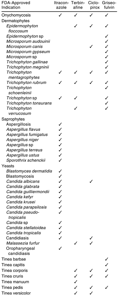
Although itraconazole, terbinafine, griseofulvin, and ciclopirox are all FDA approved for the treatment of onychomycosis, the term onychomycosis is expansive and can pertain to a relatively broad spectrum of fungal infections. Physicians can prescribe specific oral and topical antifungal medications when a mycology laboratory identifies the individual pathogens (Table 1). Because the treatment of onychomycosis caused by dermatophytes may require long-term therapy with an oral antifungal medication with potential side effects, it is essential to diagnose the infection correctly. Although onychomycosis caused by dermatophytes is readily treatable with topical medications or oral azole and allyl antifungals, onychomycosis caused by nondermatophytes is treatable only with oral azole antifungals.[12] The correct identification of the pathogens causing a fungal infection is therefore important to accurate clinical diagnosis and appropriate treatment of onychomycosis.
Table 1.
Comparison of Medications Prescribed for the Treatment of Specific Fungi Associated with Onychomycosis and Their FDA-Approved Indications.
Materials and Methods
The study population consisted of 85 patients older than 65 years with clinical signs and symptoms of onychomycosis who were selected at random from a nursing home population in Fort Lauderdale, Florida. Specimens were not submitted from patients who were currently receiving any type of oral or topical antifungal therapy. Specimens were obtained by first cleansing the affected toenail and surrounding skin with alcohol and then aggressively clipping the toenail as proximally as possible without causing excessive discomfort to the patient. The nail specimen and subungual debris were obtained from the proximal end of the growing edge of the suspected infection; the distal nail clippings were discarded. The specimens were placed in small, individually sealed plastic bags labeled with the patient’s name, the date of collection, and the location of the specimen source (right or left hallux). All 85 individual toenail specimens were then divided into two equal and separate sections. Half of each specimen was left in the plastic bag and sent to the Laboratory of Podiatric Pathology in Philadelphia, Pennsylvania (laboratory 1), and the other half was placed in a new sealed plastic bag, labeled in the same manner as the first specimen, and sent to Podiatric Pathology Laboratories in Baltimore, Maryland (laboratory 2).
Laboratory 1 Methodology
The mycology laboratory prepared and microscopically viewed a KOH preparation slide enhanced with a fluorescent dye, Calcofluor white. Fungal cultures were performed on two media—one using Sabouraud media with chloramphenicol and the other using Sabouraud media with chloramphenicol and cycloheximide. The culture using only chloramphenicol in conjunction with Sabouraud media allows a wide range of fungi to grow. The culture using chloramphenicol and cycloheximide in conjunction with Sabouraud media, however, is more selective and restricts growth to allow dermatophytes to flourish. On each culture, a tease preparation was performed with lactophenol blue. Potato dextrose agar was used for any subcultures and to identify yeast growth.
Laboratory 2 Methodology
The mycology laboratory prepared and microscopically viewed a KOH preparation slide enhanced with a fluorescent dye, Calcofluor white. The readings were based on the presence or absence of the fungal elements and were presented in the following format: no fungal elements, rare to few hyphae, moderate hyphae, hyphae and arthrospores, rare to few yeast cells, moderate yeast cells, many yeast cells, and yeast cells and pseudohyphae observed. Fungal cultures were then performed to determine the genus and species of each organism. The mycology culture tube media used included Mycosel agar and potato flake agar, which were screened weekly for the presence of fungal growth. The molds were identified from a Lactofuscin Scotch tape prep slide, and the yeasts were identified from the Micro Scan yeast identification panel. After 4 to 6 weeks of incubation, all of the positive and negative cultures for fungal growth were finalized.
The data from this study were compiled into a computer database with each patient’s identification number, age, sex, and the following for laboratory 1 and laboratory 2: fluorescent KOH preparation result, microscopic fungal culture examination result for each fungus in genus and species, and fungal type for each fungus (dermatophyte, saprophyte, yeast, or no growth).
Results
The study population consisted of 26 men (30.6%) and 59 women (69.4%). The median age was 81 years, and the mode was 89 years. Patient age ranged from 65 to 99 years, with a mean age of 82.8 years.
We defined four categories to compare the two mycology laboratory results: fluorescent KOH preparation, fungal type, genus, and genus and species. A perfect match was considered to occur when the two mycology laboratory reports exactly match each other when comparing fluorescent KOH preparation, fungal type, genus, or genus and species results. A partial match is when the two mycology laboratory reports share some results but fall short of a perfect match. A no match occurs when the two mycology laboratory reports completely fail to match any results. An ultimate match is when the two mycology laboratory reports perfectly match fluorescent KOH preparation, fungal type, genus, and genus and species for an individual patient sample.
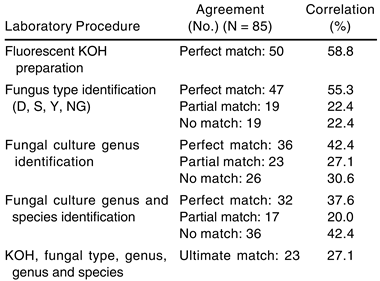
Of the 85 patients studied, the two mycology laboratories perfectly matched 50 (58.8%) patients’ fluorescent KOH preparation results (Table 2). When comparing the results of fungal type identification (dermatophyte, saprophyte, yeast, combination, or no growth) from fungal culture analysis, the two mycology laboratories perfectly matched 47 (55.3%) of the patients, partially matched 19 (22.4%) of the patients, and had no matches in 19 (22.4%) of the patients studied. The two mycology laboratories perfectly matched 36 (42.4%) of the patients, partially matched 23 (27.1%) of the patients, and had no match in 26 (30.6%) of the patients studied when comparing the results of genus identification from fungal culture analysis. When comparing the results of fungal genus and species identification from fungal culture analysis, the two mycology laboratories perfectly matched 32 (37.6%) of the patients, partially matched 17 (20.0%) of the patients, and had no match in 36 (42.4%) of the patients studied. When the fluorescent KOH preparation, fungal type, genus, and genus and species for each individual patient were compared, the two mycology laboratories ultimately matched only 23 (27.1%) of the patients studied.
Table 2.
Analysis of Agreement Between Two Mycology Laboratory Procedures.
Laboratory 1 reported 24 (28.2%) positive and 61 (71.8%) negative fluorescent KOH preparation results, 19 (22.4%) no growth results, 22 (25.9%) mixed fungal infections, and 88 total fungal organisms identified. Laboratory 2 reported 49 (57.6%) positive and 36 (42.4%) negative fluorescent KOH preparation results, 20 (23.5%) no growth results, 20 (23.5%) mixed fungal infections, and 84 total fungal organisms identified. The two mycology laboratories perfectly matched 19 patients with positive fluorescent KOH preparation results, 31 patients with negative fluorescent KOH preparations, 12 patients with no growth, and 12 patients with mixed infections.
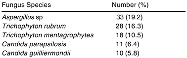
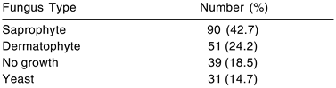
There were 172 individual fungal organisms identified from the 85 patients studied when the results from both mycology laboratories were combined. Aspergillus sp was reported most frequently, followed by T rubrum, T mentagrophytes, Candida parapsilosis, and Candida guilliermondii (Table 3). Between the two different mycology laboratories, the total identified fungal type growth was 90 (42.7%) saprophytes, 51 (24.2%) dermatophytes, 39 (18.5%) no growth, and 31 (14.7%) yeast (Table 4).
Table 3.
Top Five Organisms of 172 Identified.
Table 4.
Total Identified Fungal Type Growth.
Discussion
The average age of the total population was 82.8 years, demonstrating a geriatric population. When the fluorescent KOH preparation results for this study were compared, the two independent mycology laboratories reported the same results for 58.8% of the patients. The results of the fluorescent KOH preparations represent the highest correlation between the two mycology laboratories for the population data. As reported in previous studies, a negative fluorescent KOH preparation result does not preclude the presence of fungal infection; it only indicates that fungal hyphae were not observed in the sampled tissue, and fungi may still be grown and identified on microscopic fungal culture examination.[1] We were unable to determine why laboratory 1 had 28.2% positive fluorescent KOH preparation results while laboratory 2 had 57.6% positive fluorescent KOH preparation results for the 85 patients studied.
When comparing a relatively broad category such as fungal type (dermatophyte, saprophyte, yeast, or no growth) from microscopic examination of a fungal culture to identify the nature of the causative organisms, the two mycology laboratories reported similar results slightly less frequently than for the fluorescent KOH preparation results. The two laboratories perfectly matched fungal type 55.3% of the time, partially matched 22.4% of the time, and had no matches 22.4% of the time. This similarity has been reported in the past, with studies claiming that fluorescent KOH preparation results usually correlate with fungal culture; however, the fluorescent KOH preparation results are often inconsistent.[12]
When comparing the genus identification from fungal culture analysis, the two mycology laboratories were able to perfectly match only 42.4% of the patients and partially match 27.1% of the patients; they were unable to match any of the genus results 30.6% of the time. When comparing the combined genus and species identification from the two mycology laboratory fungal culture analyses with genus identification alone, an even lower correlation was observed. The two mycology laboratories were able to achieve a genus and species perfect match only 37.6% of the time and a partial match 20.0% of the time; they failed to match 42.4% of the genus and species results. This represents an 11.8-percentage-point increase in the inability to match any results when comparing genus and species identification with genus identification only. We attributed this finding to the relative difficulty in discriminating between the various species in a genus that are relatively similar in morphology. Mycology laboratories do not use technology such as scanning electron microscopy for routine clinical diagnoses of onychomycosis owing to cost.[2] Referring to charts used to diagnose onychomycosis in culture, there are similarities between T rubrum and T mentagrophytes when comparing hyphae of the two fungi.[13] When they are cultured and analyzed in a mycology laboratory according to the laboratory 1 and laboratory 2 methodologies, it is possible that during observation of the cultures a technician can confuse T rubrum and T mentagrophytes if reproductive elements are not seen. Previous studies have reported that genus and species cannot be identified in culture if the organism produces sterile hyphae and not conidia.[3]
When determining the ultimate match between the two mycology laboratories, the lowest correlations in this study were observed. The two mycology laboratories were able to match only 27.1% of the patient results when all four categories were compared. The ultimate match represents the strictest comparison of the two mycology laboratory results and is necessary for a clinician to be confident that a discrepancy does not exist in the reports. Previous studies have confirmed that treatment of onychomycosis is often unsuccessful for a number of reasons, often owing to incorrect identification of the infecting organism.[14] Many of the FDA-approved medications available for the treatment of onychomycosis have various specificities toward the pathogens that cause fungal infections (Table 1). According to the two mycology laboratory fungal culture analyses, genus and species matching correlated with a perfect match only 37.6% of the time. Having a disagreement of 62.4% of the fungal culture reports of the two mycology laboratories leaves the clinician with the possibility of prescribing a treatment regimen of topical and/or oral medications that may not be effective against the patient’s fungal infection.
When comparing the two reports of the mycology laboratory fungal culture analysis, the possible FDA-approved pharmacologic treatments differed for 37 (43.5%) of the 85 patients studied. The discrepancy of pharmacologic treatments is high owing to the strict guidelines used during comparison of results and the nature of the criteria used to assess treatment planning. The FDA-approved indications for various medications listed in Table 1 were interpreted literally during comparison of laboratory reports, and non–FDA-approved treatments were not included in this study.[15] One patient’s results were T mentagrophytes (laboratory 1) and T rubrum (laboratory 2); this discrepancy would not affect the pharmacologic treatment because all four medications listed are effective against both pathogens. Another patient’s results, however, were Aspergillus sp (laboratory 1) and no growth (laboratory 2); this would result in different pharmacologic treatments depending on which mycology laboratory report was used. A discrepancy in the results C parapsilosis (laboratory 1) and T rubrum (laboratory 2) would have also resulted in different pharmacologic treatments because terbinafine, although FDA approved for T rubrum, is listed as sensitive to but not FDA approved for the treatment of C parapsilosis.
In our study, a lack of agreement between the two mycology laboratories occurred most frequently when comparing all of the mycology tests combined: fluorescent KOH preparation, fungus type, genus, and genus and species identification. There are a number of possible sources of error in this study. Although utmost care was taken in obtaining and packaging a toenail sample for analysis by the mycology laboratories, the samples may have contained disproportional amounts of fungal elements on the toenail specimen, specimens may have become contaminated during packaging, and biopsy may have resulted in sufficient fungal elements for examination by fluorescent KOH preparation but not for fungal culture. Although the mycology laboratories used selective media to reduce bacterial contamination, bacteria or other fungal elements, such as opportunistic molds, may have contaminated the testing environment. Furthermore, fungal culture results are often read by a technician rather than by a pathologist specializing in fungal culture analysis. Other tests, such as toenail biopsy with surgical pathology diagnostic testing, are interpreted by pathologists and may lead to greater sensitivity and specificity.[12] Additionally, previous studies have reported that genus and species cannot be identified in a fungal culture if the organism fails to produce conidia, indicating that biopsy of areas containing small amounts of reproductive elements may lead to error in diagnostic reports.
Conclusion
The objective evaluation of the results from two different mycology laboratories for fluorescent KOH preparations and microscopic examination of fungal cultures of the same patients allowed for a direct comparison between the two mycology laboratories. The purpose of this study was not to determine whether one laboratory was correct and the other incorrect; it was designed only to compare the mycology laboratory results from the perspective of a clinical podiatric physician obtaining a toenail sample from a geriatric patient with suspected onychomycosis. This study demonstrated that two independent mycology laboratories were unable to achieve adequate agreement in a majority of their results for a geriatric patient population with suspected onychomycosis in South Florida. Additionally, the possible FDA-approved pharmacologic treatments based on the mycology laboratory reports differed for 43.5% of the patients in the study.
From the perspective of a clinical podiatric physician comparing two different mycology laboratories, we determined that the 58.8% correlation of the fluorescent KOH preparation between the laboratories may be unreliable when attempting to confirm a clinical diagnosis of onychomycosis in a geriatric population. We also determined that fungal type matching is as unreliable as fluorescent KOH preparation results, illustrating that the laboratory results agreed 55.3% of the time. On stricter examination, the two mycology laboratories agreed only 42.4% of the time when determining the genus and only 37.6% of the time when determining both the genus and species of the specimens.
When comparing the combined fluorescent KOH preparation results and fungal culture analysis for the entire 85-patient population, the two independent mycology laboratories were able to ultimate match only 27.1% of the patients. Although many managed-care providers require positive fluorescent KOH preparation and/or positive fungal culture results to approve treatment of onychomycosis, the results of this study indicate that other mycology tests may be required to properly diagnose onychomycosis in a geriatric population. In this study, 73.0% of the patients had some type of discrepancy between the two mycology laboratory reports when a toenail sample was sent for analysis.
Accurate identification of fungal species is essential for proper diagnosis and treatment planning of onychomycosis. The cost of a mycology laboratory fluorescent KOH preparation combined with fungal culture analysis and questionable reproducibility of the reports should influence the diagnosis and treatment process of the podiatric physician when treating geriatric patients with onychomycosis. A 73.0% discrepancy between the two independent mycology laboratories and differing FDA-approved pharmacologic treatment plans for 43.5% of the patients leaves the clinical podiatric physician to question whether fluorescent KOH preparation and fungal culture analysis should be routinely used in geriatric patient populations for the diagnosis of onychomycosis.
References
- Scherer WP, Kinmon K: Dermatophyte test medium culture versus mycology laboratory analysis for suspected onychomycosis: a study of 100 cases in a geriatric population. JAPMA 90: 450, 2000.
- Scherer WP, Scherer MD: Scanning electron microscope imaging of onychomycosis. JAPMA 94: 356, 2004.
- Elewski BE: Diagnostic techniques for confirming onychomycosis. J Am Acad Dermatol 35: S6, 1996.
- McCarthy DJ: Origins of onychomycosis. Clin Podiatr Med Surg 12: 221, 1995.
- Evans EGV: Causative pathogens in onychomycosis and the possibility of treatment resistance: a review. J Am Acad Dermatol 38: S32, 1998.
- Kemna ME, Elewski BE: A US epidemiologic survey of superficial fungal diseases. J Am Acad Dermatol 35: 539, 1996.
- Schlefman BS: Onychomycosis: a compendium of facts and a clinical experience. J Foot Ankle Surg 38: 290, 1999.
- Summerbell RC: Epidemiology and ecology of onychomycosis. Dermatology 194: 32, 1997.
- Scherer WP, McCreary JP, Hayes WW: The diagnosis of onychomycosis in a geriatric population: a study of 450 cases in South Florida. JAPMA 91: 456, 2001.
- Baran R, Dawber RP, Tosti A, et al: A Text Atlas of Nail Disorders, p 165, Martin Dunitz, London, 1996.
- Rodgers P, Bassler M: Practical therapeutics. Am Fam Physician 63: 663, 2001.
- Weinberg JM, Koestenblatt EK, Tutrone WD, et al: Comparison of diagnostic methods in the evaluation of onychomycosis. J Am Acad Dermatol 49: 193, 2003.
- Funder S: Practical Mycology, 3rd Ed, p 79, Hafner Publishers, New York, 1968.
- Daniel CR: The diagnosis of nail fungal infection. Arch Dermatol 127: 1566, 1991.
- PDR Electronic Library, Thomson Micromedex, Greenwood Village, CO (edition expires 6/2004).

© 2004 American Podiatric Medical Association