


Abstract
Numerous hallux proximal phalanx osteotomies have been described, but the Akin-type varisation or adduction osteotomies are currently the most commonly used by foot and ankle surgeons. This article describes the hallux proximal phalanx Akin-Scarf osteotomy. This osteotomy combines the inherent stability of the Scarf-type osteotomy with the versatility of the Akin-type osteotomies to correct pathologic hallux abductus interphalangeus, hallux equinus, or an abnormal digital length pattern, and it is an invaluable tool during the global surgical approach used for hallux valgus repair.
Transverse plane, varisation, or adduction osteotomies of the hallux proximal phalanx to correct hallux abductus interphalangeus deformity are some of the most common adjunctive procedures used during surgical correction of hallux valgus deformity.
Akin,[1] in 1925, described a transverse plane, adduction, extra-articular, closing wedge osteotomy of the proximal one-third of the hallux proximal phalanx. Since that time, numerous modifications have been developed, including 1) proximal or distal location, 2) derotation, 3) shortening, and 4) dorsiflexion (ie, the Moberg-type modification).[2-6] The Akin osteotomy itself has been oriented transversely, obliquely, and horizontally, most commonly in an effort to affect the fixation options rather than to improve the correction obtained. Few articles have mentioned complications following an Akin-type osteotomy; however, I have noticed that delayed healing, secondary callus formation, iatrogenic violation of the first metatarsophalangeal joint, overcorrection, and undercorrection occur to varying degrees and with unpredictable postoperative results.
The aim of this article is to describe the hallux proximal phalanx Akin-Scarf osteotomy, a technique that combines the inherent stability of the Scarf-type osteotomy with the versatility of the Akin-type osteotomies.
Surgical Technique
The hallux is approached medially after completion of the first metatarsal corrective osteotomy, and the procedure is performed under tourniquet control using local anesthesia, most commonly with intravenous monitored anesthesia coverage. Dissection is carried directly to bone without developing tissue layers to allow for proper soft-tissue coverage of the osteotomy and first metatarsophalangeal joint corrective capsule closure at completion of the procedure. The periosteum is elevated from the dorsal, plantar, and medial aspects of the proximal phalanx, and the extensor and flexor hallucis longus tendons are identified, retracted, and protected for the remainder of the procedure. A Scarf osteotomy is outlined on the medial aspect of the hallux proximal phalanx, with a slightly inclined horizontal arm and acute dorsal distal and plantar proximal arms (Fig. 1). The osteotomy is completed, along with any necessary modifications, and the plantar capital fragment is distracted distally, adducted, and transposed medially until the desired correction is achieved, after which it is impacted into the remaining phalanx and provisionally stabilized with a threaded wire (Fig. 2).
Figure 1.
A completed Akin-Scarf osteotomy. Note the medial approach, the full-thickness skin and subcutaneous dissection, and the configuration of the osteotomy itself.
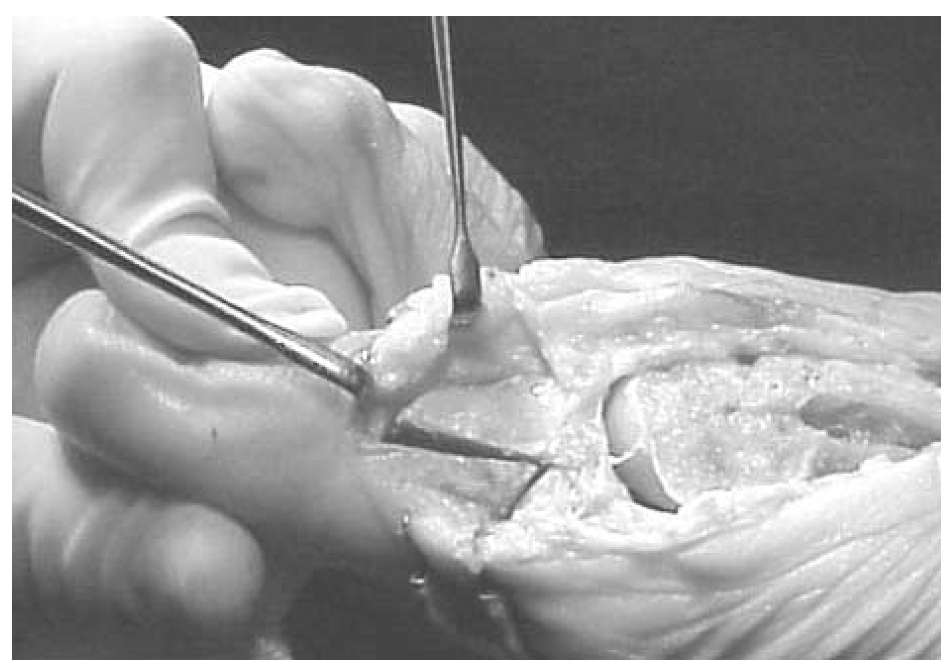
Figure 2.
A, The plantar capital fragment has been adducted, medially transposed, and impacted into the remaining hallux proximal phalanx. B, After provisional fixation of the osteotomy, rigid internal fixation of choice is performed.
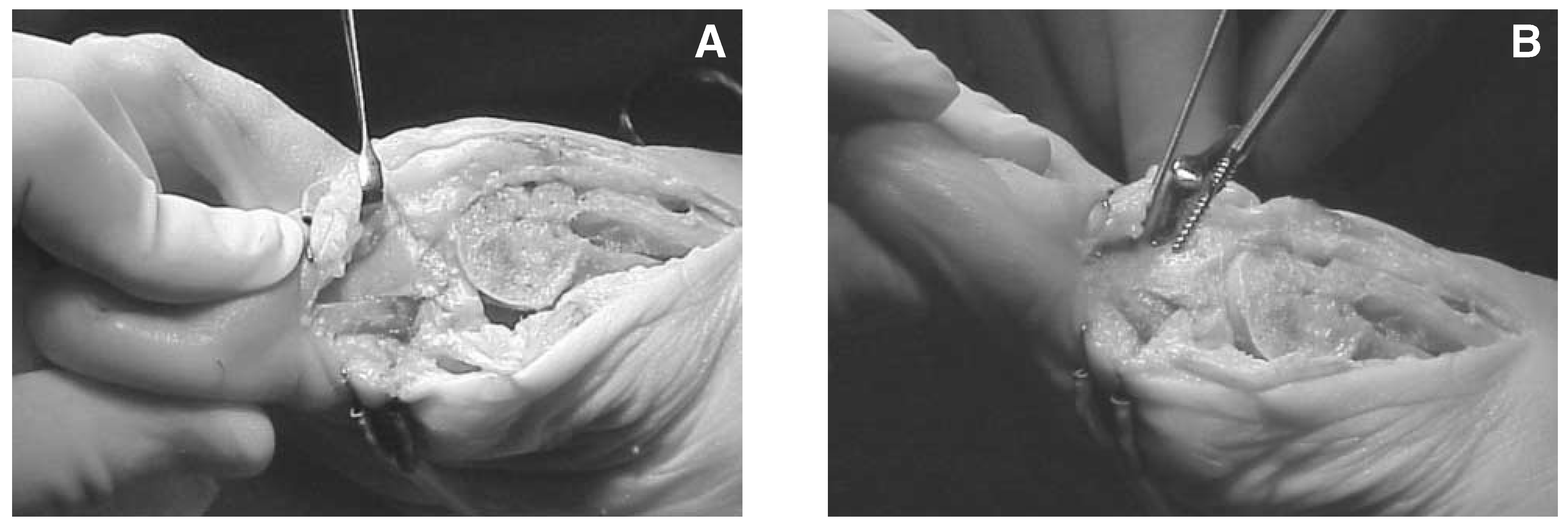
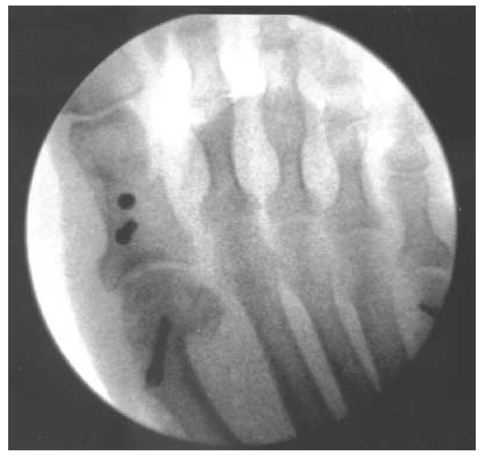
Correction is assessed clinically and radiographically, followed by application of rigid internal fixation of the surgeon’s preference (Figs. 3 and 4), although low-profile or no-profile screw fixation is ideal owing to the thin soft-tissue envelope surrounding the dorsal aspect of the osteotomy. Irrigation and capsule and skin closure are then performed according to the surgeon’s preference. The patient can walk immediately in a rigid postoperative shoe, unless other surgical procedures prevent this. The surgical dressings are removed in 7 to 10 days, with return to a roomy running shoe, and physical therapy to strengthen the plantarflexion power of the first metatarsophalangeal joint is begun, as previously described.[7]
Figure 3.
Note the correction of the hallux abductus interphalangeus deformity through adduction of the plantar capital fragment, as evidenced by the laterally rotated proximal arm and the medially rotated distal arm (A), as well as the slight medial transposition of the plantar capital fragment (B).
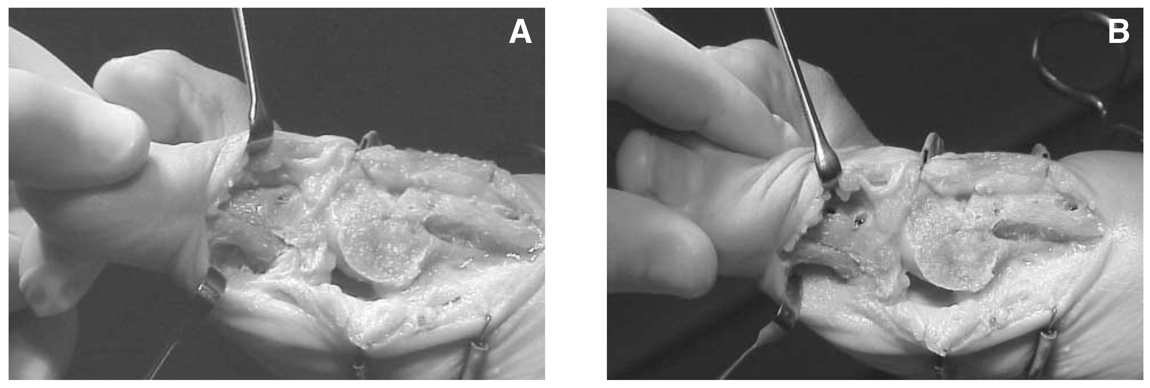
Figure 4.
Simulated weightbearing, anteroposterior, intraoperative radiograph demonstrating the correction of the hallux abductus interphalangeus deformity through adduction and medial rotation of the plantar capital fragment.
Discussion
In an effort to increase the stability while maintaining the great versatility of the Akin-type osteotomy, I have applied my current use of the Scarf osteotomy of the first metatarsal to the hallux proximal phalanx to correct an abnormal hallux abductus interphalangeus angle through combined adduction and medial transposition of the plantar capital fragment.[8] In addition, the Akin-Scarf osteotomy can readily be used to lengthen, shorten, derotate, and tilt upward the hallux proximal phalanx with predictable postoperative results. Finally, this osteotomy may be applied to the structural hallux varus deformity through a “reverse Akin-Scarf” procedure of the hallux proximal phalanx, using abduction and lateral transposition of the capital fragment to afford osseous correction (LS Weil Sr, DPM, personal communication, October 2002).
The hallux proximal phalanx Akin-Scarf osteotomy is a dependable option in the correction of pathologic hallux abductus interphalangeus, hallux equinus, or an abnormal length pattern during the global surgical approach that I use for hallux valgus repair. It provides an excellent outcome with good intermediate-term results.
References
- Akin O: The treatment of hallux valgus: a new operative treatment and its result. Med Sentinel 33: 678, 1925.
- Barouk LS: Osteotomies of the great toe. J Foot Surg 31: 388, 1992.
- Gusman DN, Messmer TE: Newell decompression procedure for hallux limitus: a preliminary report. JAPMA 85: 749, 1995.
- Hodor L, Hess T: Shortening Z-osteotomy for the proximal phalanx of the hallux using axial guides. JAPMA 85: 249, 1995.
- Moberg E: A simple operation for hallux rigidus. Clin Orthop 142: 55, 1979.
- Schwartz N, Hurley JP: Derotational Akin osteotomy: further modification. J Foot Surg 26: 419, 1987.
- Weil LS Jr, Benton-Weil W: Postoperative hallux valgus exercises. J Foot Ankle Surg 37: 355, 1998.
- Weil LS Sr: Scarf osteotomy for correction of hallux valgus: historical perspective, surgical technique, and results. Foot Ankle Clin 5: 559, 2000.
© 2004 American Podiatric Medical Association