


Abstract
A randomized, prospective study was conducted to compare the effectiveness of three individual mechanical modalities in the treatment of plantar fasciitis. Two hundred fifty-five subjects were randomly assigned to one of three treatment groups: custom-made orthoses, overthe-counter arch supports, or tension night splints. Subjects were treated for 3 months, with follow-up visits at 2, 6, and 12 weeks. No statistically significant difference was noted among treatment groups with respect to final outcomes based on first-step pain or pain felt during the day. However, there was a statistically significant difference among the three groups with respect to early patient withdrawal from the study due to continued severe pain, noncompliance, or inability to tolerate the device. Patient compliance was greatest with the use of custom-made orthoses.
Plantar fasciitis is an overuse syndrome characterized by localized inflammation of the plantar fascia at its anatomical insertion on the calcaneus. It is estimated that more than two million people receive treatment for plantar fasciitis in the United States each year.[1] Approximately 11% to 15% of adult patients who seek treatment from a podiatric physician present with a chief complaint of heel pain.[2,3] The pain, in the acute stage, is commonly localized to the origin of the medial and central bands at the calcaneus. Less frequently, the pain may progress distally along the course of the fascia.[4] The disorder is characterized by a sharp or knife-like pain upon initial standing. This first-step pain occurs after a period of nonweightbearing and often is worst upon arising from bed in the morning. The acute pain initially improves with a few steps, but then usually worsens with weightbearing. However, patients who present with chronic complaints indicate that the pain may become dull or achy and constant, worsening again after subsequent periods of rest throughout the day.
Weightbearing causes relative flattening of the longitudinal arch of the foot. The plantar fascia limits the flattening and provides stability to the arch. Hicks[5,6] has stated that the plantar fascia acts as a cable that is wound around the metatarsal head, which acts as a drum, with the proximal phalanx acting as a handle and providing the winding. The cable is a fixed length because the fascia does not stretch. A result of abnormal or excessive stress placed on the plantar fascia is a localized inflammatory response from disruption of the collagen fibers of the fascia band. The healing response is then interrupted by the continued stress produced by weightbearing, resulting in chronic degenerative changes. Histologically, these changes include collagen necrosis, angiofibroplastic hyperplasia, chondroid metaplasia, and matrix calcification.[7] Pathologically, prolonged inflammatory changes in the tissue are seen initially as edema, and are seen later as thickening of the plantar fascia.[8,9,10]
Several factors may contribute to the development of plantar fasciitis. The underlying factors that have been said to precipitate the condition can be divided into anatomical, biomechanical, and environmental classifications. Anatomical factors include pes planus,[11,12,13] pes cavus,[6,13,14] obesity,[11,15,16] tarsal coalitions,[17] unequal leg length,[14,17] and fat-pad atrophy.18 Biomechanical factors include equinus,[12,13,14] weak plantar flexor muscles,[17] weak intrinsic musculature,[17] and excessive subtalar joint pronation.[12,14,17] Environmental factors include trauma,[13] an increase in activity,[11,12] hard surfaces,[1,11,12] going barefoot, poor footwear,[11,12] prolonged weightbearing,[11] and inadequate stretching.[19] In most cases, a combination of these factors leads to the development of plantar fasciitis.
Historically, a broad range of conservative therapies have been used to treat plantar fasciitis. Modalities used include rest,[11,12] eliminating aggravating factors, nonsteroidal anti-inflammatory drugs,[15,20] oral or injectable steroids,[21] application of ice,[11,12,14,22] iontophoresis,[23] stretching,[22,24] shoe modifications,[12,22,25] heel cups,[18] taping,[11,12,22] arch supports,[1,26,27] custommade foot orthoses,[28,29] night splints,[25,30,31,32,33] and casting.[34]
Success rates for conservative treatment of plantar fasciitis vary from 46% to 100%.[35] Wolgin et al[35] conducted a retrospective review of 100 patients with 133 painful heels by means of a telephone survey to assess the long-term results of conservative treatment of plantar fasciitis. The average follow-up time was 47 months. Patients were classified by their symptoms. Eighty-two percent had no pain and no symptoms following a variety of conservative treatment modalities. The authors stated that no statistical comment could be made because treatments were not applied independently and any patient could have received a multitude of modalities at any given time. Although their population was small, the study revealed a high success rate with mechanical therapies: rigid orthoses and night splints were 100% successful and custom-made, cushioned inserts were 83% successful.
Campbell and Inman,[28] in 1974, were the first authors to describe success with mechanical therapy using arch supports. They treated 33 patients with University of California Biomechanics Laboratory (UC-BL) inserts and retrospectively reported a 94% success rate. O’Brien and Martin,[36] in 1985, performed a retrospective telephone survey of 41 patients with 58 painful heels. Excellent and good results were recorded for 96.7% of the patients, most of whom received multiple therapies. Subjectively, the patients stated that orthoses were the most successful treatment modality.
Scherer and the Biomechanics Graduate Research Group for 198829 performed a prospective study in which they treated 73 patients with 118 painful heels with taping, nonsteroidal anti-inflammatory drugs, injections, and rigid orthoses (98% received these orthoses). The study showed that, within 6 weeks, approximately 84% of the patients had at least 80% relief of symptoms. This study also identified a subgroup of 43 heels (27 patients) that received only mechanical therapy with taping or orthoses. Of this group, 90% had more than 80% relief of symptoms. The authors concluded that mechanical control of the midtarsal joint was the most successful treatment modality for plantar fasciitis.
Pfeffer et al,[1] in a recent multicenter prospective study of 236 patients, compared five treatment modalities: stretching alone, a viscoelastic heel lift, a felt insert, a heel cup, and custom-made orthoses. They reported that casting of the patients was done on the basis of information from a videotape furnished by the orthotic company, but no details were reported with respect to the biomechanical parameters or posting of the device. They concluded that prefabricated devices and stretching gave greater benefit than did custom-made orthoses for the initial treatment of plantar fasciitis of less than 8 weeks’ duration. However, the authors did state that orthoses with more shock-absorptive characteristics may be beneficial in the treatment of chronic pain.
A recently published study by Lynch et al,[37] conducted at the present authors’ institution, concluded that mechanical control of the foot was more effective than anti-inflammatory therapy or accommodative therapy in the conservative treatment of plantar fasciitis. That prospective study of 85 painful heels compared the outcomes of three groups: patients treated with nonsteroidal anti-inflammatory drugs and steroid injections, patients treated with an accommodative viscoelastic heel lift, and patients treated with mechanical therapy using a custom-made orthosis. Seventy percent of those patients treated with custom-made orthoses had an excellent or fair outcome, significantly better than the 33% and 30% rates for the anti-inflammatory and accommodative groups, respectively. Success was determined by the same visual analog scales and definition of final outcomes that will be described in the present study.
Other authors have also stated that mechanical therapy is important in treating plantar fasciitis, but some debate remains regarding the most effective form of mechanical treatment.[29,37] The purpose of this prospective, randomized study was to evaluate the effectiveness of three different mechanical modalities—over-the-counter arch supports, custom-made orthoses, and night splints—used in the treatment of plantar fasciitis.
Materials and Methods
Two hundred fifty-five patients with plantar heel tenderness, a history of pain on arising in the morning or after rest (first-step pain), and no history of trauma to the heel within the previous 3 months were enrolled in a study to evaluate the efficacy of three different treatment regimens in the management of plantar fasciitis. Patients were excluded if they were determined to have other types of heel pain consistent with other diagnoses such as bursitis, tendinitis, or neurologic pain. Patients were also excluded if they had treated themselves or received professional treatment within 1 month of being evaluated in the podiatry clinic. These treatments included rigid overthe-counter arch supports, injections, and nonsteroidal anti-inflammatory drugs. Radiographic examination of all of the subjects who were enrolled in the study revealed no heel abnormalities, except for the occasional presence of an infracalcaneal spur.
Patients ranged in age from 21 to 70 years, with an average of 47 years. Sixty-five percent of the patients were women. Prior to treatment, the median duration of symptoms was 20 weeks. The average number of hours per day for which a patient was on his or her feet was 10.
After study eligibility was determined, the subjects were randomly assigned to one of three groups. All of the subjects gave their informed consent, as approved by an institutional review board. If the plantar fasciitis was bilateral, only the most symptomatic foot was entered into the study.
Following the initial examination, follow-up visits to the podiatry clinic were made by all treatment groups at 2, 6, and 12 weeks. Evaluations at each visit included specific questions pertaining to first-step pain; the effect of pain on leisure activities, exercise activities, or work activities; the type and amount of exercise per week; and the number of hours the patient was on his or her feet each day. Evaluations also included scores on two visual analog scales. One evaluation assessed the amount of daily discomfort on a scale of 0 to 10, with 0 indicating no pain and 10 indicating severe pain. A second scale was used to assess first-step pain on the same scale. The reliability and validity of visual analog scales in the measurement of pain have been documented in the literature.[38] Furthermore, the strengths and weaknesses of such scales have been critically reviewed.[39]
All patients underwent a biomechanical examination on the initial visit to assess four positional relationships. These included the forefoot-to-rearfoot position, which determines the relationship of the plantar forefoot to the posterior heel bisection with the foot held in the subtalar joint neutral position. Position was determined as varus, valgus, or perpendicular. Ankle equinus was determined by assessing the relationship of the foot to the leg with the foot loaded in subtalar joint neutral position and with the knee flexed. Equinus was defined as the inability to dorsiflex the foot to 90°, or perpendicular to the leg. The resting calcaneal stance position was determined by evaluating the position of the posterior calcaneus to the posterior leg bisection while standing. This position was determined at the initial visit in all patients, and also at the 2-week visit for those patients using the custom-made orthoses and the over-the-counter arch supports. The calcaneal position was also evaluated during nonweightbearing for all patients.
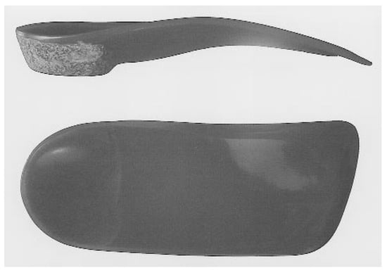
Group 1 (n = 85) received custom-made orthoses (Figure 1). On the initial visit, each patient was placed in the supine position while the foot was held with the subtalar joint neutral and the midtarsal joint fully pronated. The plantar contour of the foot underwent scanning by a laser beam and the resultant image was transmitted to a computer. The information was processed in a prescription format and sent to Bergmann Orthotic Laboratory in Northfield, Illinois. All orthoses were made of a rigid 5-mm Polydur (Atlas International, Sacramento, California) plastic material. The orthoses were intrinsically balanced in the forefoot-to-rearfoot neutral bisection. They were posted using a noncompressible composition cork material. The orthoses were constructed with 4° of rearfoot varus and with intrinsic forefoot posting, which allows the heel to remain inverted 4° at heel contact and allows 4° of motion so that the heel rocks in 4° on forefoot contact. No top covers were used. During the time the patients were awaiting the arrival of the orthoses, approximately 2 weeks, the foot was taped using a low-Dye technique.
Figure 1.
The custom-made orthoses created for patients in this study.
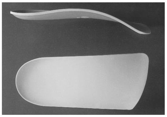
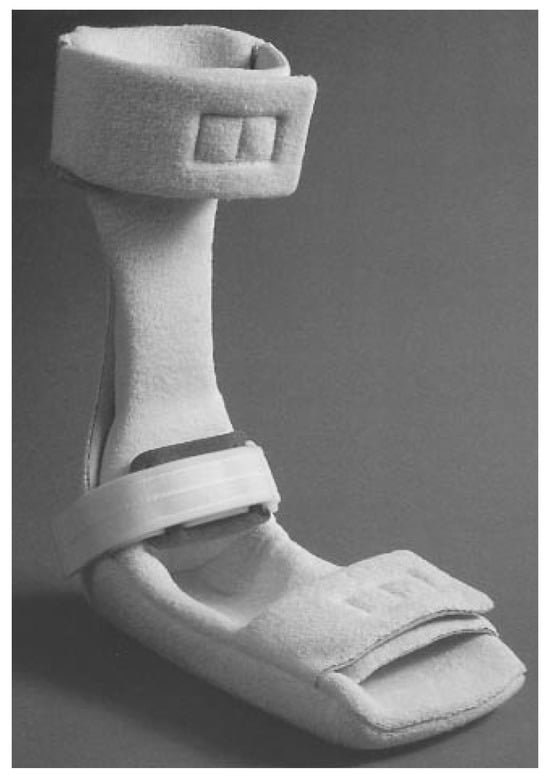
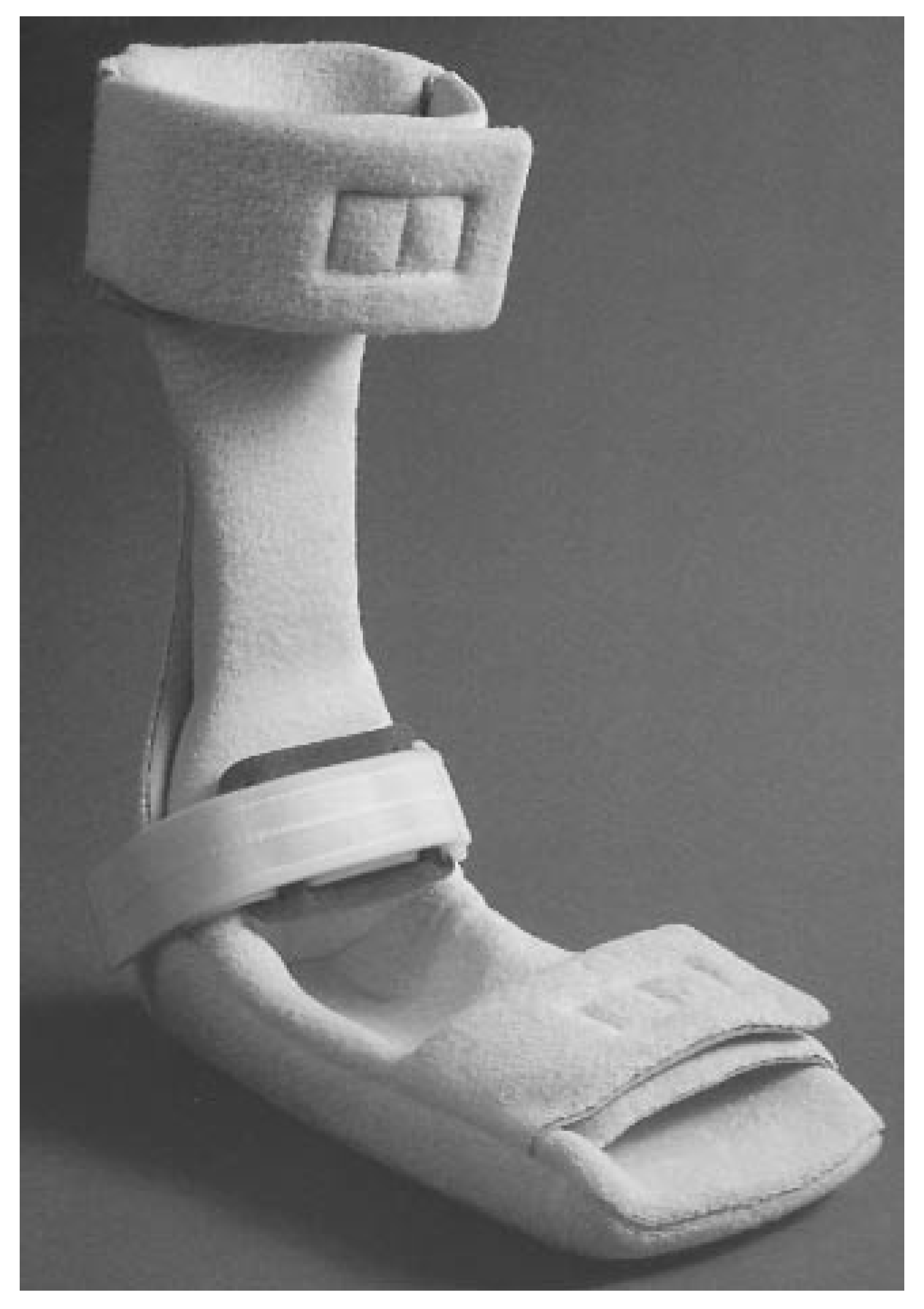
Group 2 (n = 85) received over-the-counter arch supports (Foot Soldiers, Professional Footcare International, Valencia, California) that are made of a rigid plastic (Figure 2). The foot was taped using a low-Dye technique for 2 weeks, to match the time period of group 1, after which the arch supports were dispensed. Group 3 (n = 85) received a posterior tension night splint, a prefabricated device set at 5° of ankle dorsiflexion and produced by AliMed, Inc (Dedham, Massachusetts) (Figure 3). It was dispensed at the initial visit and instructions were given to use it only at night. No taping was performed in this group because it does not represent a group receiving functional biomechanical control.
Figure 2.
The over-the-counter arch supports used in this study.
Figure 3.
The posterior tension night splint worn by patients in this study.
The effect of the heel pain on the three types of activities—leisure, work, and exercise—was defined as follows: 1) “none” meant that the patient answered no to the questions regarding all three activities, 2) “minimal” meant that the patient reported an effect on one category of activity, 3) “occasional” meant that the patient reported an effect on two categories of activity, and 4) “constant” meant that the patient reported an effect on all three categories of activity. The complaint of first-step pain was defined as follows: 1) “none” meant that the patient never complained of first-step pain, 2) “minimal” meant that the patient complained of first-step pain several times a month, 3) “occasional” meant that the patient complained of first-step pain several times a week, and 4) “constant” meant that the patient complained of first-step pain daily.
At the end of the 12 weeks, each patient reported for a final follow-up visit and the patients were categorized into outcome groups of excellent, good, and poor. An excellent outcome was defined as a visual analog scale score of 0 to 2, minimal-to-no effect on activities, and minimal-to-no first-step pain. A good outcome was defined as a visual analog scale score of 3 to 5, an occasional effect on activities, and occasional first-step pain. A poor outcome was defined as a visual analog scale score of 6 to 10, constant effect on activities, and constant first-step pain. A patient was also considered to have a poor outcome if he or she had to withdraw from the study because of severe pain, inability to tolerate the device, or noncompliance.
The areas of greatest interest in the results of this study were the final treatment outcome and the visual analog scale score over time. The final outcome was analyzed using the chi-square test. The pain scale was analyzed using repeated-measures analysis of variance. Baseline characteristics were compared using analysis of variance for continuous variables and the chi-square test for categorical variables. A P value of less than .05 was considered to indicate statistical significance.
Results
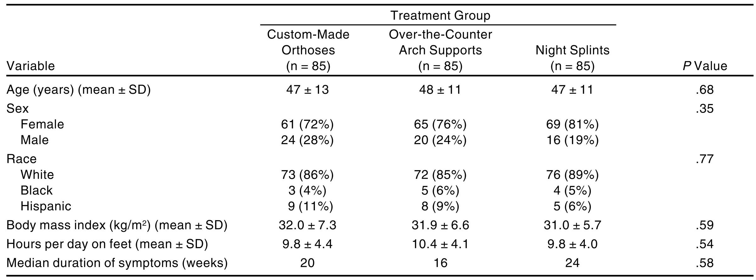
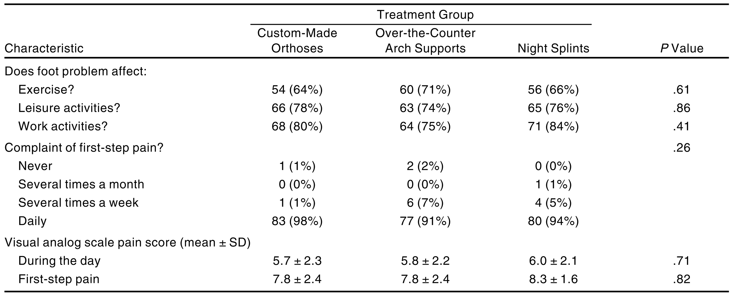
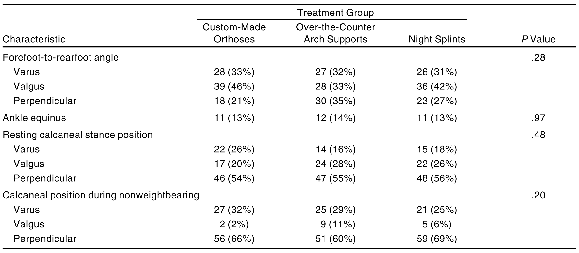
No statistically significant difference was noted among treatment groups with respect to demographic variables, which are summarized in Table 1. Nor were any statistically significant differences found among treatment groups with respect to subjective or objective baseline characteristics. Subjectively, this included whether the pain affected exercise, leisure, or work activities and whether a complaint of first-step pain occurred never, several times a month, several times a week, or daily. Visual analog scales were used to measure pain felt during the day and first-step pain in the morning. Objectively, four angular relationships were examined, as defined in “Materials and Methods.” The results are summarized in Table 2 and Table 3.
Table 1.
Summary of Demographic Variables of the Three Treatment Groups.
Table 2.
Summary of Subjective Baseline Characteristics of the Three Treatment Groups.
Table 3.
Summary of Objective Baseline Characteristics of the Three Treatment Groups.
Of the 255 patients enrolled in the study, 193 patients (76%) were seen at the 12-week visit for complete follow-up. A statistically significant difference was noted among the three treatment groups with respect to follow-up. Incomplete follow-up was due to the loss of patients to follow-up because they did not return for their appointments or to the withdrawal of patients from the study. Patients withdrawn from the study included those who requested to be withdrawn because of continued pain, those who did not use the devices properly, and those who could not tolerate the devices. The patients in the groups using overthe-counter arch supports and tension night splints had the poorest compliance rates and had the highest proportions of patients withdrawn, with 21% and 26%, respectively. The group using custom-made orthoses lost the most patients to follow-up (9%), but had relatively few who withdrew from the study (7%). Patients who withdrew from the study and therefore had no 3-month return visit were classified as poor outcomes. The follow-up information is summarized in Table 4.
Table 4.
Summary of Follow-up Information.
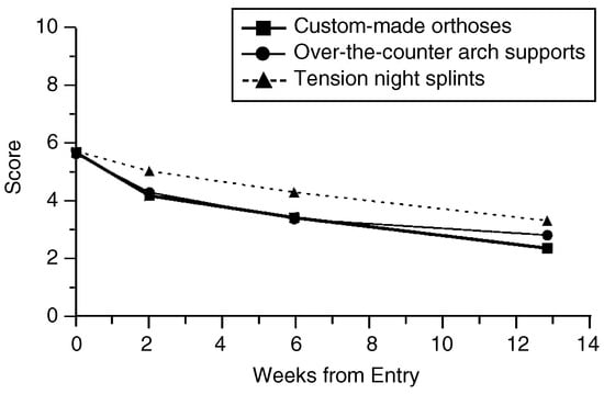
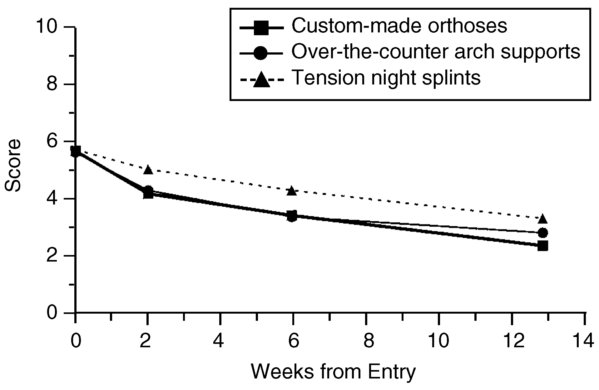
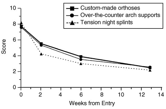
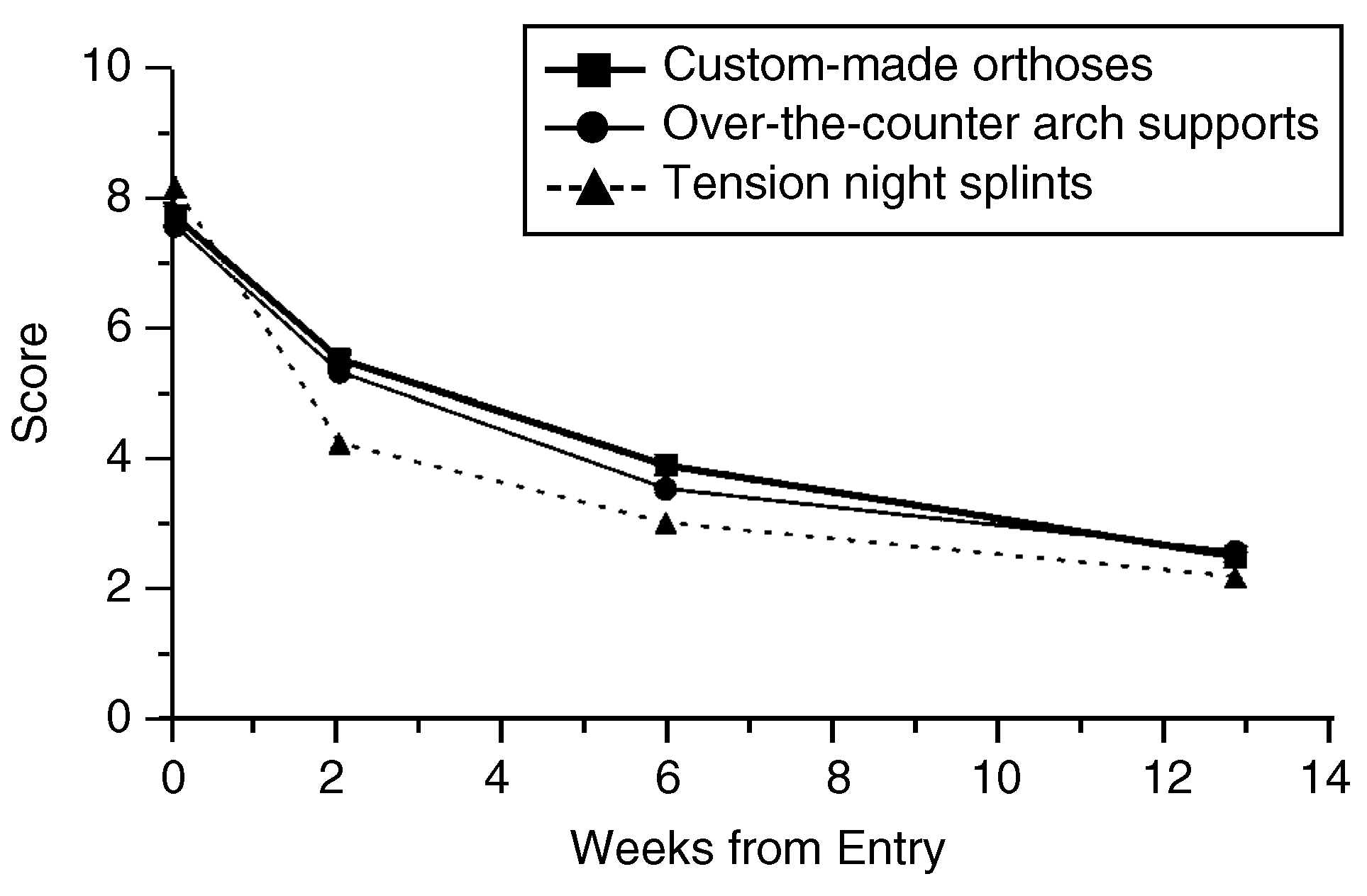
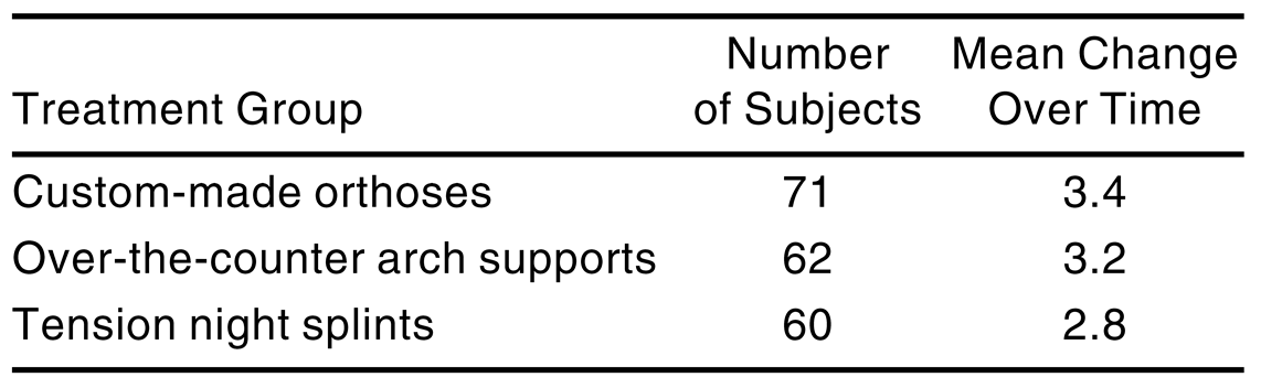
No statistically significant difference was noted among treatment groups with respect to the initial visual analog scale score of pain felt during the day. However, the mean change over time showed a slight difference. The group using custom-made orthoses showed the greatest improvement over time. These results are summarized in Table 5 and Figure 4.
Table 5.
Mean Change in Visual Analog Scale Score for Pain Felt During the Day.
Figure 4.
The visual analog scale scores for pain during the day for patients with complete follow-up.
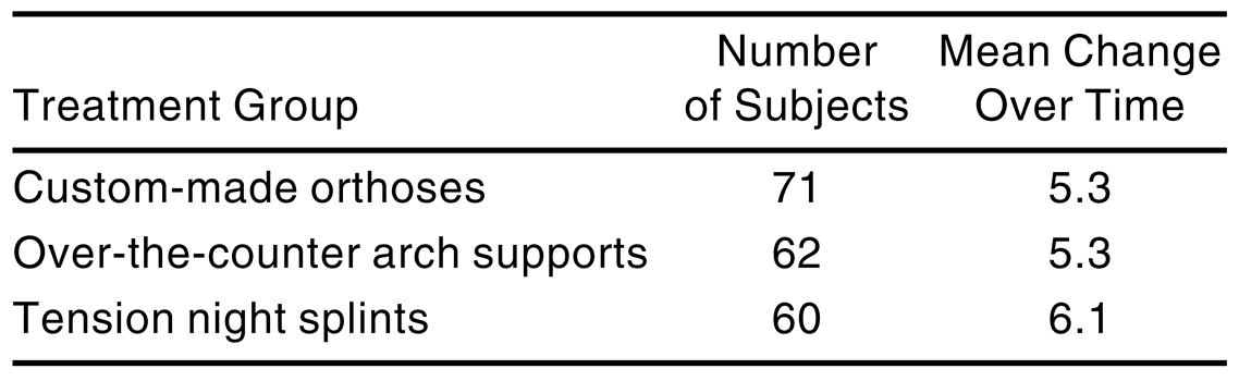
Neither was there any statistically significant difference noted among treatment groups with respect to the initial visual analog scale score of first-step pain. However, the mean change over time demonstrated that use of tension night splints had a greater effect than the other two modalities in reducing symptoms. These results are summarized in Table 6 and Figure 5.
Table 6.
Mean Change in Visual Analog Scale Score for First-Step Pain.
Figure 5.
The visual analog scale scores for first-step pain for patients with complete follow-up.
No statistically significant difference was found among treatment groups with respect to a final outcome of excellent or good versus a poor outcome based on daytime pain or first-step pain. Based on pain felt during the day, an excellent or good outcome was reported by 68% of the patients using custom-made orthoses, 57% of the group using over-thecounter arch supports, and 51% of the group using night splints. Based on first-step pain, an excellent or good outcome was reported by 61% of the group using custom-made orthoses, 57% of the group using over-the-counter arch supports, and 57% of the group using night splints.
Discussion
The present study shows that there were no statistically significant differences among the treatment groups in overall effectiveness during 12 weeks of treatment for plantar fasciitis. Tension night splints demonstrated a greater effect than the other two modalities in reducing symptoms, based on mean change over time for first-step pain, but overall, the groups were comparable in final outcome. The overall success rate of treatment in the present study is lower than rates in studies in which multiple modalities were used.[1,28,29,35,40,41,42] The aim of the present study was to compare the outcomes of groups of patients who used a single treatment modality; the overall success rate could be improved with the use of adjunctive treatment modalities. Clinical examinations as performed in this study may produce results that differ from those of other methods, such as telephone surveys or questionnaires.
Conclusion
Mechanical control of the foot is a successful method of treating plantar fasciitis. Custom-made orthoses, over-the-counter arch supports, and tension night splints are all effective as initial treatments for plantar fasciitis. Patients in the present study demonstrated the best compliance with the use of custom-made orthoses, which may indicate that orthoses provide the best long-term results.
References
- PFEFFER, G.; BACCHETTI, P.; DELAND, J.; et al. Comparison of custom and prefabricated orthoses in the initial treatment of proximal plantar fasciitis. Foot Ankle Int 1999, 20, 214. [Google Scholar] [CrossRef] [Scilit]
- MCCARTHY, D.J.; GORECKI, G.E. The anatomical basis of inferior calcaneal lesions: a cryomicrotomy study. JAPA 1979, 69, 527. [Google Scholar] [CrossRef] [Scilit] [PubMed]
- DAILEY JM: Differential diagnosis and treatment of heel pain. Clin Podiatr Med Surg 1991, 8, 153. [CrossRef] [Scilit]
- KWONG, P.K.; KAY, D.; VONER, R.T.; et al. Plantar fasciitis: mechanics and pathomechanics of treatment. Clin Sports Med 1988, 7, 119. [Google Scholar] [CrossRef] [Scilit] [PubMed]
- HICKS JH: The function of the plantar aponeurosis. J Anat 1951, 85, 414.
- HICKS JH: Mechanics of the foot: plantar aponeurosis and arch. J Anat 1954, 88, 25.
- SNIDER, M.P.; CLANCY, W.G.; MCBEATH, A.A. Plantar fascia release for chronic plantar fasciitis in runners. Am J Sports Med 1983, 11, 215. [Google Scholar] [CrossRef] [Scilit]
- BERKIOWITZ, J.F.; KIER, R.; RUDICEL, S. Plantar fasciitis: MR imaging. Radiology 1992, 182, 285. [Google Scholar] [CrossRef] [Scilit]
- CARDINAL, E.; CHHEM, R.K.; BEAUREGARD, C.G. ET AL: Plantar fasciitis: sonographic evaluation. Radiology 1996, 201, 257. [Google Scholar]
- WALL, J.R.; HARKNESS, M.A. CRAWFORD A: Ultrasound diagnosis of plantar fasciitis. Foot Ankle 1993, 14, 465. [Google Scholar] [CrossRef] [Scilit]
- SCHEPSIS, A.A.; LEACH, R.E.; GORZYCA, J. Plantar fasciitis: etiology, treatment, surgical results, and review of the literature. Clin Orthop 1991, 266, 185. [Google Scholar] [CrossRef] [Scilit]
- TANNER, S.M. HARVEY JS: How we manage plantar fasciitis. Physician Sportsmed 1988, 16, 39. [Google Scholar] [CrossRef] [Scilit]
- WARREN, B.L.; JONES, C.J. Predicting plantar fasciitis in runners. Med Sci Sports Exerc 1987, 19, 71. [Google Scholar] [CrossRef] [Scilit] [PubMed]
- WARREN, B.L. Anatomical factors associated with predicting plantar fasciitis in long-distance runners. Med Sci Sports Exerc 1984, 16, 60. [Google Scholar] [CrossRef] [Scilit] [PubMed]
- LAPIDUS, P.W.; GUIDOTTI, F.P. Painful heel: report of 323 patients with 364 painful heels. Clin Orthop 1965, 39, 178. [Google Scholar] [CrossRef] [Scilit]
- HILL, J.J.; CUTTING, P.J. Heel pain and body weight. Foot Ankle 1989, 9, 254. [Google Scholar] [CrossRef] [Scilit]
- KIBLER, W.B.; GOLDBERG, C.; CHANDLER, T.J. Functional biomechanical deficits in running athletes with plantar fasciitis. Am J Sports Med 1991, 19, 66. [Google Scholar] [CrossRef] [Scilit] [PubMed]
- SNOOK, G.A. CHRISMAN OD: The management of subcalcaneal pain. Clin Orthop 1972, 82, 163. [Google Scholar] [CrossRef] [Scilit]
- RIZZO, T.D. Plantar fasciitis: overcoming a nagging pain in the arch. Physician Sportsmed 1991, 19, 129. [Google Scholar] [CrossRef] [Scilit]
- FUREY, J.G. Plantar fasciitis: the painful heel syndrome. J Bone Joint Surg Am 1975, 57, 672. [Google Scholar] [CrossRef] [Scilit]
- BLOCKEY NJ: The painful heel: a controlled trial of the volume of hydrocortisone. BMJ 1956, 1, 1277.
- ROY S: How I manage plantar fasciitis. Physician Sportsmed 1983, 11, 127. [CrossRef] [Scilit]
- GUDEMAN, S.D.; EISELE, S.A.; HEIDT, R.S., JR.; et al. Treatment of plantar fasciitis by iontophoresis of 0.4% dexamethasone: a randomized, double-blind, placebo-controlled study. Am J Sports Med 1997, 25, 312. [Google Scholar] [CrossRef] [Scilit]
- BOYD HS: Plantar fasciitis. Orthop Review 1992, 21, 116.
- MIZEL, M.S.; MARYMONT, J.V. TREPMAN E: Treatment of plantar fasciitis with a night splint and shoe modification consisting of a steel shank and anterior rocker bottom. Foot Ankle Int 1996, 17, 732. [Google Scholar] [CrossRef] [Scilit]
- GOULET MJ: Role of soft orthosis in treating plantar fasciitis: suggestion from the field. Phys Ther 1984, 64, 1544. [CrossRef] [Scilit]
- ROSE GK: Painful heel. BMJ 1955, 2, 831.
- CAMPBELL, J.W.; INMAN, V.T. Treatment of plantar fasciitis and calcaneal spurs with the UC-BL shoe insert. Clin Orthop 1974, 103, 57. [Google Scholar] [CrossRef] [Scilit] [PubMed]
- Scherer PR, BIOMECHANICS GRADUATE RESEARCH GROUP FOR 1988: Heel spur syndrome: pathomechanics and nonsurgical treatment . JAPMA 1991, 81, 68.
- POWELL, M.; POST, W.R.; KEENER, J.; et al. Effective treatment of chronic plantar fasciitis with dorsiflexion night splints: a crossover prospective randomized outcome study. Foot Ankle Int 1998, 19, 10. [Google Scholar] [CrossRef] [Scilit] [PubMed]
- WAPNER, K.L. SHARKEY PF: The use of night splints for treatment of recalcitrant plantar fasciitis. Foot Ankle 1991, 12, 135. [Google Scholar] [CrossRef] [Scilit] [PubMed]
- RYAN J: Use of posterior night splints in the treatment of plantar fasciitis. Am Family Phys 1995, 52, 891.
- BATT, M.E.; TANJI, J.L. SKATTUM N: Plantar fasciitis: a prospective randomized clinical trial of the tension night splint. Clin J Sports Med 1996, 6, 158. [Google Scholar]
- CHANG, C.C. MILTNER LJ: Periostitis of the oscalcis. J Bone Joint Surg 16, 355, 1934.
- WOLGIN, M.; COOK, C.; GRAHAM, C. ET AL: Conservative treatment of plantar heel pain: long-term follow-up. Foot Ankle Int 1994, 15, 97. [Google Scholar] [CrossRef] [Scilit]
- O’BRIEN, D. MARTIN WJ: A retrospective analysis of heel pain. JAPMA 1985, 75, 416. [Google Scholar] [CrossRef] [Scilit]
- LYNCH, D.M.; GOFORTH, W.P.; MARTIN, J.E. ET AL: Conservative treatment of plantar fasciitis: a prospective study. JAPMA 1998, 88, 375. [Google Scholar]
- FLANDRY, F.; HUNT, J.P.; TERRY, G.C. ET AL: Analysis of subjective knee complaints using visual analog scales. Am J Sports Med 1991, 19, 112. [Google Scholar] [CrossRef] [Scilit] [PubMed]
- WEWERS ME, LOWE NK: A critical review of visual analogue scales in the measurement of clinical phenomena. Res Nurs Health 1990, 13, 227. [CrossRef] [Scilit]
- MARTIN, R.L.; IRRGANG, J.J.; CONTI, S.F. Outcome study of subjects with insertional plantar fasciitis. Foot Ankle Int 1998, 19, 803. [Google Scholar] [CrossRef] [Scilit]
- GILL, L.H.; KIEBZAK, G.M. Outcome of nonsurgical treatment for plantar fasciitis. Foot Ankle Int 1996, 17, 527. [Google Scholar] [CrossRef] [Scilit] [PubMed]
- DAVIS, P.F.; SEVERUD, E.; BAXTER, D.E. Painful heel syndrome: results of nonoperative treatment. Foot Ankle Int 1994, 15, 531. [Google Scholar] [CrossRef] [Scilit] [PubMed]


© 2001 American Podiatric Medical Association