


To the Editor:
There is overwhelming agreement that bullous lesions associated with carbon monoxide poisoning typically appear within 24 hours and are found primarily on the palmar surface of the hands and the plantar surface of the feet [1,2,3]. Any areas of pressure or friction are vulnerable to the development of such lesions. These areas include the posterior scalp, wrists, knees, legs, ankles, and dorsa of the feet [3,4]. No recent reports have directly associated bullous lesions with carbon monoxide poisoning. The case described herein is an unusual presentation of a secondary manifestation of carbon monoxide poisoning.
Case Report
A 37-year-old man presented to the emergency department after attempted suicide by carbon monoxide inhalation. After medical stabilization, the patient was transferred to the mental-health unit. Twenty-four hours after admission, the authors were consulted for evaluation and treatment of two painful bullae located on the left heel.
The patient stated that he had a history of depression and multiple suicide attempts. He had had a cerebral vascular accident secondary to a suicide attempt 4 years previously with no residual effects. He was taking the following medications: fluoxetine hydrochloride, trazodone hydrochloride, thiamine hydrochloride, and acetaminophen. The patient denied the use of alcohol, tobacco, or illicit drugs. He had no known drug allergies. He recalled no history of trauma or any occurrence of a similar lesion.
Upon physical examination, the patient was found to be alert, oriented to person, place, and time, and in no apparent distress. His vital signs were stable, and he was afebrile. The neurovascular status of both lower extremities was intact. The dermatologic examination revealed an 8 × 9-cm bulla on the plantar aspect of the heel as well as a 2 × 3-cm bulla beneath the cuboid, both on the left foot. A localized rim of erythema surrounded each bulla. Otherwise, skin temperature, texture, and turgor were normal.
Attention was directed to the bullae; the overlying skin was noted to be tense. Upon drainage, both bullae were found to be multilocular. The fluid was serous in nature. Cultures and Gram’s stain performed at this time were negative.
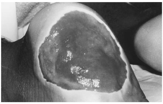
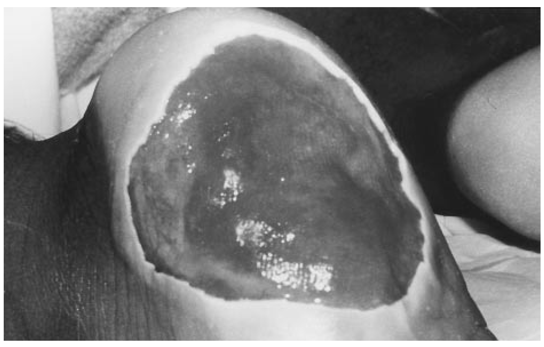
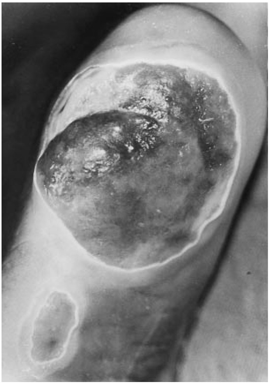
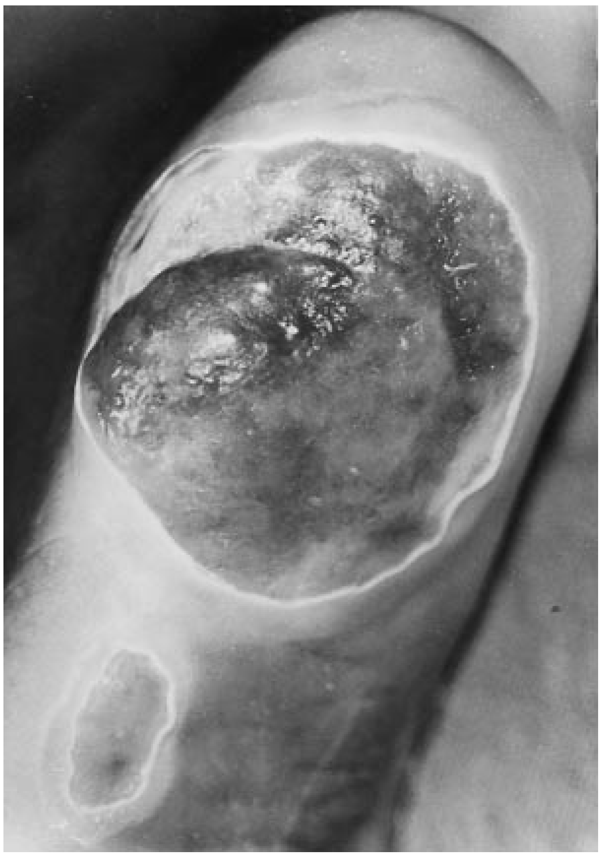
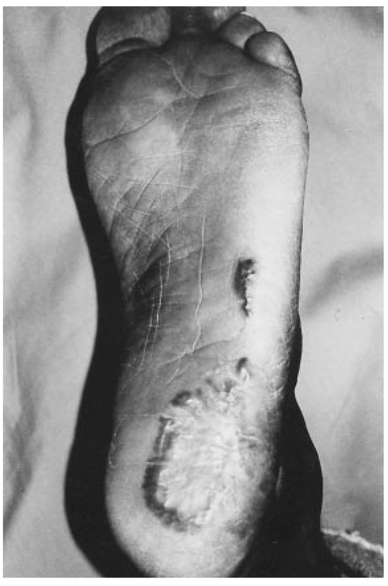
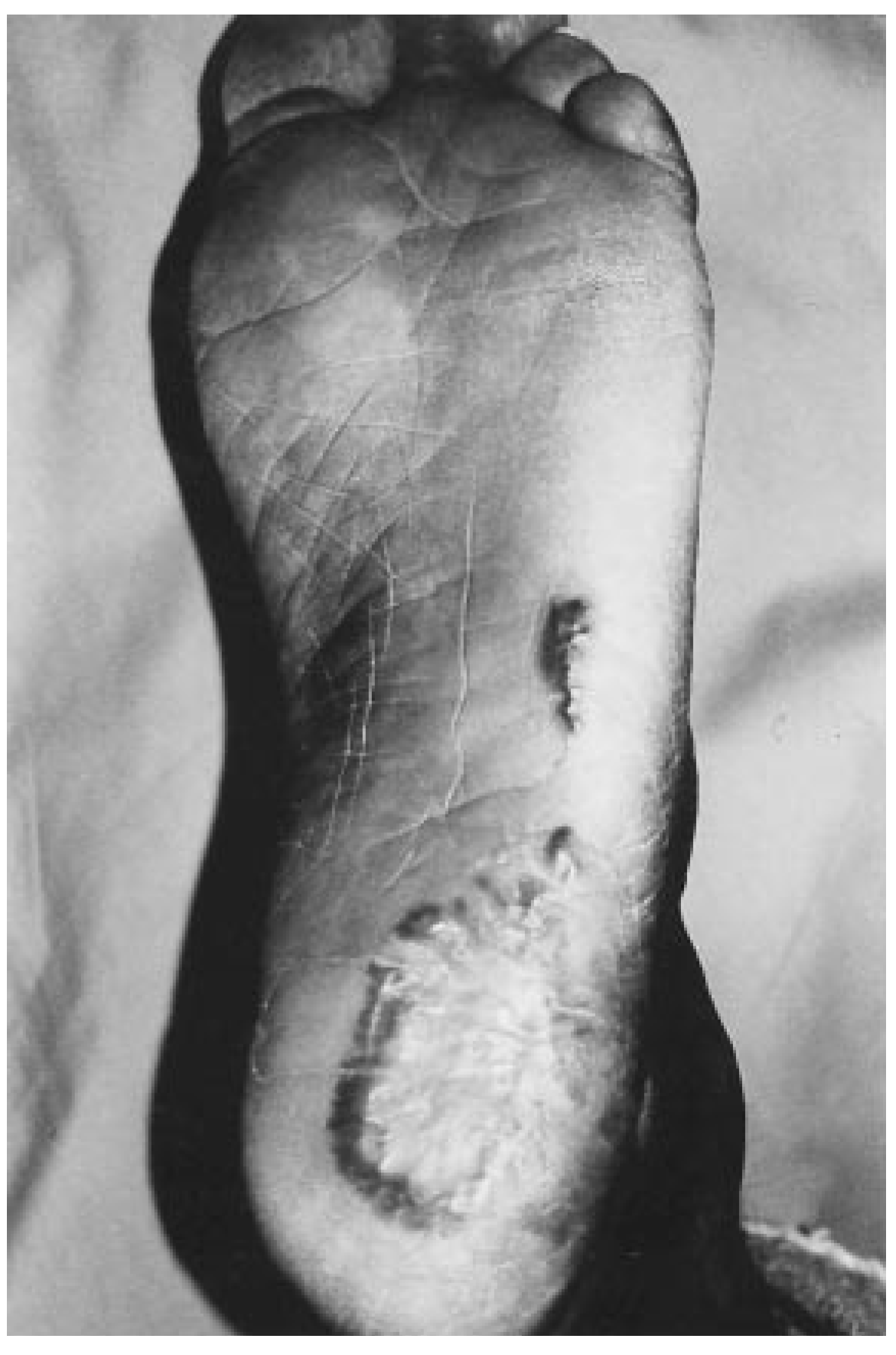
Local wound care consisted of a dry, sterile dressing. The patient remained nonweightbearing with crutch-assisted ambulation. The bullae were de-roofed, revealing contents of an “apple jelly” color and consistency. No granular base was noted. There were no signs of infection. During the next 24 hours, there was copious serous drainage requiring large bulky dressings to be changed as many as ten times (Fig. 1). An estimated 4 L of serous fluid drained from the wounds. Profound epidermal and dermal lysis occurred beneath the initial bullae. Over the following week, the lesions continued to drain profusely and required well-padded dressings. Intradermal and subdermal bullae continued to form at the two sites during this period (Fig. 2).
Figure 1.
Day 3. Larger bulla (8 × 9 cm) fills the plantar aspect of the left heel and is actively draining a serous fluid.
Figure 2.
Day 5. Bullae are observed on the lateral aspect of the left foot. Continued intradermal and subdermal blistering is apparent.
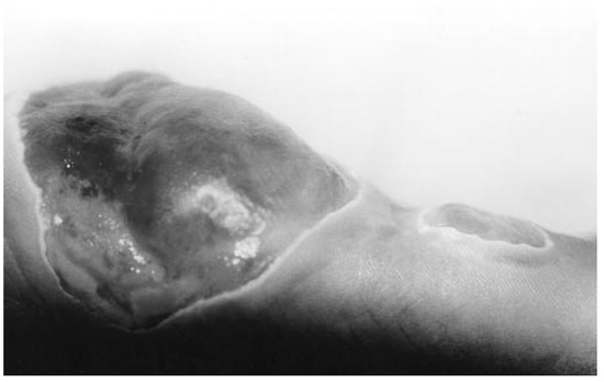
Laboratory tests consisting of a complete blood count, chemistry profile, urinalysis, and wound cultures were performed, and the results were found to be within normal limits. The patient remained afebrile and stable. The wounds remained benign in appearance over time. As the wounds stabilized and the drainage decreased, wound care was modified (Fig. 3). Aluminum chlorohydrate soaks, followed by application of gentamicin 0.1% ointment, were instituted three times per day. The patient was discharged after 2 weeks with instructions for local wound care.
Figure 3.
Day 14. New blister formation has ceased and drainage is greatly decreased.
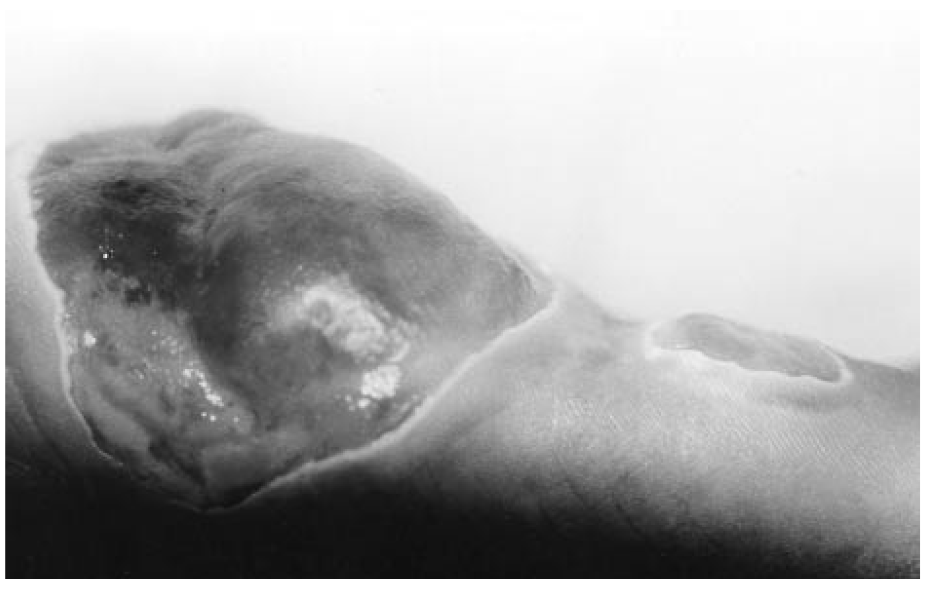
The patient was seen weekly in the outpatient clinic and a gradual, uneventful course of wound healing was noted. The patient remained nonweight-bearing with crutch-assisted ambulation during the healing process. The wounds developed granular bases, which gradually epithelialized with complete resolution after approximately 8 months (Fig. 4).
Figure 4.
Eight months. The lesions have healed and the patient is able to bear full weight without pain.
Discussion
The authors found no recent case reports directly associating bullous lesions and carbon monoxide poisoning. The literature on dermatologic lesions associated with carbon monoxide poisoning is scant. Three types of skin lesions have been described: 1) tense vesicular lesions with an erythematous base; 2) dusky, erythematous plaques; and 3) large, tense bullae bordered by a rim of localized erythema [1]. There have been very few reported cases of this latter type of bullae formation from toxic poisoning. These types of cutaneous manifestations have been found primarily in patients who lost consciousness after overdoses from barbiturates, methadone hydrochloride, hydrocodone bitartrate, meprobamate, imipramine hydrochloride, or glutethimide [1,2,5,6]. It has been reported that only approximately 5% of patients with toxic poisoning develop associated blisters or bullae [5].
Multiple hypotheses on the etiology of bullous formation in conjunction with carbon monoxide poisoning have been proposed. Four general theories predominate: 1) local hypoxia and trauma [1]; 2) pure toxic effect [7]; 3) pure local friction [2]; and 4) hypoxia, local pressure, and hyperthermia [8].
No scientific research exists proving or disproving any of these theories. Biopsies of these lesions show significant and dramatic alteration and necrosis of the eccrine sweat glands within the epithelium.1 Subepidermal bullae demonstrate intraepidermal spongiosis and vesiculations located peripherally about intraepidermal sweat ducts [1]. Interestingly, in the reported cases, reestablishment of apparently normal sweat glands occurred within 10 to 14 days [1,3,5].
Conclusion
An unusual and rarely encountered clinical entity has been presented. The initial lesions, although locular, appeared benign in nature. An infectious process was considered unlikely. The lesions were initially thought to have been caused by localized trauma. It was not until the bullae began to form deeper layers of blistering, with increased drainage, that an extensive differential diagnosis including carbon monoxide–related bullae formation was considered. This case report aims to heighten the physician’s awareness of the possibility of bullae formation due to toxic poisoning.
References
- Mandy, S.; Ackerman, A.B. Characteristic traumatic skin lesions in drug-induced coma. JAMA 1970, 213, 253. [Google Scholar] [CrossRef] [Scilit] [PubMed]
- Sorensen, B.F. Skin symptoms in acute narcotic intoxication. Dan. Med. Bull 1963, 10, 130. [Google Scholar] [PubMed]
- Leavell, U.W.; Farley, C.H.; Mcintyre, J.S. Cutaneous changes in a patient with carbon monoxide poisoning. Arch. Dermatol. 1969, 99, 429. [Google Scholar] [CrossRef] [Scilit] [PubMed]
- Long, P.I. Dermal changes associated with carbon monoxide intoxication. JAMA 1968, 205, 120. [Google Scholar] [CrossRef] [Scilit] [PubMed]
- Varma, A.J.; Fisher, B.K.; Sarin, M.K. Diazepam-induced coma with bullae and eccrine sweat gland necrosis. Arch. Intern. Med. 1977, 137, 1207. [Google Scholar] [CrossRef] [PubMed]
- Beveridge, G.W.; Lawson, A.A.H. Occurrence of bullous lesions in acute barbiturate intoxication. BMJ 1965, 1, 835. [Google Scholar] [CrossRef] [Scilit] [PubMed]
- Holton, C. Cutaneous phenomena in acute barbiturate poisoning. Acta Dermatovener 1951–1952, 27, 162. [Google Scholar]
- Achten, G.; Ledoux-Corbusier, M.; Thys, J.P. Carbon monoxide poisoning and cutaneous lesions [in French]. Ann. Dermatol. Syphiligr (Paris) 1971, 98, 421. [Google Scholar] [PubMed]
© 1999 American Podiatric Medical Association