


The diagnosis of pyoderma gangrenosum is a difficult one to make because of the condition’s ability to mimic other ulcerative lesions of the foot and its lack of specific laboratory and pathologic findings. A high index of clinical suspicion can lead to a definitive diagnosis. This article presents a case report and a discussion of the evaluation and management of pyoderma gangrenosum. (J Am Podiatr Med Assoc 89(3): 141-142, 1999)
Pyoderma gangrenosum can appear anywhere on the body but is most commonly seen in the trunk and lower extremities [1]. The condition is often difficult to diagnose because it can mimic other ulcerative conditions that may also affect the foot, including erythema nodosum, erythema multiforme, folliculitis, bacterial infection, fungal infection, spider bites, and vascular lesions [2]. Diagnosis is based on the clinical appearance of the lesion, coexisting systemic disease, and the exclusion of other causes of ulceration [3].
Pyoderma gangrenosum may be idiopathic or may be associated with inflammatory bowel disease, rheumatoid arthritis, IgA gammopathy, or myeloproliferative disorders [4]. The earliest symptom of pyoderma gangrenosum may be pain in the area of the impending lesion. The lesion begins as a pustule often following minor trauma. The pustule rapidly evolves into a hematoma-like lesion, progressing to a sterile abscess that spreads concentrically to rapidly undermine adjacent areas of viable skin, and finally to an enlarging, painful ulcer. The ulcer is characterized by a purulent necrotic center surrounded by a violaceous undermined border that fades to erythema more peripherally [5].
Pyoderma gangrenosum is not associated with specific laboratory and microscopic findings [6]. Histologically, necrosis and ulceration of epidermis and dermis along with heavy infiltration of polymorphonuclear leukocytes may be seen. There may be dense inflammatory cell infiltrates, abscess or granuloma formation, and loss of elastic tissue fibers. Abnormal laboratory findings may include elevated erythrocyte sedimentation rate, leukocytosis, low serum iron, hyperglobulinemia, or hypoglobulinemia.
Corticosteroid therapy is the primary form of treatment [7]. Some patients may benefit from the use of dapsone, sulfapyridine, and sulfasalazine.
Case Report
A 75-year-old woman presented to Jamaica Hospital Medical Center with a painful ulcer on the dorsal aspect of her right foot of 2 months’ duration. The patient could not remember sustaining trauma to the area or any activity that could have precipitated the appearance of the lesion.
The patient’s medical history was significant for ulcerative colitis diagnosed 20 years previously. She had also experienced pyoderma gangrenosum on the other foot 1 year previously. She had not had any previous surgeries. She was taking cephalexin, which had been prescribed 1 month previously by her primary- care physician, who thought her ulcer might be infected. The patient’s vital signs were all within normal limits, and review of vascular, neurologic, and musculoskeletal systems was unremarkable.
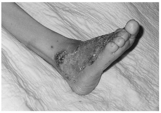
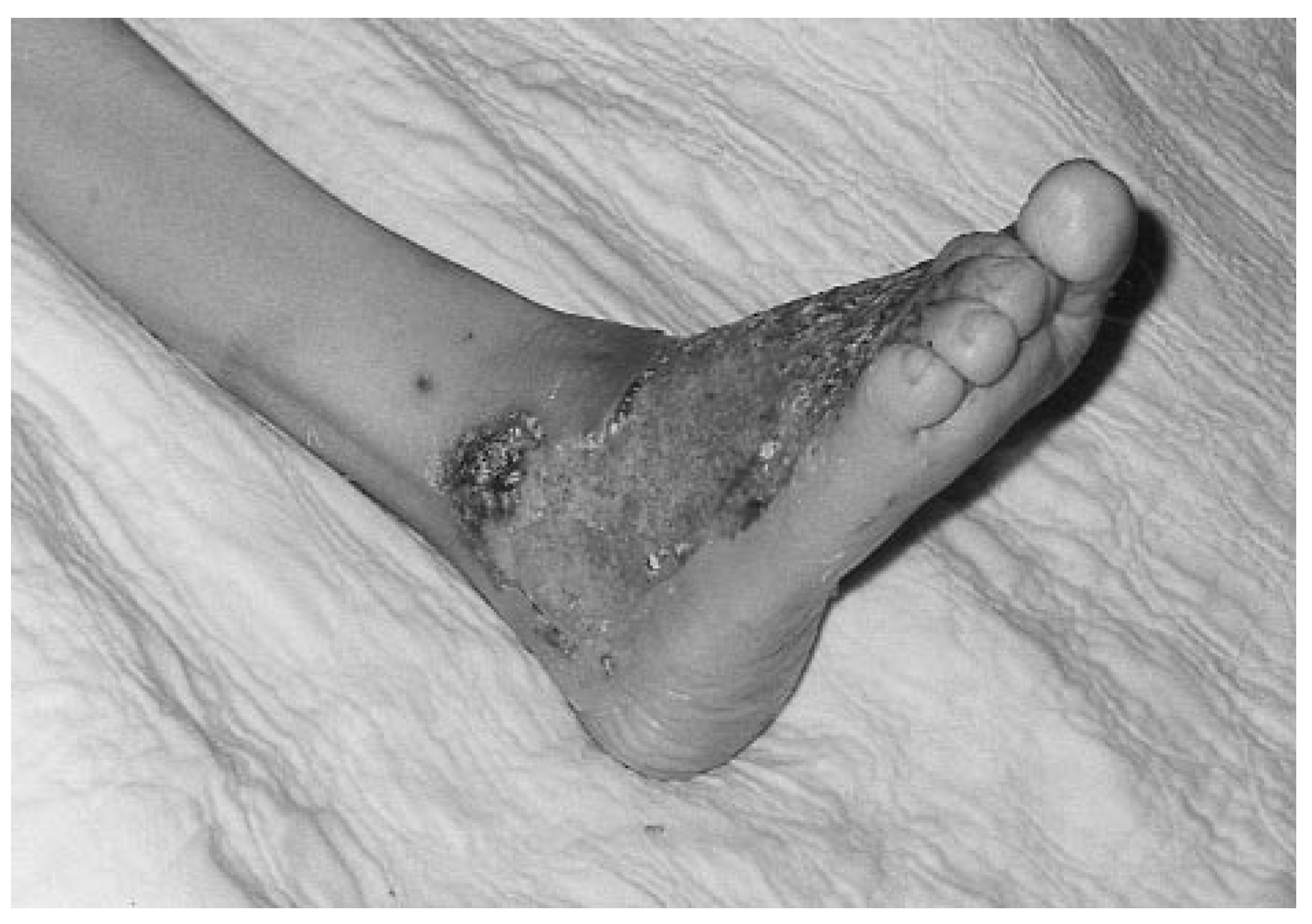
Physical examination revealed a superficial ulcer covering the dorsolateral aspect of the right foot and extending to the lateral malleolus (Figure 1). The ulcer measured approximately 5 × 6 cm. It was irregular in shape, and its borders were surrounded by a halo of erythema. A yellowish exudate was noted draining from the lesion, and there were areas of necrotic tissue along the edges of the ulcer. The ulcer was exquisitely tender on palpation.
Figure 1.
Pyoderma gangrenosum of the right foot at initial presentation.
Diagnostic testing consisted of a complete blood count, blood chemistry profile, x-rays, and a threephase bone scan, all of which were unremarkable. The only abnormal finding was an elevated erythrocyte sedimentation rate of 85 mm/h.
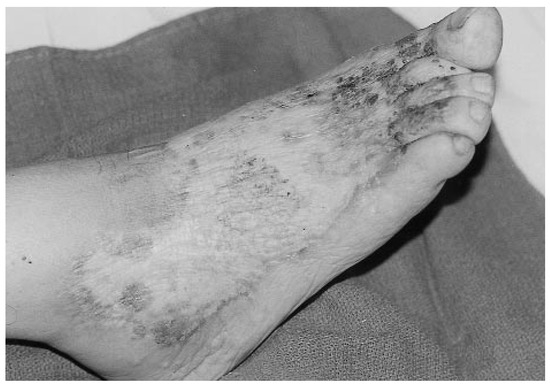
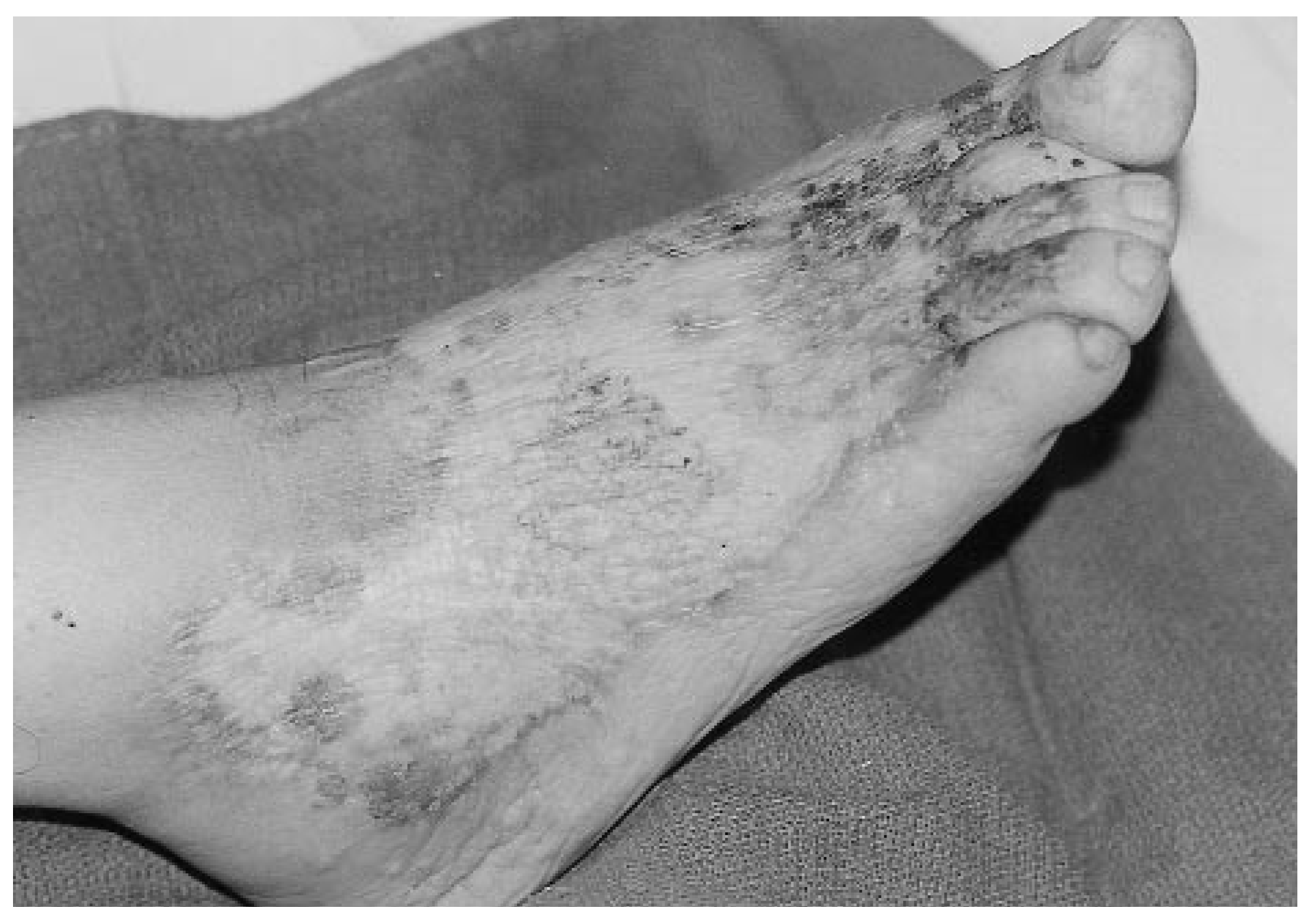
The patient was admitted to the hospital with a diagnosis of pyoderma gangrenosum and was begun on a regimen of intravenous nafcillin, 1 g every 12 hours. At the recommendation of the podiatry department, wet-to-dry dressing changes with normal saline were begun, along with application of hydrocortisone 1% cream once daily. The intravenous nafcillin and hydrocortisone cream were then discontinued and the patient was started on prednisone, 10 mg once daily, and sulfasalazine, 500 mg twice daily; this regimen was continued for 10 days, at which time the patient was discharged from the hospital. At discharge, the patient was instructed to continue local wound care and was prescribed a maintenance drug regimen of sulfasalazine, 500 mg three times daily. At follow-up 8 weeks later, the lesion had healed completely (Figure 2). A cribriform scar remained at the site of the outer region of the ulcer.
Figure 2.
Pyoderma gangrenosum lesion at 8-week follow-up; complete healing is evident.
Discussion
The diagnosis of pyoderma gangrenosum is often difficult owing to the disorder’s ability to mimic other ulcerative conditions. Diagnosis is based on the clinical appearance of the lesion and coexisting systemic disease. In the case reported here, ulcerative colitis was the underlying systemic condition. The diagnosis of pyoderma gangrenosum was made after other possible causes of nonhealing ulcers such as infection, diabetes mellitus, vascular insufficiency, and neurologic pathology had been ruled out.
Traditionally, all nonhealing ulcers are biopsied to rule out malignancy [8]. In this case, biopsy of the lesion was not performed because it was feared that debridement of the lesion might be traumatic and cause new lesions.
Once the patient was placed on corticosteroid therapy, the condition improved and the lesion healed uneventfully.
References
- Seitzinger, J.W. Pyoderma gangrenosum. N. J. Med. 1989, 86, 465. [Google Scholar] [PubMed]
- Hodges, R.E. Nutrition and the Integument, Including Mucous Membranes. In Nutrition in Medical Practice; Hodges, R., Ed.; WB Saunders: Philadelphia, PA, USA, 1980; p. 265. [Google Scholar]
- Lerner, J.; Lerner, M.; Pasternackoot, W.A. Pyoderma gangrenosum. J. Foot Ankle Surg. 1993, 32, 569. [Google Scholar] [PubMed]
- Callen, J.P. Pyoderma gangrenosum and related disorders. Adv. Dermatol. 1989, 4, 51. [Google Scholar] [CrossRef] [Scilit] [PubMed]
- Walling, A.D.; Sweet, D. Pyoderma gangrenosum. Am. Fam. Physician 1987, 35, 159. [Google Scholar] [PubMed]
- Wolf, K.; Stingl, G. Pyoderma Gangrenosum. In Dermatology in General Medicine, 4th ed.; Fitzpatrick, T.B., Eisen, A.Z., Wolf, K., Eds.; McGraw-Hill: New York, NY, USA, 1993; p. 1171. [Google Scholar]
- Hickman, J.G.; Lazarus, G.S. Pyoderma Gangrenosum: New Concepts in Etiology and Treatment. In Dermatology Update: Review for Physicians; Moschella, S.L., Ed.; Elsevier: New York, NY, USA, 1979; p. 325. [Google Scholar]
- White, J.W. Hypersensitivity and Miscellaneous Inflammatory Disorders. In Dermatology, 2nd ed.; Moschella, S.L., Hurley, H.J., Eds.; WB Saunders: Philadelphia, PA, USA, 1985; p. 478. [Google Scholar]
Disclaimer/Publisher’s Note: The statements, opinions and data contained in all publications are solely those of the individual author(s) and contributor(s) and not of MDPI and/or the editor(s). MDPI and/or the editor(s) disclaim responsibility for any injury to people or property resulting from any ideas, methods, instructions or products referred to in the content. |
© 1999 American Podiatric Medical Association