


Abstract
The authors review the ankle and pantalar fusion literature. The authors performed a retrospective review on 42 ankle and pantalar fusions, emphasizing the role of internal fixation. The Podiatry Institute technique for internal fixation of ankle fusions is described and experience with the technique is reviewed. Complications included delayed union and nonunion at the fusion site or the repaired fibular osteotomy site and tibial fractures at screw stress riser sites. Modifications to reduce these complications are discussed. Experience with internal fixation in ankle and pantalar fusions, both in this study and in current literature, has been positive.
Ankle fusion has been practiced as a surgical procedure for more than 100 years [1]. There have been at least 40 techniques and modifications described to effect fusion [2]. Arthrodesis itself is a technique designed to achieve bony union across a joint. Despite the development of artificial joint implants for many areas of the body, arthrodesis remains a primary procedure for severe diseases and disorders of the ankle [3,4]. The authors describe a technique of internal fixation for ankle and pantalar fusions and their experience with this technique over 10 years.
That so many procedures and modifications of procedures have been described suggests that no one procedure has been found to be universally acceptable in all situations for ankle or pantalar fusion.
Indications
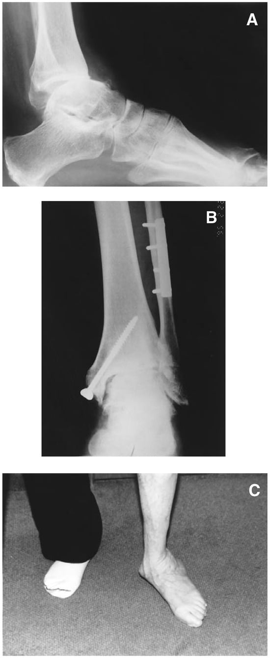
Arthrodesis of the ankle is primarily indicated in patients with severe pain and deformity (Figure 1). Individual indications include post-traumatic arthrosis, rheumatoid arthritis, talar collapse, drop foot, tumors with joint invasion, neuromuscular deformities, and failed ankle joint prostheses.
Figure 1.
Examples of indications for ankle and pantalar arthrodesis. (A), Lateral view showing anterior subluxation of the talus on the tibia with coexisting degenerative joint disease. (B), Anteroposterior ankle view showing degenerative joint disease following pronation-external rotation ankle fracture. (C), Peritalar collapse of the left foot following tibialis posterior dysfunction.
The most common indication for ankle fusion is post-traumatic arthrosis following malleolar or pilon fractures [5]. Degeneration of the ankle joint following fracture of the ankle is associated with many factors, most notably lateral shifting of the talus or shortening of the fibula. Failure to open reduce certain types of fractures, or inadequate reduction or fixation, can result in joint malalignment leading to degenerative joint disease. Ramsey and Hamilton [6] showed that the initial 1-mm lateral displacement of the talus in the ankle mortise after fracture results in a 42% decrease in tibiotalar contact. Since a decrease in contact area means that a greater force is transmitted per unit area, it is reasonable to see how fractures with small amounts of lateral shifting can result in severe degenerative joint disease of the ankle.
Paralytic deformities are also an indication for ankle or pantalar fusion, particularly when there are no useful muscles to transfer [7]. Although poliomyelitis was once the most common paralytic indication for ankle fusion, it is less common today, except in adult patients suffering post-poliomyelitis syndrome and children from developing countries. Other neuromuscular deformities that may result in ankle fusion include nerve injuries, deformities and paralysis following compartment syndrome, spinal cord trauma, and myelodysplasias. Many neuromuscular deformities require pantalar rather than ankle fusion because of severe deformities affecting the foot [8]. In its more advanced form, Charcot-Marie-Tooth disease can be an indication to treat drop foot deformity with ankle fusion (if there is insufficient muscle power available for transfer) or pantalar fusion when there is significant varus or adductus deformity.
Patients with rheumatoid arthritis who have severe pain and deformity may benefit from ankle fusion [9]. In ankle fusion, the shifting of load-bearing stresses to more distal aspects of the foot may increase the deterioration of the joints and reduce the ability of those joints to compensate for the ankle fusion. Some patients with rheumatoid arthritis may be poor candidates for ankle fusion because of the vasculitis, mononeuritis multiplex, osteoporosis, and skin atrophy associated with the disease [10].
Other indications for ankle fusion include septic destruction of the joint, erosion of the joints secondary to avascular necrosis, tumors, or crushing injuries, or failure of a total ankle implant.
Essentially, the indications for ankle or pantalar arthrodesis can be reduced to the following: patients with significant pain caused by deterioration of the joint; severe instability of a joint, often caused by neuromuscular deformities; and severe deformity that may be congenital or associated with neuromuscular deformities.
The subjective indications for performance of an ankle fusion include daily pain, significant limitation of activity, the desire to function free of braces, or the desire to reduce the need for specialized shoes because of severe deformity in the foot and ankle. In addition, those patients selected for ankle fusion should be those for whom it would be reasonable to expect a significant reduction in pain, decrease in deformity, and increase in activity. Because this is a joint-destructive procedure, the surgeon should give the patient the alternative to choose, when feasible, nonoperative modes of therapy such as pharmacologic management of the pain and inflammation, hockeybracing, modified shoes, and limited surgical procedures where appropriate.
Preoperative Evaluation
Before performing an ankle fusion, there are several aspects of the patient that must be evaluated. These include:
Tissue Integrity. The patient should be assessed in order to determine that there is adequate vascular status and adequate skin coverage over the areas for the proposed surgical approach. The patient’s bone stock should be evaluated by conventional radiographs in order to decide that there is adequate bone stock for fusion.
Adjacent Joints. It is critical to learn whether any deterioration has occurred at adjacent joints. As with any fusion, the compensation for the fusion will occur in surrounding joints. It is particularly important to determine whether degenerative joint disease has already occurred in the subtalar joint. Performance of ankle fusion may accelerate any joint deterioration already present in the subtalar joint [11]. Plain radiographs may provide the initial suspicion that there is some degenerative joint disease in the subtalar joint. Additional assessment can be done through tomography, arthrography, technetium scans, and selective anesthetic blocks of the subtalar joint, besides assessing the quality and quantity of motion. Ahlberg and Henricson [12] have reported that two thirds of the patients in their series of ankle fusions reported pain in the area of the subtalar joint at longterm follow-up visits. Because the ankle is usually fused at a right angle to the leg, compensation for heel height must come from plantarflexion at the midtarsal and tarsometatarsal joints. It is therefore incumbent upon the surgeon to evaluate the relationship of the forefoot to the rearfoot, both in a static and dynamic sense, preoperatively.
Superstructural Considerations. Gait analysis and biomechanical examination should be performed preoperatively in patients being considered for ankle fusion. Before deciding the eventual position of the fusion, the patient’s superstructural position should be noted. It is important to detect whether there are existing deformities in the hip, knee, tibia, and foot. Stability at the knee joint is particularly important for those patients having pantalar fusion. Even for patients having a solitary ankle fusion, there have been reports of increased medial collateral ligament instability at the knee, as a result of external rotatory compensation forces [13]. Tibial varum, genu recurvatum, and metatarsus adductus are all factors that must be evaluated and compensated for in the eventual fusion position.
Other Factors. Other factors that may affect the outcomes of the surgical procedure include the patients’ age and weight, their general health status, their sensory status in the limb, the patients’ expectations, and their psychological ability to deal with loss of motion at the ankle.
Surgical Approaches and Techniques for Ankle Fusion
Surgical Approaches. A variety of surgical approaches has been described for ankle and pantalar fusions. It is worthwhile to review these approaches because individual circumstances do not always allow a uniform approach to all patients. There are two anterior approaches that have been described for ankle fusion. Charnley [14] initially described a transverse anterior approach that provides excellent access to the talus and lower surface of the tibia. His approach involved severing the extensor tendons, and the anterior neurovascular bundle. This approach provides excellent exposure to the joint, but at a significant neurovascular cost. Charnley [15] claimed good results with this approach. The transverse skin approach creates an increased risk of nerve entrapment and is mentioned for historical purposes.
A midline longitudinal approach has also been described for anterior access to the ankle. This gives good exposure to the lower end of the tibia. The approach has limitations in the exposure to the malleoli, and the posterior aspect of the ankle joint.
Medial malleolar approaches have been described that give good exposure to the medial, anteromedial, and posteromedial aspects of the joint [16]. This approach uses a medial malleolar osteotomy, where the malleolus can be either repaired or used for bone graft, or discarded.
Anteromedial and anterolateral approaches have been described, which give good exposure to the anterior ankle and the associated malleoli [17]. Posterior approaches to the ankle joint have also been described, although they have limited indications [18]. The posterior approach can be performed through a longitudinal incision parallel to the Achilles tendon. This approach is useful when performing a subtotal fusion in patients with poor anterior skin coverage. The disadvantages of the posterior approach involve poor visualization of the majority of the ankle surface, and the risk of unintended injury to the subtalar joint.
A lateral approach has been described, which is the usual approach for the technique described in this article. Previous authors have described the lateral transfibular approach, which begins with a hockey-stick incision over the lateral aspect of the fibula, crossing over the lateral aspect of the neck of the talus, and ending at the base of the fourth metatarsal [19]. This approach involves osteotomy and reflection of the fibula, and provides excellent exposure of the ankle joint, except medially. Much of the dissection is subperiosteal, although it is important to use due caution around the perforating peroneal artery. The approach may require an ancillary medial incision for removal of cartilage from the medial malleolus.
The surgical approach to be used in any given situation is dictated by the exposure necessary to achieve complete removal of the joint, visualize the alignment of the ankle, and achieve fixation. Overlying skin conditions and certain types of deformities will further dictate the surgical approach to be used.
Techniques and Modifications. There is a large number of techniques and modifications available for ankle fusion. It is helpful to review the major categories of these techniques in order to understand the advantages and limitations of the particular technique described by the authors. Many previously reported techniques can be grossly simplified into several categories: articular wedging, with or without bone grafting, articular wedging combined with malleolar osteotomy, anterior arthrodesis with inlay grafting, dowel and other subtotal fusions, compression arthrodesis, arthroscopic resection, and a few miscellaneous techniques.
Arthrectomy. The simplest technique involves excision of all cartilage in the joint to achieve boneto- bone contact. Packing with bone graft may or may not be used to supplement the technique. Goldthwait [20] performed this technique through a U-incision, and Hallock [21] added the use of a tibial bone graft. The technique of Chuinard and Peterson [22] used joint arthrectomy with the insertion of an autogenous iliac crest interpositional graft. This technique was designed to create a distraction compression without causing damage to open epiphyses in young children.
Anterior Bone Graft. The technique that is most commonly described throughout the literature uses an anterior bone graft across both sides of the ankle joint in order to facilitate fusion [23]. The technique is advantageous because it provides good quality bone across the fusion site. The graft itself, however, does not provide significant stability to the fusion site. The belief of Wescott [24] that this graft would provide sufficient stability to obviate the need for a cast, would certainly seem to be unrealistic. The most common form of this technique uses a graft slid down from the distal tibial metaphysis into the talus.
Malleolar Osteotomy. A third category is the use of a malleolar osteotomy combined with joint resection. Osteotomy of either malleolus facilitates the resection of cartilage, and the malleolus itself can then be used either as a strut graft across the joint, or as filler bone graft to promote bone-to-bone contact. Glissan [16] used a medial malleolar osteotomy for his technique. Most of the techniques in this category have involved the transfibular approach and fibular osteotomy. Horwitz [19] divided the fibula in half longitudinally and applied half anteriorly as a graft, and the other half as an onlay graft to the lateral side of the talus and tibia. Wilson [25] used a bimalleolar approach with a tibial graft medially, and the lateral half of the longitudinal hemisection of the fibula as an onlay graft anterolaterally. Verhelst et al [26] advocated complete removal of the distal fibula. Some other approaches have been described including those by Anderson [27], Staples [28], Mead [29], Marcus et al [30], Stewart [31], and Bingold [32], who all described modifications involving malleolar osteotomies.
Subtotal. The fourth category of ankle fusion techniques is the subtotal techniques. These techniques have limited, but important indications. They accomplish fusion without total resection of the ankle joint, and are usually performed when there are restrictions on the amount of surgical dissection and operating time that the individual can sustain, or in those situations where there is no deformity, and the ankle can be fused in the existing position. Because of the nature of this type of fusion, extensive articular wedging is not possible. One of the better examples of this category of fusion is that described by Pridie [33]. His technique is performed through a medial approach with removal of the medial malleolus. A large auger bit is used to ream the central portion of the joint, and the cavity is then filled with bone chips. The Pridie technique was modified by Hone, who cut a dowel from anterior to posterior and rotated the dowel 90° to place intact bone across the joint [34]. Similar kinds of procedures were described by Baciu and Filibiu [35], Graham [36], and Ottolenghi et al [37].
Compression. The fifth category of ankle fusion is compression arthrodesis. This technique was popularized by Charnley [14], who is the acknowledged father of compression arthrodesis. Charnley believed that there were two clear advantages to compression arthrodesis: (1) to eliminate all shearing strains, and (2) to prevent any gap, and to promote bone-to-bone contact between the cut bone surfaces. Charnley [14,15] described compression arthrodesis with the use of various external fixation devices. Anderson and other authors have also described external fixation techniques [28] A technique for compression arthrodesis using internal fixation is that described by Morgan et al [39]. Their technique used crossing bone screws into the talus, one from the medial malleolus, and one from the lateral malleolus. This procedure is different in a few important ways from the procedure currently described. This will be discussed in greater detail in the “Discussion” section. Most current literature reports on ankle fusion involve using compression techniques, with an increasing number of reports of internal fixation techniques.
The last category of technique includes those miscellaneous procedures not falling into the above-listed categories, such as tibiocalcaneal fusions.
Regardless of the technique used, all techniques should be measured against Glissan’s four requirements for a successful fusion [16]. These are as follows: (1) complete removal of all cartilage, fibrous tissue, and any other material that may prevent contact of bone to bone; (2) accurate and close fitting of the fusion surfaces; (3) optimal position of the ankle joint; and (4) maintenance of the bone apposition in an undisturbed fashion until the fusion is complete.
Fixation. It is unquestionably critical to maintain bone-to-bone contact in close apposition until healing of the fusion surfaces occurs. Although many techniques have been described for this purpose, the fixation modalities for ankle fusion can be broken down into three categories:
(1) Bone Graft. Bone graft stabilization with sliding inlay grafts, or dowel-type grafts. These types of bone grafts provide little stability for an ankle fusion. Cancellous bone matter does not have sufficient mechanical strength to resist forces in any direction. Cortical bone, however, has some inherent mechanical strength, but begins its healing by resorbing, thus resulting in an initial decline in mechanical strength. In addition, any bone graft with any significant cortical material will take so long to incorporate that the patient will need to maintain nonweightbearing status for an unacceptable length of time.
(2) External Fixation. External fixation devices have been described by some authors [38,40]. Both the four-pin Mueller and the Calandruccio devices must be used carefully in feet where the talus is small [40]. Since both of those devices require two pins in the talus, it is possible to compromise the medial neurovascular bundle. In addition, it may also be necessary to resect the posterior inferior aspect of the fibula in order to prevent pin impingement, when the talus and the tibia are compressed. The small two-pin Mueller device is more cosmetically acceptable to patients, although it does not provide as much rigidity as the larger devices. In addition, because it is aligned in only one plane, it does not have significant ability to resist rotary stresses. With any of the external devices, an aiming device can be useful in order to ensure that the pins are placed parallel to each other, and that they are placed at an angle of approximately 15° of external rotation. In addition, it is important that pin placement does not occur through an intact malleolus, since that would obviously prevent the translational motion necessary to generate compression between the talus and the tibia. The inferior pin should also not be placed through the calcaneus, unless a subtalar fusion is being performed in conjunction with the ankle fusion. The fixators are generally removed under anesthesia (general or local), 6 to 10 weeks following the initial surgery. In the interim, the pins must be meticulously cleansed in order to prevent pin-track infections.
The Ilizarov technique has also been described for ankle fusion, particularly for revisional surgery or cases requiring multiplanar correction [41].
(3) Internal Fixation. Two types of internal fixation have been described: T-plate internal fixation, as described by Scranton [42], and the lag screw technique as described by Morgan et al [39]. Scranton et al [43] devised the T-plate compression device because of the unsuccessful results from external fixation. In addition, Scranton et al [43] did extensive research on the mechanical stability of a variety of fixation modes for ankle fusion. Their technique uses a T-plate, which is generally applied medially. Two cortical screws are used to attach the plate to the talus, and three screws are used proximally on the tibia. A modification has been described with two plates, one medial and one lateral.
The technique described by Morgan et al [39] has been described earlier. It consists of crossing screws that are over drilled through the tibia and the fibula. Exposure for this technique is performed through the anterolateral approach to the ankle and foot. The collateral ligaments are released and the foot pulled down, in order to effect exposure. This gives good exposure for all aspects of the tibiotalar area, although it does not provide exposure for the syndesmotic area. Neither of the malleoli is osteotomized using this technique. In 101 fusions with a mean 10-year follow-up period, 90% of the patients had a good to excellent result, with a radiographic union rate of 95% [39].
Materials and Methods
Thirty-five patients who underwent either ankle or pantalar fusion at Doctors Hospital in Tucker, GA, between 1978 and 1987 were included in this study. Seven other patients were included who underwent similar procedures performed by one author at Mount Sinai Medical Center in Cleveland from 1985 until 1990. Forty-two procedures were reviewed.
Patient evaluation was carried out by chart and xray review on all 42 patients. Preoperative and postoperative x-rays were available for all patients. In order to be included in the study, x-rays needed to be available for at least 6 months postoperatively. Most sets of x-rays included preoperative, intraoperative, immediate postoperative, postoperative views every 4 to 6 weeks until healing was complete, and views at 6-month and 12-month intervals. A physician questionnaire was completed by the surgeon on 31 of the 42 patients. Twenty of the 42 patients responded to a patient questionnaire that subjectively measured their opinion about the surgery and its effect on their activity level.
A pantalar arthrodesis was performed on 13 of the patients and an ankle fusion was performed on 29 of the patients. Seven patients who underwent an ankle fusion had a previous triple arthrodesis. Follow-up evaluation ranged from 1 to 13 years with an average follow-up of 7.43 years. Twelve surgeons performed the procedures.
The surgical technique used in these procedures is as described below. The method of fixation varied. The majority of the patients had the tibiotalar arthrodesis site fixated with two crossing 6.5-mm screws. In addition, two or more 4.0-mm screws were usually used to secure the fibular onlay graft. Three patients were stabilized with pin fixation and four with external fixation techniques.
Surgical Technique of Ankle Arthrodesis
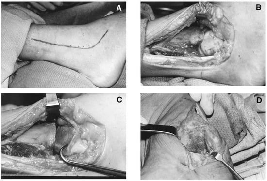
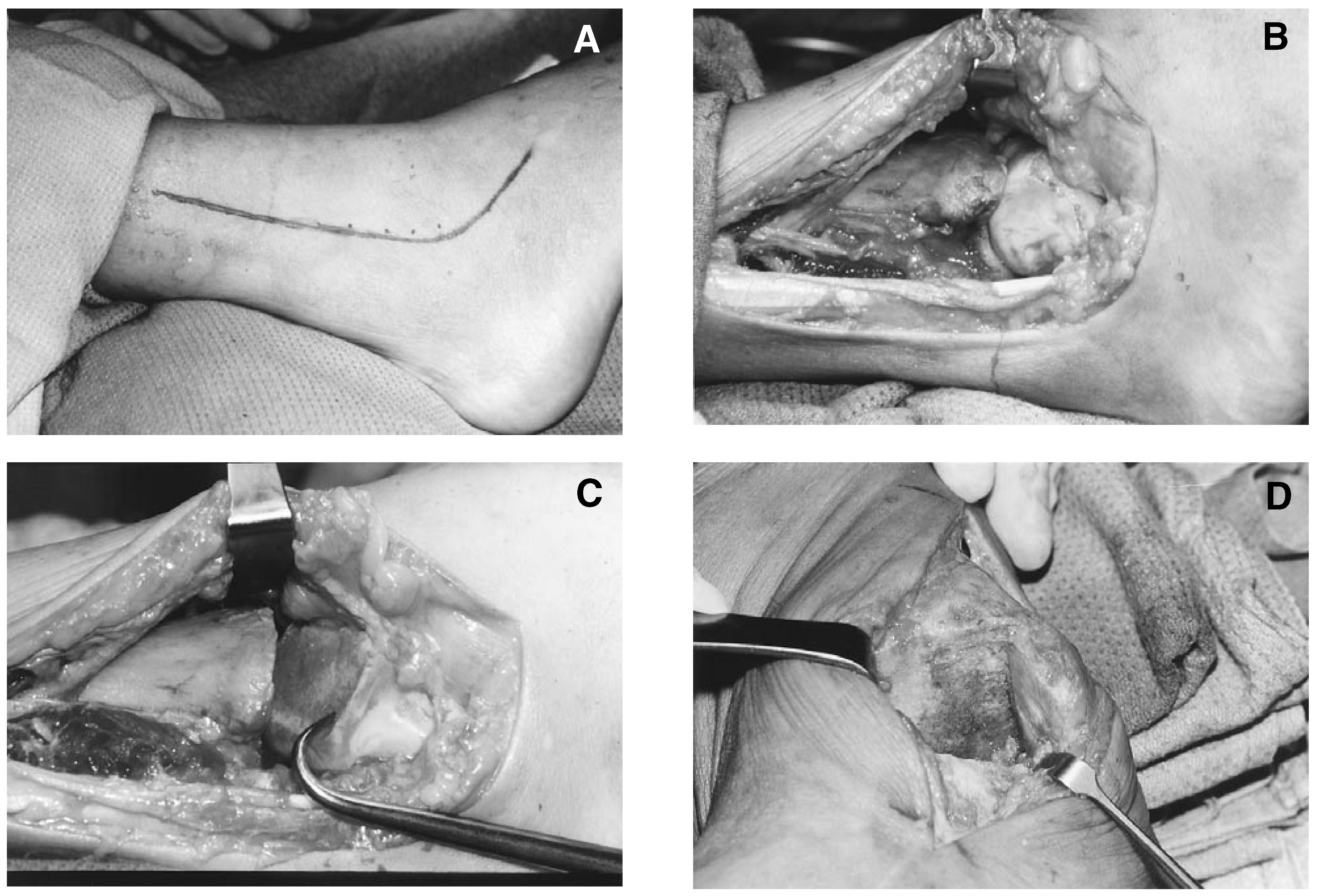
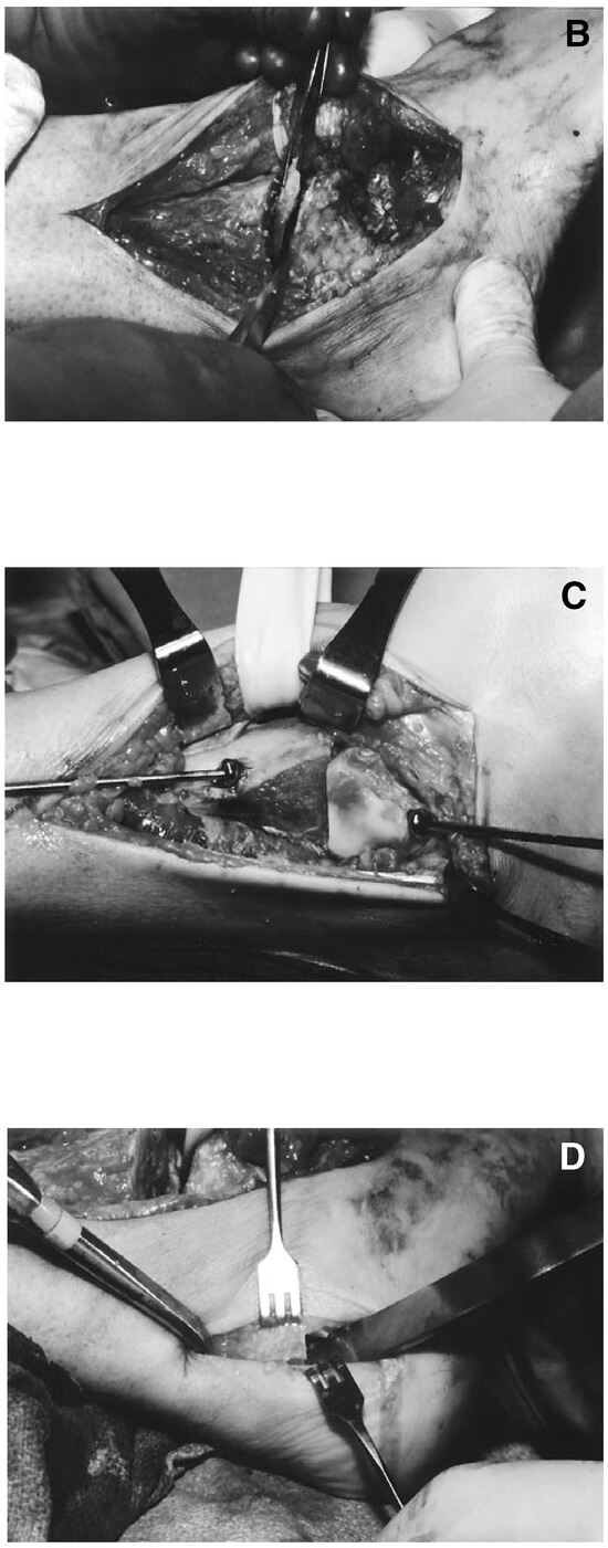
The standard surgical approach for the Podiatry Institute technique involves two incisions, one medial and one lateral. The lateral incision begins over the distal third of the fibula and curves distally over the inferior aspect of the sinus tarsi, paralleling the peroneal tendons (Figure 2).
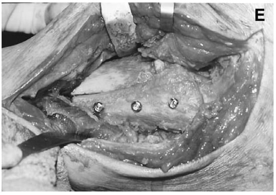
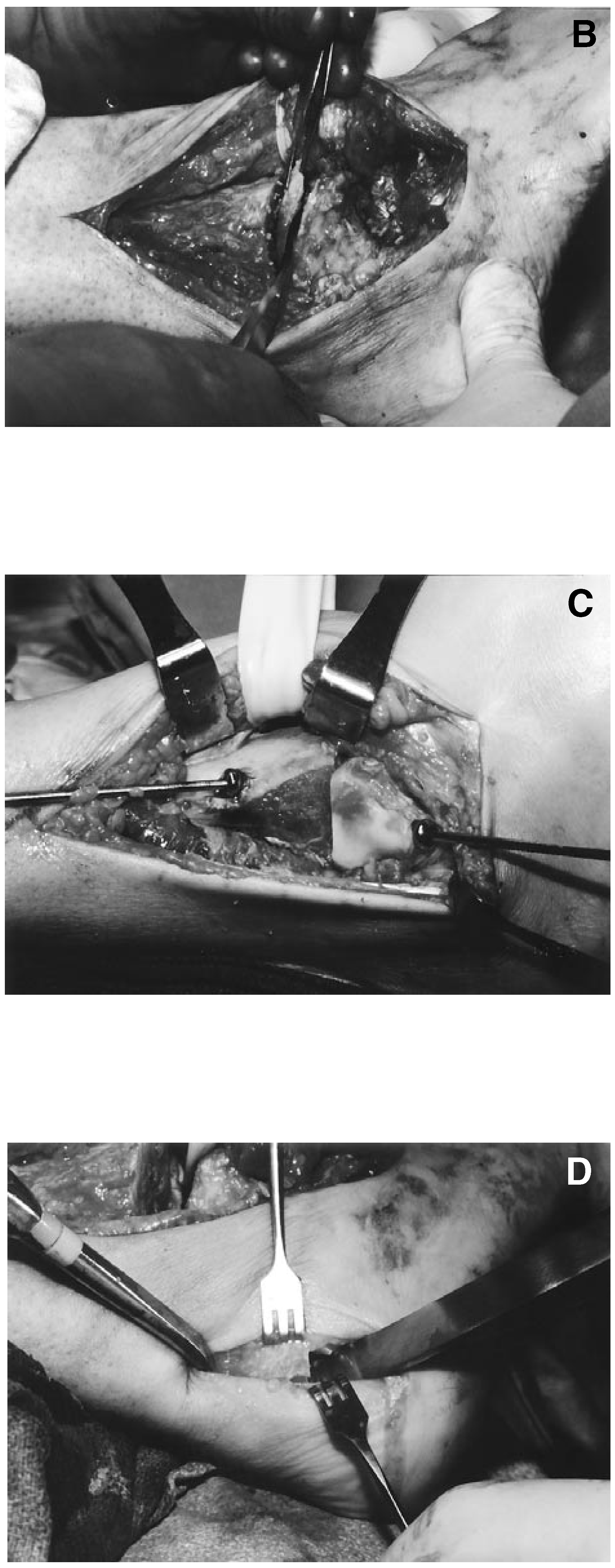
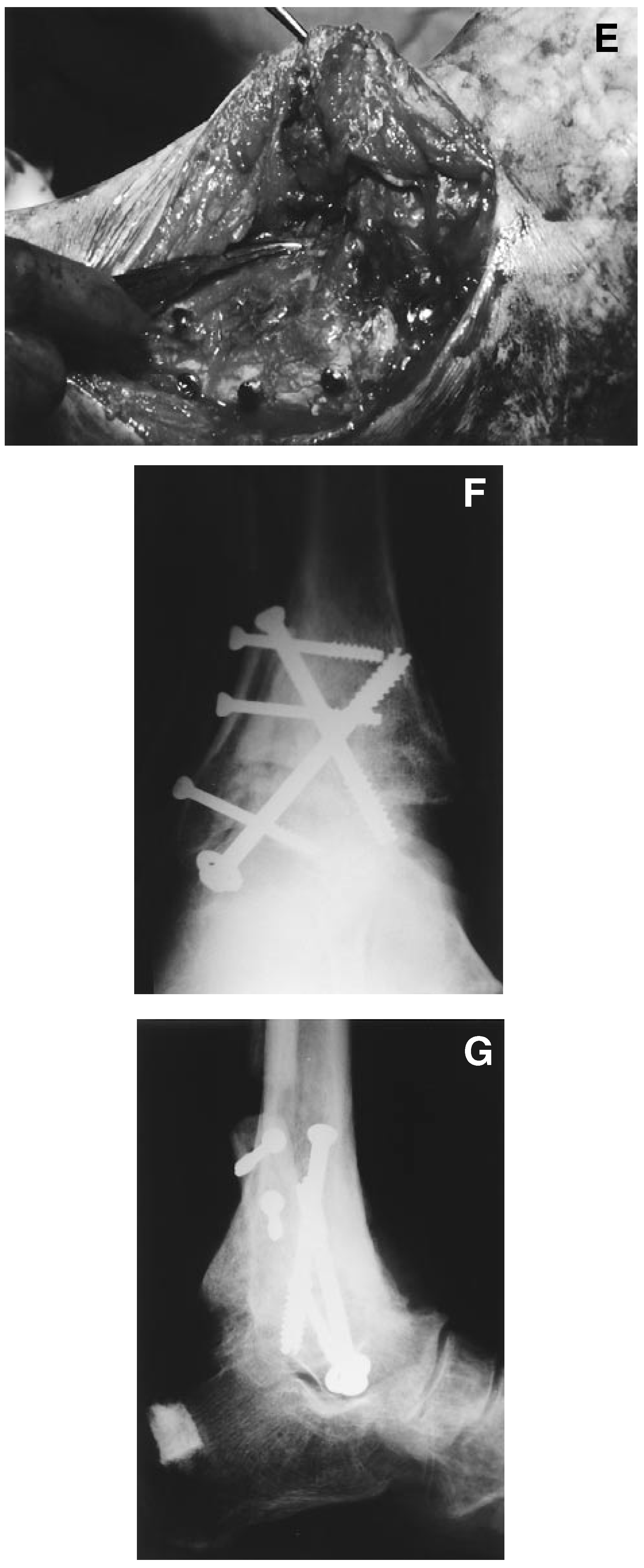
Figure 2.
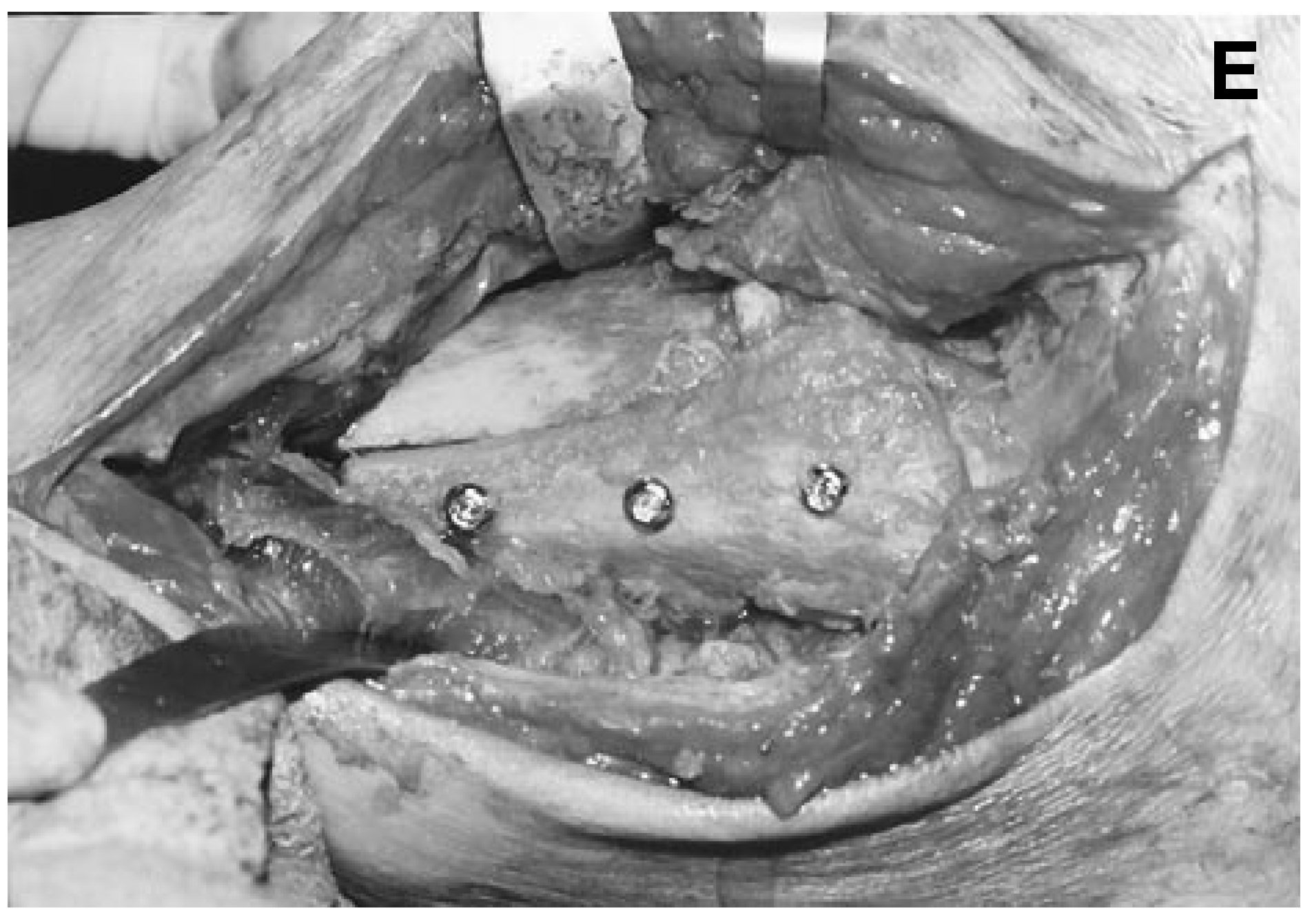
Ankle arthrodesis technique as described in the “Materials and Methods” section. (A), Lateral view with skin marker showing incisional approach. (B), Lateral view after removal of the distal portion of fibula. (C), Lateral view after table-top bone cuts from talar and tibial surfaces. The bone hook holds down talus to show extent of bone removal. (D), View of ancillary medial gutter approach with excision of cartilage off of the medial malleolus. (E), Lateral view after tibiotalar fixation and application of fibular strut.
As the periosteum is reflected from the fibula to the lateral and anterior aspect of the tibia, the perforating peroneal artery is encountered. Every attempt should be made to retract or if necessary, clamp and ligate this vessel to minimize intraoperative and postoperative blood loss. If the ligaments of the distal anterior tibiofibular joint have not already been dissected along with the periosteum, they will need to be transected in order to reflect the fibula posteriorly.
The medial incision begins over the distal fourth of the tibia, passes over the medial malleolus, and terminates at the level of the talonavicular joint complex. The saphenous nerve and vein can be found at the anterior border of the medial malleolus and are carefully retracted. The deep fascia and periosteum are incised in a linear manner and subperiosteal dissection completed. The amount of exposure of the medial side will depend on the surgeon’s preference and experience. At the very least, approximately 3 to 5 cm of periosteum should be reflected proximal to the ankle joint to allow insertion of the medial fixation screw(s). The medial and lateral wounds should then meet subperiosteally allowing elevation of all vital structures including tendons, nerves, arteries, and veins crossing the anterior aspects of the ankle joint and dorsum of the foot.
The ankle joint is then exposed through a transfibular approach. Various osteotomies of the fibula have been described. A through-and-through transection osteotomy approximately 5 to 8 cm proximal to the ankle joint can also be performed. A section of the fibula measuring approximately 2.5 cm in length can be resected proximally. This bone is preserved and used as an autogenous graft if necessary to fill in remaining deficits following fixation. The fibula can then be retracted posteriorly after release from the soft tissue attachments. These attachments consist of the interosseous membrane and the inferior distal tibiofibular ligaments. In some cases, the fibula may be temporarily removed in toto and later replaced as an onlay graft.
At this point, the ankle joint is carefully examined. Free ossicles of bone are excised. Old or fractured fragments, loose cartilage, and hypertrophic synovium are also excised.
The ankle joint is then placed in its desired position in the sagittal plane. The surgical assistant maintaining the position of the ankle joint is located at the distal end of the extremity. Using an oscillating power saw, the articular surfaces of the talar dome and tibial plafond are resected from lateral to medial. Appropriate wedge resections of bone are performed to correct for undesirable frontal plane varus or valgus deformity. Care must be taken when resecting the tibial plafond to avoid inadvertent transection through the medial malleolus. This can be accomplished by inserting a retractor or osteotome along the lateral wall of the medial malleolus itself.
The two surfaces are then placed in contact and the position of the ankle joint and the sagittal and frontal planes assessed. Abduction (external rotation) and adduction (internal rotation) are easily corrected by rotating the surfaces against each other. This should not result in any loss of contact between the two surfaces. Depending on the surgeon’s preference, the articular cartilage of the medial malleolus and the medial surface of the talus are resected using a power saw, rongeurs, bone curette, or power burrs. Intimate contact of these two surfaces may not be easily achieved and will likely require bone grafting with cancellous chips. Reciprocal planing of this area will help achieve intimate contact between bone surfaces.
Temporary fixation is then obtained. Two large Kirschner wires or, more frequently, small Steinmann pins are used. Clinical assessment is performed. Intraoperative x-rays are then obtained. These should consist of an anteroposterior and lateral view and a calcaneal axial view. The forefoot should be loaded when taking the lateral x-ray to adequately assess the final position in the sagittal plane. If alterations in the position of fusion are necessary, additional intraoperative x-rays should be obtained to confirm satisfactory alignment and position.
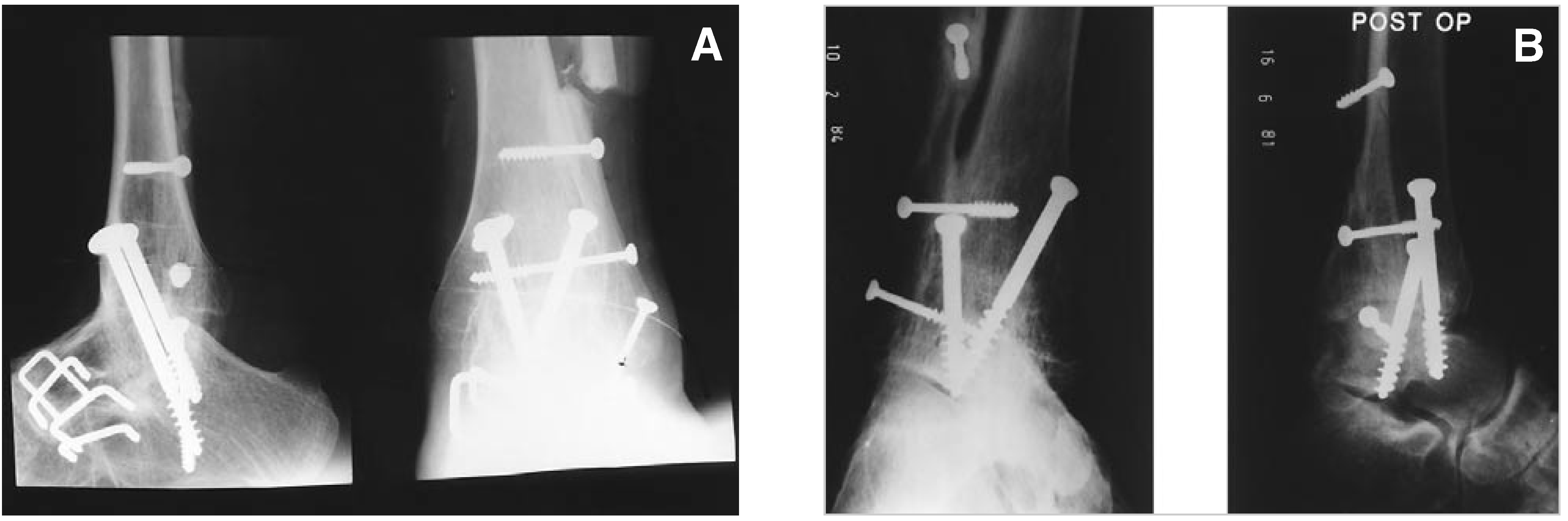
Final fixation is obtained using standard AO/ASIF technique with 6.5-mm short-thread cancellous screws. Compression across the ankle joint should be seen (Figure 3). It is again critical to confirm that the screws have not crossed the subtalar joint. This can be done intraoperatively by manipulation of the subtalar joint, direct visualization, and additional intraoperative x-rays if necessary.
Figure 3.
(A), Lateral and anteroposterior radiographic views 3 years after pantalar fusion. Note crossing 6.5-mm cancellous screws traversing both ankle and subtalar joints to effect fusion. Fibular strut has been reapplied to tibiotalar bone. (B), Lateral and anteroposterior radiographic views of ankle fusion technique.
Finally, the fibula is replaced as an onlay graft to the lateral surface of the tibia and talus. The inner surface of the fibula should be denuded of its cartilage and cortical bone to promote consolidation. Likewise, the corresponding surface of the talus and tibia may also require remodeling to accept the fibula as an onlay graft. This is typically accomplished using bone rongeurs and coarse-cutting power burrs. The fibula is easily fixated to the tibia and talus by use of 4.0-mm cancellous screws, or 4.5-mm self-tapping malleolar screws. The fibula can be replaced with its full thickness or as a hemisection. Remaining portions of resected bone are then used as autogenous cortical and cancellous grafts. Cancellous pieces of bone are inserted about areas of the tibiofibular joint, talofibular joint, and the medial aspect of the ankle joint between the talus and medial malleolus.
With the introduction of cannulated screw systems, the internal fixation process can be achieved more efficiently. Other modifications have also been introduced. One modification is to insert one of the screws from the lateral aspect of the talus into the medial tibia, thus reducing the risk of tibial stress risers.
After the tourniquet is released and adequate hemostasis has been obtained, two closed suction drains are inserted, one medially and one laterally. Subcapsular and subperiosteal closure are then completed using 2-0 absorbable synthetic suture in a continuous manner. The subcutaneous tissues are then reapproximated with a running stitch of 3-0 and 4-0 absorbable synthetic suture and skin through 5-0 absorbable or nonabsorbable subcuticular stitch. The incisions are then reinforced with wound closure strips. A dry sterile compressive dressing is applied followed by the application of a Jones’ cylinder compression cast.
Technique of Pantalar Arthrodesis
The surgical approach for the Podiatry Institute technique of pantalar arthrodesis (triple plus ankle arthrodesis) requires some incision modifications to provide adequate exposure to the subtalar, calcaneocuboid, and talonavicular joints. The lateral incision extends distally to the base of the fourth and fifth metatarsocuboid articulation. The medial incision extends to the base of the first metatarsal.
Similar dissection to that previously described is performed through the subcutaneous tissue with emphasis on protection of the cutaneous nerves and neurovascular structures. Laterally, it will be necessary to reflect the extensor digitorum brevis muscle dorsally and distally. The contents of the sinus tarsi are reflected dorsally or excised in toto. If not already done previously, the calcaneofibular ligament is transected. Subperiosteal and subcapsular dissection is then completed over the head and neck of the talus to communicate with the lateral incision. This again allows the surgeon to completely elevate all vital structures across the dorsum of the foot.
The technique and sequence of the joint resections will vary depending on the severity of the deformity, experience of the surgeon, and personal preferences.
Temporary fixation of the subtalar joint is accomplished by inserting a 0.062 Kirschner wire through the neck of the talus across the subtalar joint complex. The talonavicular joint is temporarily fixated with a single 0.062 Kirschner wire through the navicular tuberosity, across the joint, and into the body of the talus. Temporary fixation of the calcaneocuboid joint is generally not necessary, although it can be readily performed if desired.
After clinical assessment is performed, intraoperative x-rays are obtained to confirm satisfactory alignment and positioning and placement of the internal fixation devices. Appropriate adjustments are then made as necessary and final fixation accomplished.
When performing pantalar arthrodesis, the ankle and subtalar joint can be fixated by using the previously described technique for ankle fusion. The screws now, however, will cross both the ankle and subtalar joint, effectively “sandwiching” the talus between the calcaneus and tibia. Typically, two 6.5- mm long-thread cancellous screws are used for this purpose. Additional stabilization of the subtalar joint can be accomplished by using a single 6.5-mm longthread screw directed through the neck of the talus and traversing the posterior facet of the subtalar joint. In other situations, it may be preferable to fixate the subtalar joint separately from the ankle joint arthrodesis site. Precise placement is critical to avoid “collision” of the screws as they are inserted.
The talonavicular joint is fixated using a single 6.5- mm long-thread cancellous screw. Because of the relatively small size of the navicular tuberosity in comparison to the 6.5-mm cancellous screw, it is critical that aggressive countersinking of the medial surface of the medial cuneiform is performed. When the screw has been inserted and tightened to its final position, it should rest flush with the distal surface of the navicular. A 6.5-mm long-thread cancellous screw is most commonly used for fixation of this joint and provides an effective, rigid internal compression fixation. This screw is typically 50 to 60 mm in length.
The calcaneocuboid joint is typically fixated with two large bone staples. One staple is placed at the lateral surface of the joint and a second staple from dorsal to plantar. In a majority of cases, this provides rigid, stable fixation and is more than adequate. If a compression fixation is desirable (ie, with cortical cancellous grafts), a 6.5-mm long-thread cancellous screw may be used. The screw enters from the distal articular surface of the cuboid and is well countersunk into this surface. When the screw is tightened to its final position, it should be flush with the articular cartilage.
Finally, the fibula is secured to the tibia and talus as previously described. Autogenous or cortical cancellous bone chips are then packed about the appropriate areas to fill any remaining defects.
Postoperative Management
Postoperative management following an ankle or pantalar arthrodesis consists of nonweightbearing for 12 weeks or until radiographic consolidation of the fusion sites is seen. A long leg cast is recommended for the first 4 to 6 weeks with the knee flexed approximately 25° to 30°. This will prevent inadvertent pull by the tendo Achillis on the calcaneus, which can occur when the knee is allowed to extend and flex. Following the initial period of long leg casting, the cast is removed or converted to a short leg cast for the remaining time.
Following cast removal, patients are permitted approximately 50% weightbearing. Throughout the subsequent 2 weeks, they are allowed to increase to full weightbearing and to discard any gait-assistance aids that have been used, such as crutches or a walker. Swelling is controlled by the use of an elastic compression dressing such as elastic bandages. The patient should then be fitted for Jobst® (Beiersdorf-Jobst, Inc, Charlotte, NC) stockings or a similar pressure-gradient device. A Jobst compressor pump may be helpful in resolving edema.
The use of nonsteroidal anti-inflammatory agents and low-dose diuretics may be helpful in resolving residual edema. Physical therapy consisting of hydrotherapy, muscle stimulation, and cross-fiber massage are also used for 3 to 4 months following surgery. Muscle-strengthening exercises are instituted as well.
Appropriate shoe modifications are then made. Orthotic devices are helpful in preventing degenerative arthritic changes of the remaining tarsal joints. Patients are instructed as to appropriate shoes. The shoe should be lined with an appropriate shockabsorption material with concentrated cushioning in the heel area. In some cases, it may be necessary to add an entire full-length lift to the shoe to compensate for shortening, which occurs not infrequently.
Additional serial radiographs are obtained at 6 months, 9 months, and 1 year to monitor the fusion sites and the position of the internal fixation devices. The viability of the talus must be monitored closely to prevent avascular necrosis and collapse of the talar body. Bending of the screws should alert the surgeon to incomplete fusion of one or more joints, most commonly the ankle. Outward migration of any of the internal fixation screws should also alert the physician to a potential failure or delayed fusion. Consideration should then be given to a period of immobilization and nonweightbearing. Noninvasive electrical bone stimulation may help promote a complete union.
Removal of the internal fixation devices has not been performed routinely. The authors believe, however, that large cancellous screws transfixing the ankle joint or the ankle and subtalar joint (and pantalar arthrodesis) should be removed when complete consolidation is visualized radiographically. This will allow the tibia and talus to respond in accordance with Wolff’s law to stresses across this area and hopefully decrease the probability of transverse fracture of the tibia or stress fracture.
Results
The average age of the patients was 42.8 years with a range of 23 to 70 years. The patients were 20 males and 22 females. Twenty procedures were performed on the left extremity and 22 were performed on the right.
Trauma was the most common etiology of the ankle disorder, accounting for 20 of the cases. Most commonly, these were malleolar fractures that were inadequately reduced or improperly fixated. The second most common disorder was neuromuscular problems (17 patients). This group included postpoliomyelitis patients and those with Charcot-Marie- Tooth disease and other neuromuscular disorders.
The chief complaint of the majority of patients was pain. Instability or pain and instability accounted for the remainder of the patients.
Patients’ legs were casted from 6 to 32 weeks with an average of 11 weeks. Partial weightbearing was initiated from 4 to 13 weeks, at an average of 8 weeks. Full weightbearing was begun at 6 to 36 weeks, with an average of 12 weeks.
Complications
Soft tissue complications were few. There were no postoperative infections. There were two minor dehiscences that both healed after a brief period of local care. One patient had a complaint of incisional neuritis.
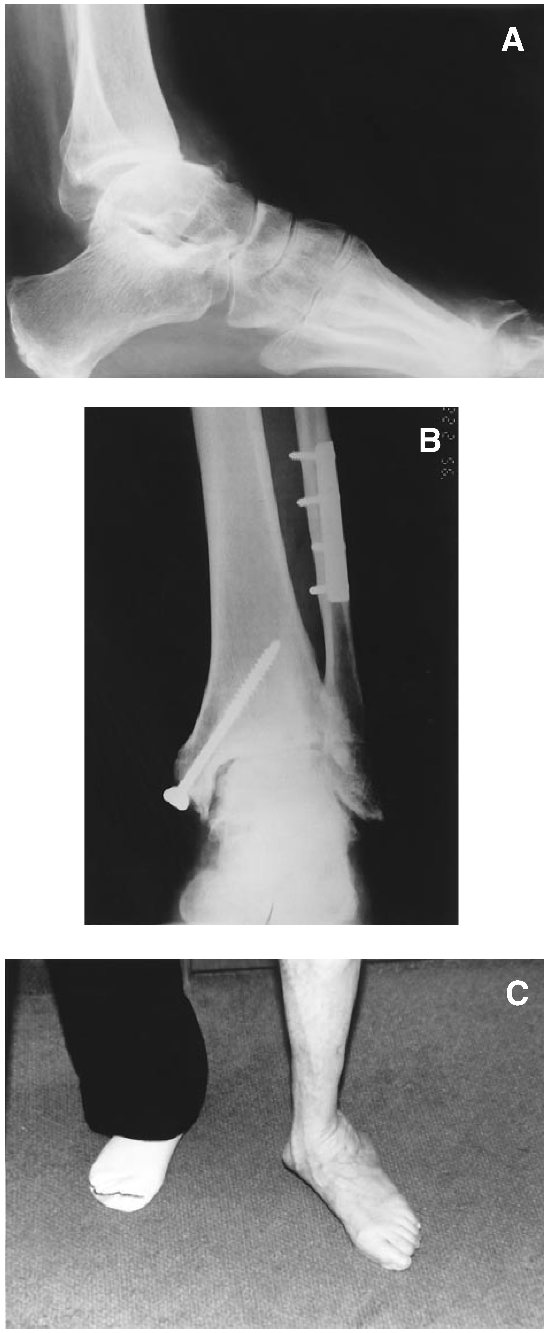
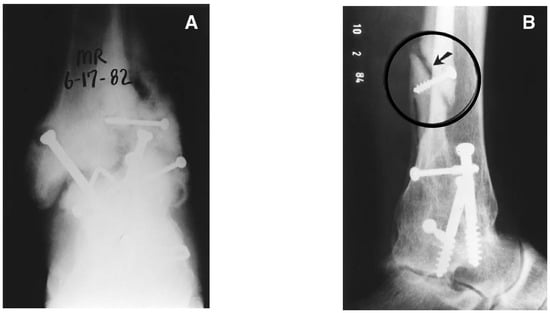
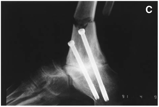
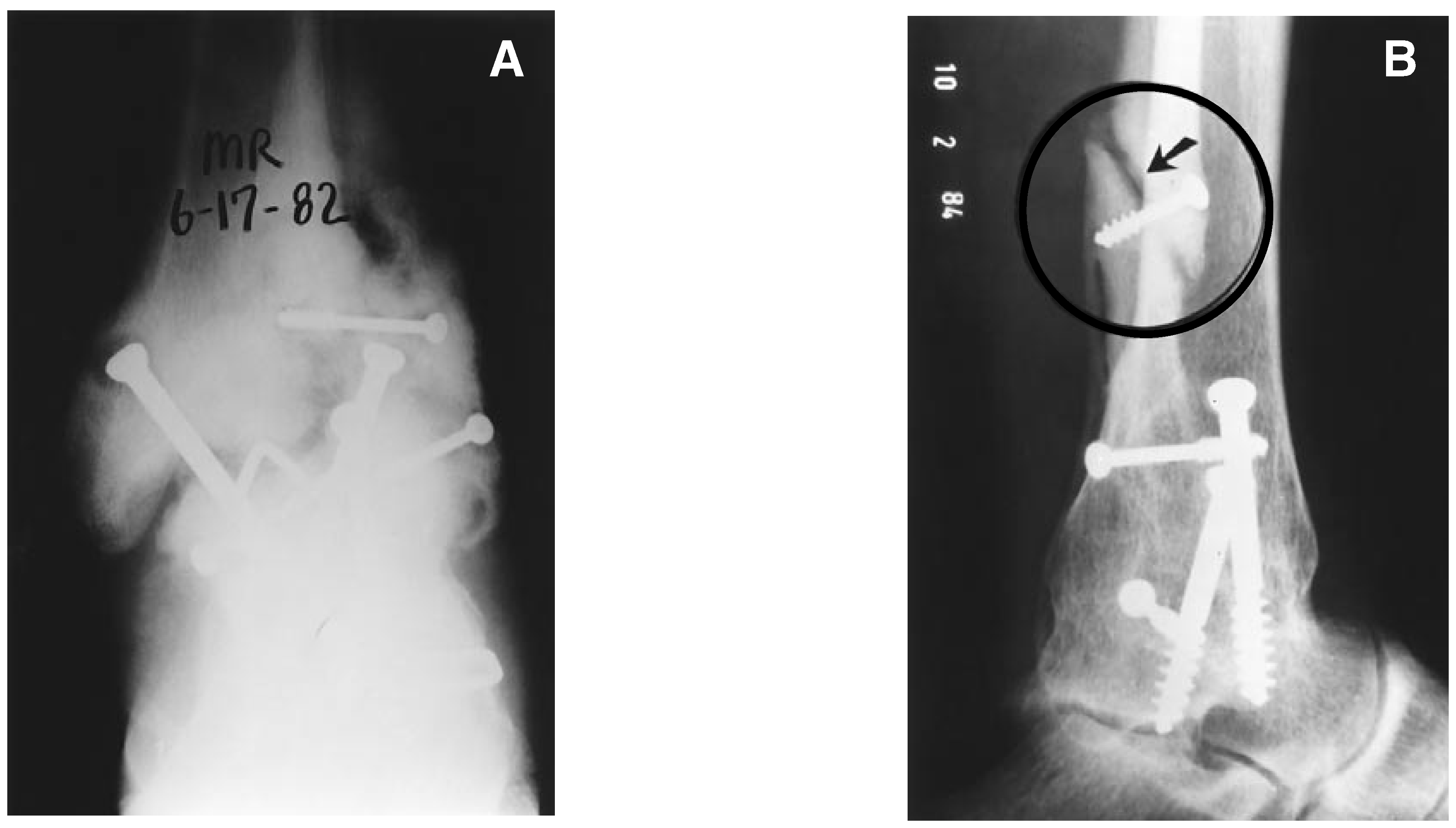
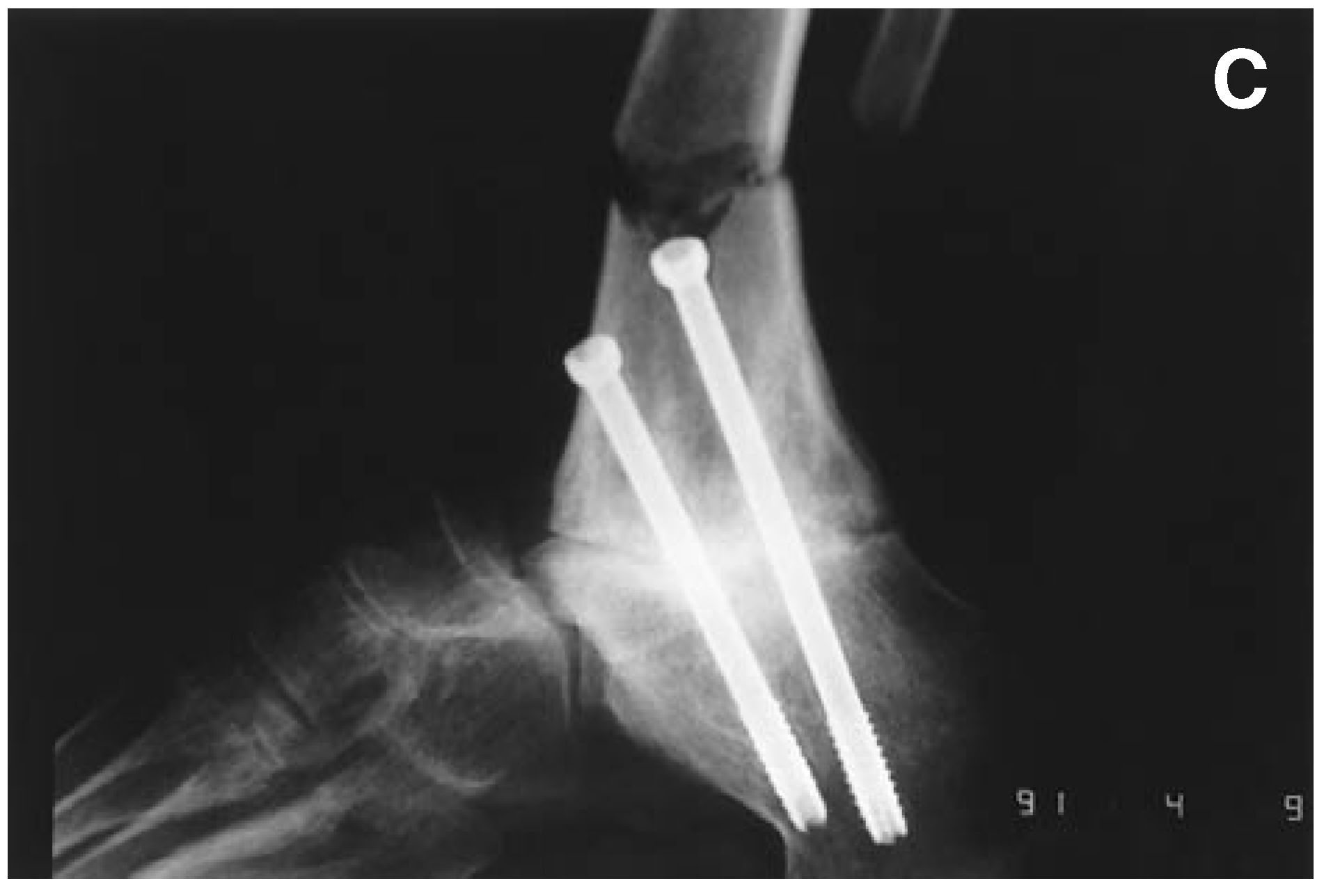
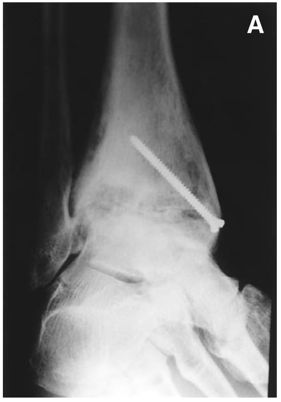
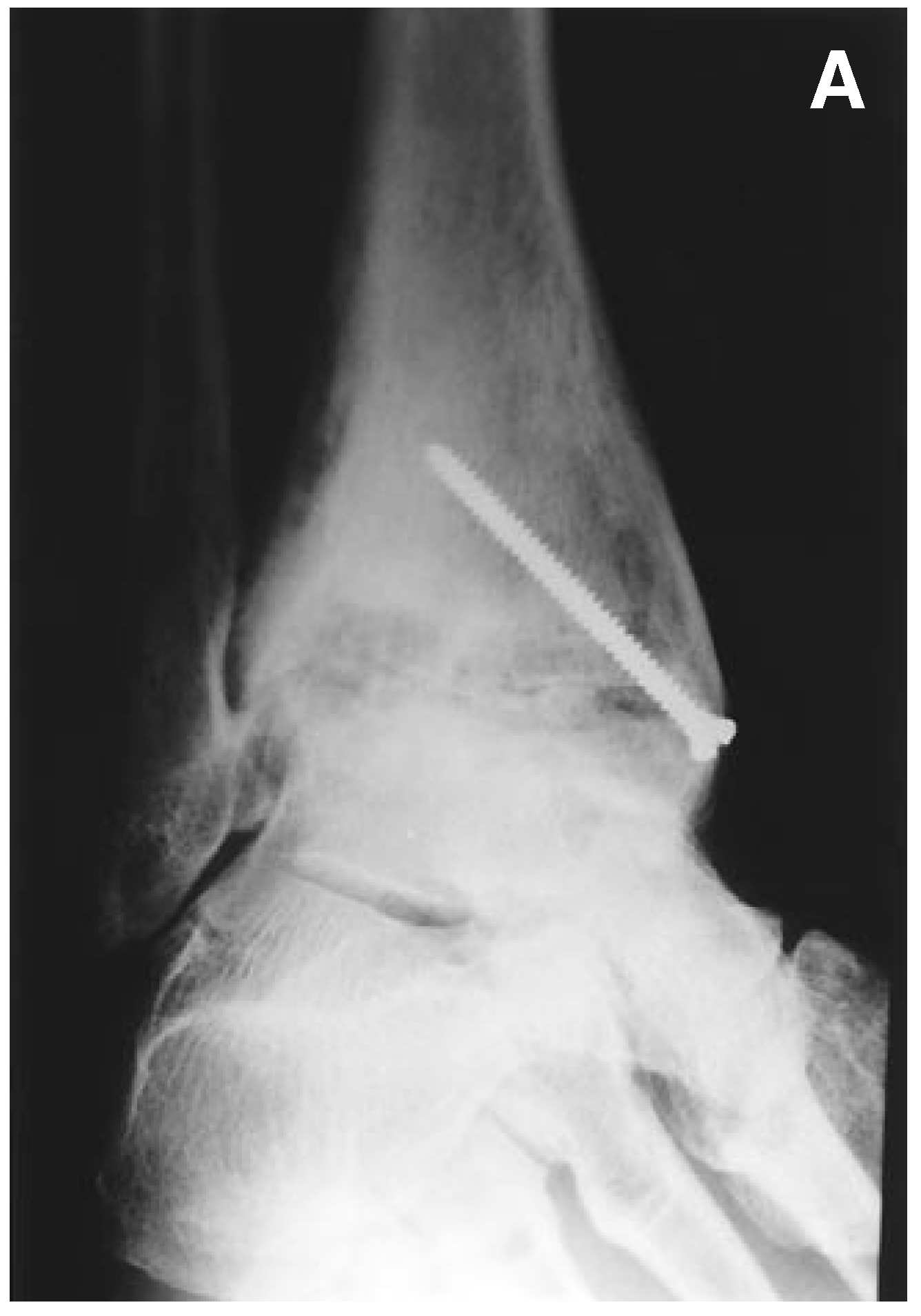
Bone complications were more numerous and more significant (Figure 4). Nonunions (no change in bone healing after 9 months) and delayed healing (completion to bone union taking more than 5 months) accounted for the majority of the bone complications. The incidence of nonunion was related to the procedure performed. Four nonunions occurred in the 13 pantalar fusions (30.8%), whereas the ankle fusion nonunions represented three out of the 29 fusions (10.3%). Only one of the three ankle fusion nonunions occurred at the tibiotalar joint; the other two occurred at the repaired fibular osteotomy site. The fibular osteotomy complications are avoidable and the technique has since been modified to prevent them. If the fibular osteotomy nonunions are excluded, the ankle fusion nonunion incidence was 3.4%. The overall nonunion incidence was 16.7% (total), or 11.9% with the fibular osteotomy site excluded. These figures are calculated based on the occurrence of a nonunion at any one site in a patient.
Figure 4.
Bone complications. (A), Anteroposterior ankle radiographic view showing hypertrophic nonunion following pantalar fusion in a neuropathic patient. (B), Lateral ankle radiographic view showing nonunion at the fibular strut repair site. This step is no longer performed as part of the technique. (C), Lateral view of tibial fracture through one of the screw sites in a tibiocalcaneal fusion.
Tibial fractures occurred in two patients. One patient had two fractures at the tibial screw sites: the first at full weightbearing at 6 months, and the second 3 years postoperative. The other patient also sustained a fracture at the tibial screw site at full weightbearing.
Patient Questionnaire
Eight of the 13 pantalar patients responded to the patient questionnaire (61.5%), a higher percentage than the ankle fusion patients (12/29 = 41.4%). The overall response rate was 47.6% (20/42). Patients reported an average duration of 6.87 years between the onset of symptoms and their surgeries. The majority of patients were satisfied that their problems were corrected. Patients were asked to give an overall rating on a scale of zero (no improvement/no correction) to ten (complete improvement/complete correction). Fifteen of the 20 patients responded with a score greater than seven. Seven of these patients responded with a ten. Five patients responded with a number less than seven, with two patients giving a response of zero. The average improvement was 6.9. Fifteen of the 20 patients were either satisfied or very satisfied with the overall results of their procedures and 17 patients said they would recommend the procedure to a friend with a similar problem.
Overall daily activity level was better than before surgery for 13 of the 20 patients, the same for two, and less than before surgery for five patients. Fifteen of 20 patients reported that they felt their ankle was fused in a comfortable position; five felt it was not. Twelve patients reported new areas of pain following surgery. Of the 11 who specified an area, the locations were hip (one), knee (four), fibula (one), front of tibia (three), ankle (one), and foot (one). In the area of surgery, ten patients reported that they have no pain, five reported minimal pain, four reported mild to moderate pain, and one patient reported continuous pain, regardless of activity.
The majority of the poor responses by patients came from the group that received solitary ankle fusion. It is unclear whether this was caused by a self-reporting bias in those ankle fusion patients who answered the questionnaire, or if this was influenced by the relative underreporting of the ankle fusion patients. In addition, patients who underwent pantalar arthrodesis had, on average, more preoperative pain and deformity. They may have been easier to satisfy in terms of their expectations. In general, those patients who had more preoperative pain, and a long and intensive course of conservative care, were most satisfied with their surgery.
Discussion
Additional Fixation Considerations
The authors’ primary method of fixation has included AO/ASIF internal fixation with the 6.5-mm cancellous screw being used most frequently. The authors’ results and subjective impressions indicate that excellent rigidity and rigid compression are readily accomplished.
The use of large cannulated screws may also be used. Many commercial brands are currently available. Their primary advantage is in the insertion of the permanent fixation device directly over the temporary fixation or guide pins. In addition, a reduction in the total operative time is seen.
In some circumstances, the bone may be extremely osteoporotic and lack cortical definition capable of supporting the large cancellous screw(s). Stable fixation in these cases can be accomplished using large Steinmann pins inserted at appropriate angles to provide rigid, stable fixation of the fusion sites. Carrier and Harris [44] described successful use of two vertical Steinmann pins for fixation of ankle fusions in patients with severe rheumatoid arthritis. These Steinmann pins can be left external to the skin or buried beneath the periosteum and capsular tissues. In either case, the authors recommend bending the tip of the pin to prevent inward migration which, in turn, would make retrieval difficult. External fixators may also be used.
Large bone staples can also be used as an alternative fixation modality. Not uncommonly, it may be necessary to predrill the holes for insertion of the staples to avoid significant shifting of the adjacent fusion surfaces. Large bone staples may be supplemented with the addition of large Kirschner wires or Steinmann pins.
Modifications to the Technique
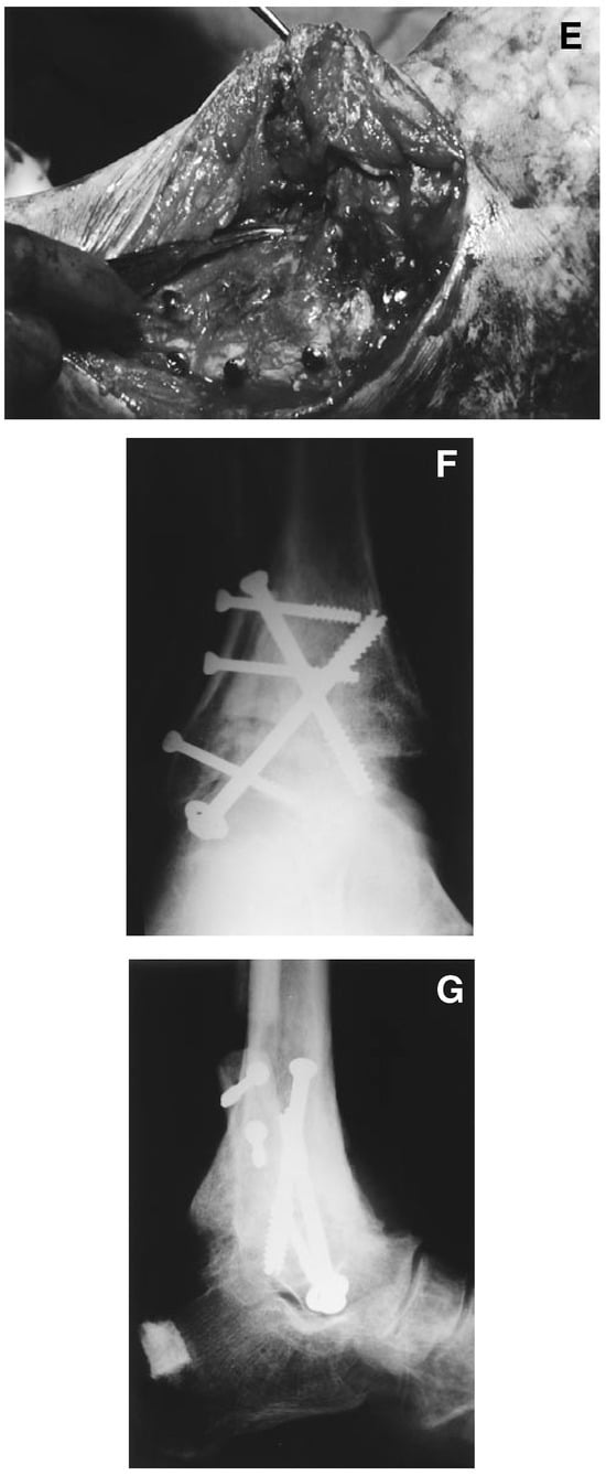
In response to this study, various modifications have been made to the original technique and postoperative care. As previously mentioned, the fibular osteotomy is no longer repaired (Figure 5). Instead, it is used as a strut graft against the tibia and talus. It can be used either whole or longitudinally split in half. This has eliminated the fibular nonunion problem.
Figure 5.
Modifications of primary technique. (A), Preoperative oblique radiograph. (B), Lateral view after fibular resection showing wedging to reduce equinus at tibiotalar articulation. (C), Lateral view showing application of cannulated large-fragment screws from lateral side of tibia and lateral process of the talus. (D), Lateral view of foot showing removal of calcaneal bone graft. (E), Lateral view of the ankle with hemostat showing location of autogenous calcaneal slot graft across tibiotalar interface. Fibular strut has been reapplied. (F), Anteroposterior ankle radiograph showing modified fixation technique. (G), Lateral ankle radiograph showing modified fixation technique. Note also application of hydroxyapatite to calcaneal donor area.
Other modifications are used as necessary. Anterior bone grafts across the talotibial interface can be used when the fusion is revisional, or if there is some other reason to be concerned about healing potential in the area. The calcaneus is an excellent source for this type of autogenous graft [45]. Implantable bone stimulators can also be used for revisional ankle fusions.
Postoperative care has been modified to include an above the knee cast for the first 4 weeks in most cases. It is difficult to neutralize internal and external rotation forces if the knee is allowed to continue to move. Total casting time lasts 4 months for most patients.
The tibial fractures that occurred have caused the authors to consider orienting one screw from the talus upward. Other authors have avoided this problem by beginning one screw in the intact fibula, although this may lose the beneficial effect of the fibular strut graft.
Technology is offering new ways of performing ankle fusions. The Ilizarov technique may be particularly advantageous for revisional fusions including infected failed fusions [46]. Arthroscopic ankle fusion (arthroscopic resection of cartilage, combined with internal fixation) shows promise for those patients in whom there is not significant deformity [47].
Comparison of Internal Versus External Fixation
There is no question that no one surgical approach, fixation technique, or osteotomy technique can be used for all patients when performing an ankle fusion. It is useful to review the advantages and disadvantages of internal and external fixation in order to identify more clearly where each technique has its best advantages. External fixation has been the most common technique used for ankle fusion: There are several reasons for this. The technique is relatively straightforward for those surgeons with experience in ankle surgery, and with external fixation devices. The fixator itself can generate significant amounts of static, interfragmentary compression, because of the perpendicular relationship of the fixation device to the fusion site. With external fixators, the amount of compression can be adjusted during the postoperative period. Usually, this is not particularly important for ankle fusions because of the relatively short duration of the application of the fixator. In various other types of long-bone trauma and long-bone sepsis, the ability to regulate compression and distraction becomes more important.
One of the major complications associated with external fixation is pin-track infection. The incidence of these infections has been variously reported in the literature, but appears to be relatively high [38,48]. Experienced surgeons familiar with the use of external fixation indicate that the problem is not significant. The prevention of these pin-track infections requires diligence on the part of the surgeon, both during the operative technique (to prevent burning of skin and bone) and also during the postoperative periods. Postoperative care for a pin site should be meticulous, and should include cleansing with hydrogen peroxide and application of an antiseptic or antibiotic ointment.
Another difficulty with external fixators is the psychological ability of patients to tolerate the sight of the devices. To most patients, these devices appear cold, unwieldy, painful, and unsightly. Good patient selection and education is vital to ensure that patients can tolerate these devices. Internal fixation is more readily accepted by patients, and does not create the problems of pin-track infections.
The real question is, which type of fixation provides a greater likelihood of achieving fusion? It is virtually impossible to answer this question because of the differences in various studies of ankle fusion patients. The variables include types of indications for fusion, the age of the patients, the surgical technique, the length of application of fixation, and the postoperative course. Moeckel et al [48] attempted to answer this question by comparing external fixation fusions to internal fixation fusions at one institution. They found a significantly higher rate of complications in the external fixation group (61%) versus the internal fixation group (28%). Almost half of the complications in the internal fixation group were stress fractures associated with the donor site for the sliding inlay graft. The external fixation group had a nonunion rate of 21%, whereas the internal fixation nonunion rate was only 5%. Maurer et al [49]. reported a 100% fusion rate (35/35) using crossing screws compared with an 83% fusion rate (10/12) using external fixation.
Scranton et al [43] reported on the mechanical stability of external fixators versus their T-plate design. They stated that although the external fixators are capable of producing a great deal of static interfragmentary compression, they lack rotatory stability. According to their mechanical studies, the Scranton T-plate device provided fixation comparable to, or superior to that achieved with a Charnley external fixator. The crossed 6.5-mm lag screws used in the Podiatry Institute technique generate substantial interfragmentary compression. The crossing angulation cancels out any shifting moment in the fusion site, and the technique is likely to resist anteroposterior forces better than the T-plate fixation of Scranton, and resists rotatory motions more so than the external fixators.
Thordarson et al [50] performed a biomechanical analysis on fresh-frozen cadaver limbs with ankle fusions performed with cancellous screw fixation and a fibular strut graft. Their study showed that crossing screws are most effective resisting medial-lateral and plantarflexion-dorsiflexion moments, less effective resisting rotation forces. They found the strut graft to be effective in providing additional stability, particularly in poor quality bone.
In a later study, Thordarson et al [51] compared cancellous screw internal fixation with Calandruccio external fixation. Based on this biomechanical study in fresh-frozen cadaver limbs, they recommended crossing screws as the fixation of choice if the bone quality is good, and a mild torque produces no visible motion. Even when bone quality is poor, if dorsiflexion- plantarflexion are of concern, screw fixation is still preferable to Calandruccio external fixation.
Considerations for Pantalar Fusions
Pantalar fusions consist of arthrodesis of the ankle, subtalar, and generally, midtarsal joints. Lorthioir [52] originally described the procedure in 1911 as fusion of the ankle, subtalar, and talonavicular joints. However, the calcaneocuboid joint is now often included in the procedure. Pantalar fusion is most often indicated when there is deterioration of the subtalar and midtarsal joints, or there is severe deformity than cannot be accommodated by fusion of the ankle alone. The procedure has been described as either a one-stage or two-stage (triple arthrodesis, ankle fusion) surgery. Although Liebolt [53] recommended that the procedure be performed in two stages because of the duration of operating time, and the difficulty in accurately correcting deformity of all joints in one sitting, the procedure is most commonly now performed at one sitting.
A more interesting distinction is between the use of the articular wedging technique versus extirpation of the talus, as described by Lorthior [52] and Hunt and Thompson [54]. The extirpation technique theoretically creates less shortening and allows more accurate bone-to-bone contact, by allowing less bone removal to create flush surfaces. The literature indicates that extirpation of the talus does not seem to create a problem in patients with sensate feet. However, the use of this procedure in insensitive feet results in a significant increase in the incidence of nonunion and avascular necrosis of the talus. One of the nonunions in this series was a neuropathic patient who had a pantalar arthrodesis using the extirpation technique. Based on the authors’ results, they have moved away from extirpation of the talus as a primary procedure in pantalar arthrodesis.
Complications and Failure
A variety of studies on ankle arthrodesis indicate a very high rate of complications. In particular, there are fusion rates reported in the literature from 100% and 94% down to 40% [49,55,56]. Most of the literature, however, seems to indicate a fusion rate somewhere around 80% to 85% [11,57]. The authors’ fusion rate is quite acceptable in these terms: three of their nonunions were in neuropathic patients (out of five neuropathic patients); one of these nonunions was at the talocalcaneal joint; the other two were in the ankle joint. One of the neuropathic patients also admitted to being noncompliant. Neuropathic patients have been identified as being particularly difficult to fuse, particularly during the acute phase of the Charcot process [56,57,58]. Stuart and Morrey [59] reported a 62% complication rate and a 62% reoperation rate in diabetic neuropathic patients. Patients who had radiographic signs of neuropathic arthropathy had only a 38% rate of satisfactory results.
Two of the authors’ patients with nonunions were at the fibular osteotomy site. This was a consistent problem early in the series when the fibula was most often repaired, usually with only one screw. In retrospect, it should have been clear that this was inadequate fixation for this type of fracture and osteotomy. This part of the technique has been modified to reduce the occurrence of this problem. The fibula is now either screwed directly to the tibia (split longitudinally or left whole) as an onlay graft, or removed.
Frey et al [60] reviewed 78 ankle fusions, reporting an overall complication rate of 56%. Delayed unions were 12%, nonunions 41%, infection 9%, and malunion in 3%. They found no correlation with the technique used, but the data suggested that those with major medical problems or open injuries were more likely to go on to nonunion. Patients with a previous subtalar or triple arthrodesis were more likely to heal than other patients. Cobb et al [61] reported that cigarette smokers have a 3.75-times greater risk of nonunion in ankle fusion than nonsmokers.
The tibial fracture problem is quite significant. Although the morbidity associated with the problem was small in this series, there is potential for more devastating fractures. In reviewing the two cases where fractures occurred, it appears that two factors that may contribute to fracture are excessive overdrilling of the tibial cortex, and the alignment of the two screw heads. In both cases where fractures occurred, the screws were placed close together and in the same plane. Offsetting the screws (one higher, one lower on the tibia), and keeping the screw heads on opposite sides of the tibia may decrease the risk of fracture. Another alternative is to cross one of the screws from the talus upward into the tibia, as described by Wagner and Pock [62] and Gschwend and Steiger [63] (Figure 5). Dennis et al [55] reviewed 16 cases where a similar technique was used and achieved a 94% fusion rate. This seems to be a reasonable approach; two of the authors have used this technique successfully on several occasions. One of the authors is now investigating the mechanical performance of various internal fixation options for ankle fusion at the Pennsylvania College of Podiatric Medicine.
Summary
Internal fixation for ankle fusions has become an increasingly popular technique. There have been very good results from a specific technique. The primary complications were osseous in nature. Nonunion and tibial fracture complications occurred, which technique modifications can reduce.
Acknowledgments
The surgeons of The Podiatry Institute who have contributed to the concepts expressed within this article.
References
- MAHAN KT, YU GV: Ankle and plantar fusion. Presented at Thirteenth Annual Comprehensive Conference in Foot Surgery, Tucker, GA, 1984.
- VOGLER H: Ankle arthrodesis: clinical and conceptual applications. Clin Podiatr Med Surg 2: 59, 1985.
- STAUFFER RN: Salvage of painful total ankle arthroplasty. Clin Orthop 170: 184, 1982.
- MCGUIRE M, KYLE R, GUSTILO R, ET AL: Comparative analysis of ankle arthroplasty versus ankle arthrodesis. Clin Orthop 226: 174, 1988.
- ABDO R, WASILEWSKI S: Ankle arthrodesis: a long-term study. Foot Ankle 13: 307, 1992.
- RAMSEY PL, HAMILTON W: Changes in tibiotalar area of contact caused by lateral talar shift. J Bone Joint Surg 58A: 356, 1976.
- NATARAJAN M: Pan-talar arthrodesis for post polio flail foot. Singapore Med J 8: 214, 1967.
- ANSART MB: Pan-arthrodesis for paralytic flail foot. J Bone Joint Surg 33B: 503, 1951.
- IWATA H, YASUHARA N, KAWASHIMA K, ET AL: Arthrodesis of the ankle joint with rheumatoid arthritis. Clin Orthop 153: 189, 1980.
- MAHAN K: Wound healing in the rheumatoid patient. Categoric Foot Rehabilitation. Presented at Fourteenth Annual Surgical Seminar, Tucker, GA, 1985.
- AARON A: Ankle fusion: a retrospective review. Orthopedics 13: 1249, 1990.
- AHLBERG A, HENRICSON A: Late results of ankle fusion. Acta Orthop Scand 52: 103, 1981.
- SCRANTON P: An overview of ankle arthrodesis. Clin Orthop 268: 96, 1991.
- CHARNLEY J: Compression arthrodesis of the ankle and shoulder. J Bone Joint Surg 33B: 180, 1951.
- CHARNLEY J: “Compression Arthrodesis of the Ankle,” in Compression Arthrodesis, E S Livingstone, London, 1953.
- GLISSAN DJ: The indications for inducing fusions at the ankle joint by operation with description of two successful techniques. Aust N Z J Surg 19: 64, 1949.
- KENNEDY JC: Arthrodesis of the ankle with particular reference to the Gallie procedure. J Bone Joint Surg 42A: 1308, 1960.
- WHITE A: A precision posterior ankle fusion. Clin Orthop 98: 239, 1974.
- HORWITZ T: The use of the transfibular approach in arthrodesis of the ankle joint. Am J Surg 60: 550, 1942.
- GOLDTHWAIT J: An operation for the stiffening of the ankle joint in infantile paralysis. Am J Orthop Surg 5: 271, 1908.
- HALLOCK H: Arthrodesis of the ankle joint for old painful fractures. J Bone Joint Surg 27: 49, 1945.
- CHUINARD E, PETERSON R: Distraction-compression bonegraft arthrodesis of the ankle. J Bone Joint Surg 45A: 481, 1983.
- HATT RN: The central bone graft in joint arthrodesis. J Bone Joint Surg 22: 393, 1940.
- WESCOTT HH: An operation for the fusion of the tibioastragaloid joint. Va Med Monthly 61: 38, 1934.
- WILSON HJ: Arthrodesis of the ankle: a technique using bilateral hemi-malleolar onlay grafts with screw fixation. J Bone Joint Surg 51A: 775, 1969.
- VERHELST MP, MULIER J, HOOGMARTENS M, ET AL: Arthrodesis of the ankle joint with complete removal of the distal part of the fibula. Clin Orthop 118: 93, 1979.
- ANDERSON R: Concentric arthrodesis of the ankle joint. J Bone Joint Surg 27: 37, 1945.
- STAPLES OS: Posterior arthrodesis of the ankle and subtalar joints. J Bone Joint Surg 38A: 50, 1956.
- MEAD NC: Arthrodesis of the ankle joint: a simple, efficient method. Bull Northwestern University Med School 25: 248, 1951.
- MARCUS R, BALOURDAS G, HEIPLE K: Ankle arthrodesis by chevron fusion with internal fixation and bone grafting. J Bone Joint Surg 65A: 833, 1983.
- STEWART M: “Arthrodesis,” in Campbell’s Operative Orthopedics, Vol 1, ed by A Edmonson, A Crenshaw, CV Mosby, St Louis, 1980.
- BINGOLD AC: Ankle and subtalar fusion by a transarticular graft. J Bone Joint Surg 38B: 862, 1956.
- PRIDIE KH: Arthrodesis of the ankle. J Bone Joint Surg 35B: 152, 1953.
- HONE MR: Dowel fusion of the ankle joint. J Bone Joint Surg 50B: 678, 1978.
- BACIU C, FILIBIU E: Rapid arthrodesis of the ankle joint via verticalisation of the joint space. Arch Orthop Trauma Surg 93: 261, 1979.
- GRAHAM CE: A new method for arthrodesis of an ankle joint. Clin Orthop 68: 75, 1970.
- OTTOLENGHI C, ANIMOSO J, BURGO P: Percutaneous arthrodesis of the ankle joint. Clin Orthop 68: 72, 1970.
- MALARKEY R, BINSKI J: Ankle arthrodesis with the Calandruccio frame and bimalleolar onlay grafting. Clin Orthop 268: 44, 1991.
- MORGAN C, HENKE J, BAILEY R, ET AL: Long-term results of tibiotalar arthrodesis. J Bone Joint Surg 67A: 546, 1985.
- WILLIAMS JE, JR, MARCINKO D, LAZERSON A, ET AL: The Calandruccio triangular compression device: a schematic introduction. JAPA 73: 536, 1983.
- HAWKINS B, LANGERMAN R, ANGER D, ET AL: The Ilizarov technique in ankle fusion. Clin Orthop 303: 217, 1994.
- SCRANTON P: Use of internal compression in arthrodesis of the ankle. J Bone Joint Surg 67A: 550, 1985.
- SCRANTON PE, FU FH, BROWN T: Ankle arthrodesis: a comparative clinical and biomechanical evaluation. Clin Orthop 151: 234, 1980.
- CARRIER D, HARRIS C: Ankle arthrodesis with vertical Steinmann’s pins in rheumatoid arthritis. Clin Orthop 268: 10, 1991.
- MAHAN KT: Calcaneal donor bone grafts. JAPMA 84: 1, 1994.
- JOHNSON E, WELTMER J, LIAN G, ET AL: Ilizarov ankle arthrodesis. Clin Orthop 280: 160, 1992.
- MYERSON M, QUILL G: Ankle arthrodesis: a comparison of an arthroscopic and an open method of treatment. Clin Orthop 268: 84, 1991.
- MOECKEL B, PATTERSON B, INGLIS A, ET AL: A comparison of internal and external fixation. Clin Orthop 268: 78, 1991.
- MAURER J, CIMINO W, COX C, ET AL: Trans-articular crossed screw fixation: a technique of ankle arthrodesis. Clin Orthop 268: 56, 1991.
- THORDARSON D, MARKOLF K, CRACCHIOLO A: Arthrodesis of the ankle with cancellous-bone screws and fibular strut graft. J Bone Joint Surg 72A: 1359, 1990.
- THORDARSON D, MARKOLF K, CRACCHIOLO A: Stability of an ankle arthrodesis fixed with cancellous-bone screws compared with that fixed by an external fixator. J Bone Joint Surg 74A: 1050, 1992.
- LORTHIOIR J: Huit ras d’arthrodese du pied avec exturoatuib temporaire de l’astragala. Ann Soc Belge Chir 11: 184, 1911.
- LIEBOLT FL: Pantalar arthrodesis in poliomyelitis. Surgery 6: 31, 1939.
- HUNT WS, THOMPSON HA: Pantalar arthrodesis: a onestage operation. J Bone Joint Surg 36A: 349, 1954.
- DENNIS D, CLAYTON M, WONG D, ET AL: Internal fixation compression of the ankle. Clin Orthop 253: 212, 1990.
- SAMILSON R, SANKARAN B, BERSANI F, ET AL: Orthopedic management of neuropathic joints. Arch Surg 78: 115, 1959.
- PAPA J, MYERSON M: Pantalar and tibiotalocalcaneal arthrodesis for post-traumatic osteoarthrosis of the ankle and hindfoot. J Bone Joint Surg 74A: 1042, 1992.
- PAPA J, MYERSON M, GIRARD P: Salvage, with arthrodesis, in intractable diabetic neuropathic arthropathy of the foot and ankle. J Bone Joint Surg 75A: 1056, 1993.
- STUART M, MORREY B: Arthrodesis of the diabetic neuropathic ankle joint. Clin Orthop 253: 209, 1990.
- FREY C, HALIKUS N, VU-ROSE T, ET AL: A review of ankle arthrodesis: predisposing factors to nonunion. Foot Ankle Int 15: 581, 1994.
- COBB T, GABRIELSEN T, CAMPBELL D, ET AL: Cigarette smoking and nonunion after ankle arthrodesis. Foot Ankle 15: 64, 1994.
- WAGNER H, POCK HG: Die verschraubungsarthrodese der Sprunggelenke. Unfallheilkunde 85: 280, 1982.
- GSCHWEND N, STEIGER U: Stable fixation in hindfoot arthrodesis: a valuable procedure in the complete RA foot. Rheumatology 11: 114, 1987.
© 1997 American Podiatric Medical Association