


Abstract
The authors describe the anatomical necessity and functional significance of torsion of the tendon of tibialis posterior. A wooden model of the right lower extremity has been constructed which includes representations of the ankle, subtalar, and oblique midtarsal joints. The model is used to show that the presence of torsion within the tendon of tibialis posterior eliminates the need for any longitudinal slippage between individual tendon fibers during triplane movement of the joints of the lower extremity.
Several authors have used models of a lower extremity as a visual aide during the discussion of three-dimensional concepts. [1,2,3] These models have included representations of the osseous components, the individual triplanar axes of motion of the joints present within the leg and foot, and in some cases, the tendons that act on them. For example, Inman1 used a series of wooden models, which included representations of the ankle and subtalar joints, to explain the mechanical advantage and functional significance of the 90° lateral twist present within the fibers of the Achilles tendon.
In a previous study, the authors documented the presence of torsion within the tendon of tibialis posterior. [4] In each of the 17 cadaver specimens studied, a continuous torsion within the tendon of tibialis posterior proximal to the medial malleolus was observed. The mean torsion was 47.5° with a range from 21° to 62°. The direction of torsion that the fibers of the tendon of tibialis posterior undergo, from proximal to distal, is counterclockwise on the right and clockwise on the left. [4]
The purpose of this study is to discuss the anotomical necessity and functional significance of torsion of the tendon of tibialis posterior. A wooden model incorporating the average accepted values for the axes of motion of the ankle, subtalar, and oblique midtarsal joints has been constructed. The model is intended to illustrate the anatomical necessity and functional significance of the torsion present withing the tendon of tibialis posterior.
Ankle Joint
Anatomically, the ankle joint consists of the dorsal surface (trochlea) of the body of the talus and its articulation with the lateral surface of the medial malleolus, the tibial plafond, and the medial surface of the lateral malleolus. The dorsal surface of the talus is wedge shaped, being wider anteriorly than posteriorly, and possesses a medially directed concavity. [5,6] Because of this unusual configuration, some authors have questioned whether the ankle joint functions around a single axis of motion, allowing both dorsiflexion and plantarflexion, or about two separate axes of motion. [3,6,7,8,9] In a detailed study of 107 cadaver limbs, Inman [1] concluded that the ankle joint functions as a hinge joint around a single-fixed axis.
The axis of the ankle joint passes obliquely through the talus just proximal to the talocalcaneal articulation, from posterior-inferior-lateral to anterior-superior-medial, and can be approximated by a line connecting the distal tips of both malleoli. [1,5,6]
The ankle joint axis deviates 12° from the transverse plane, with a range of 2° to 25°, and 23° from the frontal plane, with a range of 15° to 40° (Figs. 1 and 2). [1,2,5,10,11,12,13,14,15,16,17] The latter of these deviations has been termed tibial torsion, and occurs because there is an angular difference between the proximal and distal ends of the tibia around its longitudinal axis. The ankle joint axis deviates from each of the cardinal planes; therefore, motion of the foot at the ankle consists of the triplanar motions of supination and pronation.
The tendon of tibialis posterior passes posterior and medial to the axis of the ankle joint. Because of this position, muscular contraction of tibialis posterior produces slight inversion, adduction, and plantarflexion of the foot at the ankle joint. [1,5,10]
Subtalar Joint
The functional subtalar joint is composed of the anatomical subtalar joint (ie, the articulation between the posterior calcaneal facet of the talus and the posterior talar facet of the calcaneus) and the talocalcaneal portion of the talocalcaneonavicular joint (ie, the articulation between the anterior and middle calcaneal facets of the talus and the anterior and middle talar facets of the calcaneus). The three articulations between the talus and calcaneus essentially move in unison about a common axis of motion. The axis passes obliquely, from the posterior-inferior-lateral margin of the calcaneus through the sinus tarsi to the anterior-superior-medial border of the neck of the talus. The articulations allow rotation between the foot and leg to occur. [3,5,7,18,19,20,21,22,23,24,25,26]
Figure 1.
A view of the medial aspect of the left lower extremity. The deviation of the ankle joint axis from the transverse plane is 12°. The deviation of the subtalar joint axis from the transverse plane is 42°. The longitudinal midtarsal joint axis deviates 15° from the transverse plane. The oblique midtarsal joint axis deviates 52° from the transverse plane. Note the location of the tendon of tibialis posterior as it crosses these joints (redrawn from Root ML, Orien WP, Weed JH: Normal and Abnormal Functions of the Foot, Vol II, p 205, Figs. 8-22, Clinical Biomechanics, Los Angeles, 1977. Reprinted by permission).
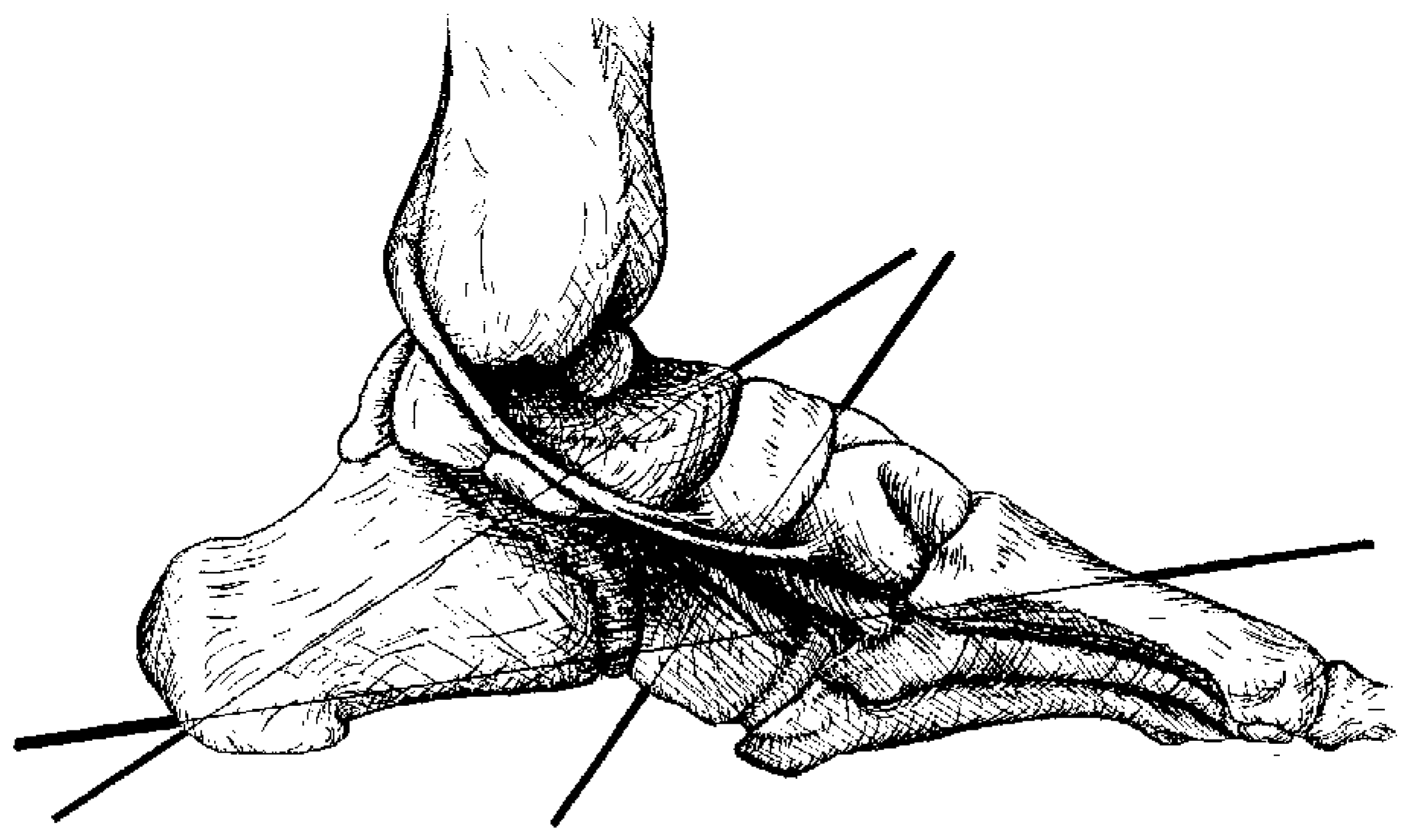
The average subtalar joint deviates 42° from the transverse plane, with a range of 20° to 68°, and 17° to the medial side of the sagittal plane, with a range of 4° to 29° (Figs. 1 and 2). Since the subtalar joint axis acts as an oblique hinge joint, the plane of motion is angular and occurs within the three cardinal planes. This imparts a triplane movement to the bones (ie, some movement of the talus or calcaneus occurs in the frontal, sagittal, and transverse planes as the joint moves). [1,3,5,10,20,21,22,23,24,25,26]
The tendon of tibialis posterior passes posterior and medial to the axis of the subtalar joint. Therefore, muscular contraction of tibialis posterior across the subtalar joint axis allows for approximately equal amounts of inversion and adduction of the foot to occur during supination of the foot. [5]
Midtarsal Joint
The midtarsal joint consists of the calcaneocuboid joint and the talonavicular portion of the talocalca-neonavicular joint. [2,3,5,21] The midtarsal joint contains two separate axes of motion, a longitudinal axis and an oblique axis. Both of these axes pass obliquely through the foot, and can be used independently of one another or together simultaneously. [3,5]
Figure 2.
A view of the plantar aspect of the left lower extremity. The deviation of the ankle joint axis from the frontal plane is 23°. The deviation of the subtalar joint axis from the medial side of the sagittal plane is 17°. The deviation of the longitudinal midtarsal joint axis to the medial side of the sagittal plane is 9°. The deviation of the oblique midtarsal joint axis to the medial side of the sagittal plane is 57°. Note the location of the tendon of tibialis posterior as it crosses these joints (redrawn from Root ML, Orien WP, Weed JH: Normal and Abnormal Functions of the Foot, Vol II, p 206, Figs. 8-23, Clinical Biomechanics, Los Angeles, 1977. Reprinted by permission).
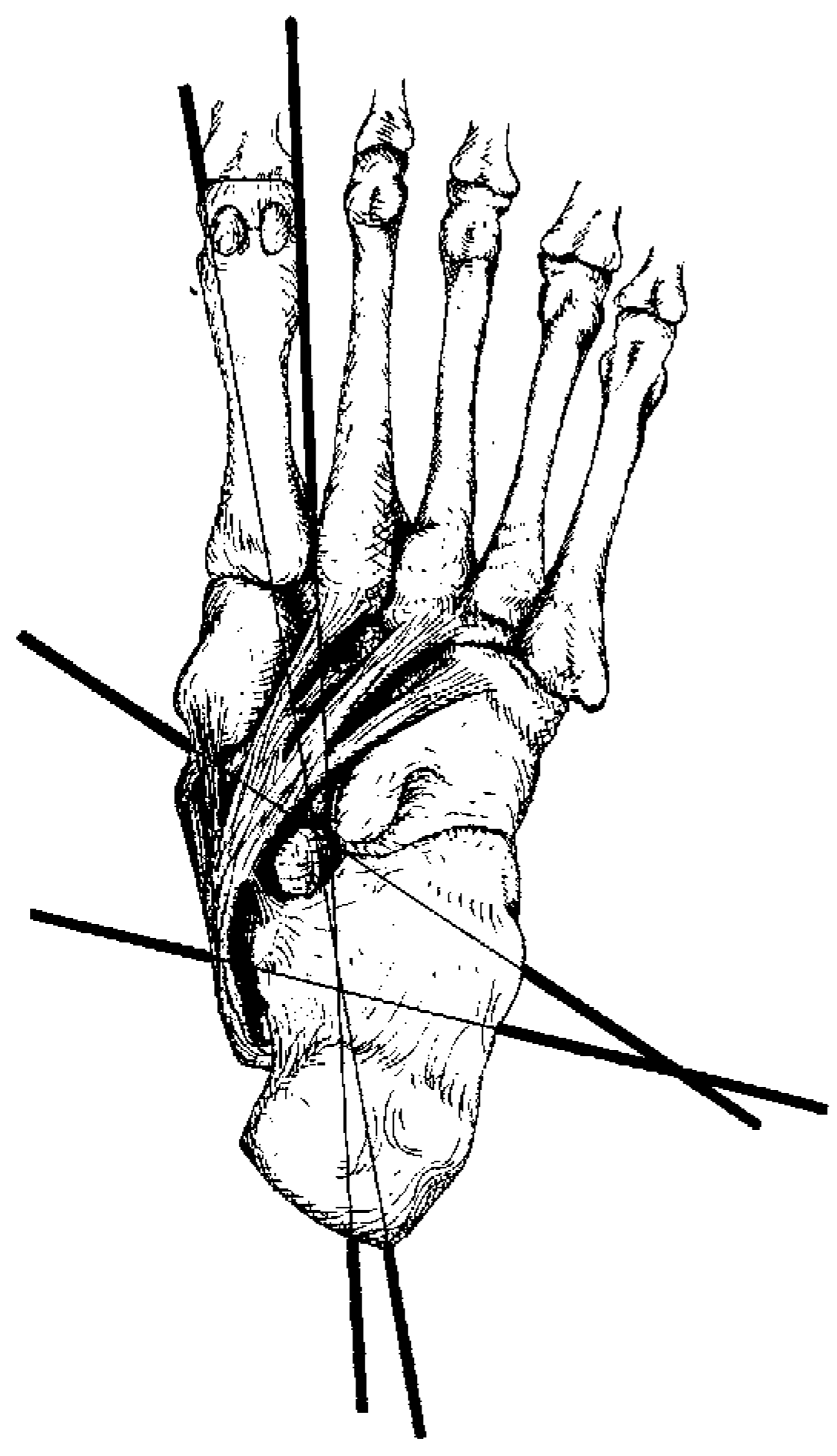
The average longitudinal midtarsal joint axis deviates 15° from the transverse plane, and 9° to the medial side of the sagittal plane (Figs. 1 and 2). The longitudinal axis angles from the superior aspect of the navicular to the lateral aspect of the calcaneus, slightly superior to the subtalar joint axis. Because the longitudinal axis deviates from the three cardinal planes, the motion produced is triplanar, and is seen mostly as inversion and eversion of the forefoot of the rearfoot. [3,5,7]
The average oblique midtarsal joint axis deviates 52° from the transverse plane, and 57° to the medial side of the sagittal plane (Figs. 1 and 2). The oblique axis angles from the superomedial aspect of the head of the talus to the lateral aspect of the calcaneus, slightly anterior to the subtalar joint axis. Because the oblique axis deviates from the three cardinal planes, the motion produced at the oblique midtarsal joint is triplanar, and allows for major amounts of dorsiflexion and abduction and plantarflexion and adduction of the forefoot on the rearfoot. [2,5,7,21]
The tendon of tibialis posterior travels parallel to the longitudinal midtarsal joint axis, and perpendicular to the axis of the oblique midtarsal joint. Because of this course, tibialis posterior can exert a considerable plantarflexion and adduction force around the oblique midtarsal joint axis, and essentially no force about the longitudinal midtarsal joint axis. [5]
Anatomy and Function
From its muscular origin on the posterior aspects of the tibia and fibula, tibialis posterior gives rise to a tendon that passes posterior and inferior to the medial malleolus and partially inserts on the plantar aspect of the navicular at the navicular tuberosity. [27,28,29,30]
Tensile forces transmitted through the tendon of tibialis posterior must pass through three joints: the ankle, subtalar, and oblique midtarsal joints. The axes of these three joints are oriented differently, and, as previously described, allow for different types of movement.
Tibialis posterior functions during the stance phase of the gait cycle, beginning contraction just after heel contact, and relaxing at, or shortly after, heel-off. [5,31,32] Tibialis posterior is frequently regarded as the primary invertor of the foot; however, by pulling the navicular medially and inferiorly, it is also a powerful adductor and plantarflexor of the foot. [27,32,33] When all of these actions are combined, supination of the foot is produced.
The medial malleolus functions as a pulley, around which the tendon of tibialis posterior turns. [5,27,28,29,30] This orientation established an effective angle of approach for the tibialis posterior’s functions at both the ankle and subtalar joints.
Tibialis posterior can produce a strong supinatory force at the subtalar joint. [5,19] At heel contact, and during the early part of midstance, tibialis posterior eccentrically contracts, which decelerates internal rotation of the tibia and subtalar joint pronation. During the late part of midstance, tibialis posterior accelerates subtalar joint supination and external tibial rotation. [5,32]
Distal to its partial insertion on the navicular tuberosity, the main tendon spreads out in an anterolateral direction, and inserts onto the plantar aspects of the intermediate and lateral cuneiforms, the cuboid, and bases of the second, third, fourth, and, sometimes, fifth metatarsals. [5,30]
Materials and Methods
A wooden model of the right lower extremity was constructed. The portion of the model representing the right tibia and fibula consists of a segment of wood measuring 19 × 3 × 11/2 inches. The distal end has been constructed with a 12° deviation from the transverse plane. A solid brass hinge has been attached to this end to simulate the ankle joint axis.
The portion of the model representing the right talus consists of a segment of wood that is 31/2 × 3 × 11/2 inches. The dorsal end has been constructed with a 42° deviation from the transverse plane. A solid brass hinge has been attached to this end to simulate the subtalar joint axis.
The portion of the model representing the right foot consists of a segment of wood measuring 10 × 3 × 11/2 inches. This segment has been divided into two sections, consisting of the calcaneus and fore-foot. The cut was made with a 52° deviation from the transverse plane, and a 57° deviation to the medial side of the sagittal plane. These two sections were connected with a solid brass hinge to simulate the oblique axis of the midtarsal joint. The most distal end of the portion of wood representing the talus was attached to the dorsal surface of the proximal portion of the wood segment representing the calcaneus. It was connected at an angle of 17° to the medial side of the sagittal place, and held in position by two wood screws.
The ankle and subtalar joints, along with their respective axes of motion, are represented by two crossed hinges, riveted together in an approximately normal relationship. Each of these values corresponds with the average accepted values for the individual joint’s axial arrangement. The model has been constructed in this manner in order to reproduce lower extremity movements as accurately as possible. Two nylon ball-bearing roller pulleys were attached to the distal medial aspect of the segment of wood representing the tibia and fibula. The pulleys were meant to represent the medial malleolus.
Two separate, 1/8 × 24-inch wire cables were used to represent the medial and lateral fibers of the tendon of tibialis posterior. The wire cables were held in place by two U-shaped staples positioned over the tracks of the pulleys. These staples reduce extraneous motion of the cables, similar to the function the flexor retinaculum has on the tendon of tibialis posterior in a human limb. The most distal ends of the wire cables were glued into their own predrilled 2-inch deep apertures. The apertures are parallel to each other and located at the proximal medial portion of the forefoot segment. This location coincides with the primary insertion of the tendon of tibialis posterior on the navicular.
Each of the joints in the model can function independently from one another. The isolated motions when tension was applied to the cables were dorsiflexion and plantarflexion at the ankle joint; eversion and abduction and inversion and adduction at the subtalar joint; and dorsiflexion and abduction and plantarflexion and adduction at the oblique midtarsal joint.
All three joints in the model could also function simultaneously. In this manner, the model could undergo the triplanar motions of supination and pronation about the combined axes of motion. Two separate trials were conducted for each of these motions, one with the cables oriented parallel to each other and one with the cables twisted approximately 45° on each other. From their parallel position, the cables were removed from their respective U-shaped staples positioned over the tracks of the pulleys, twisted on each other, and reinserted into the opposite U-shaped staple. This transposition resulted in approximately 45° of total torsion between the two cables, with the actual crossing of the cables occurring below the level of the pulley. From proximal to distal, this directs the medial cable posteriorly and the lateral cable anteriorly.
Results
The ankle, subtalar, and oblique midtarsal joints have been included in the model of the right lower extremity in order to simulate open kinetic chain motion through a pull of the cables representing the tendon of tibialis posterior. An arrow was fixed to each cable to show any unequal excursion of the cables that might occur during motion of the joints. It should be noted that each of the following photographs was taken from a view that would show the maximal difference between the excursions of the cables as the motions of each of the joints were produced.
With the ankle joint motion isolated and the cables side by side, a slight difference in the excursion of the two arrows (ie, the medial and lateral cables) can be noted during motion from plantarflexion to dorsiflexion (Fig. 3). The arrow attached to the medial cable moves further proximally during plantarflexion, and further distally during dorsiflexion, than the arrow attached to the lateral cable.
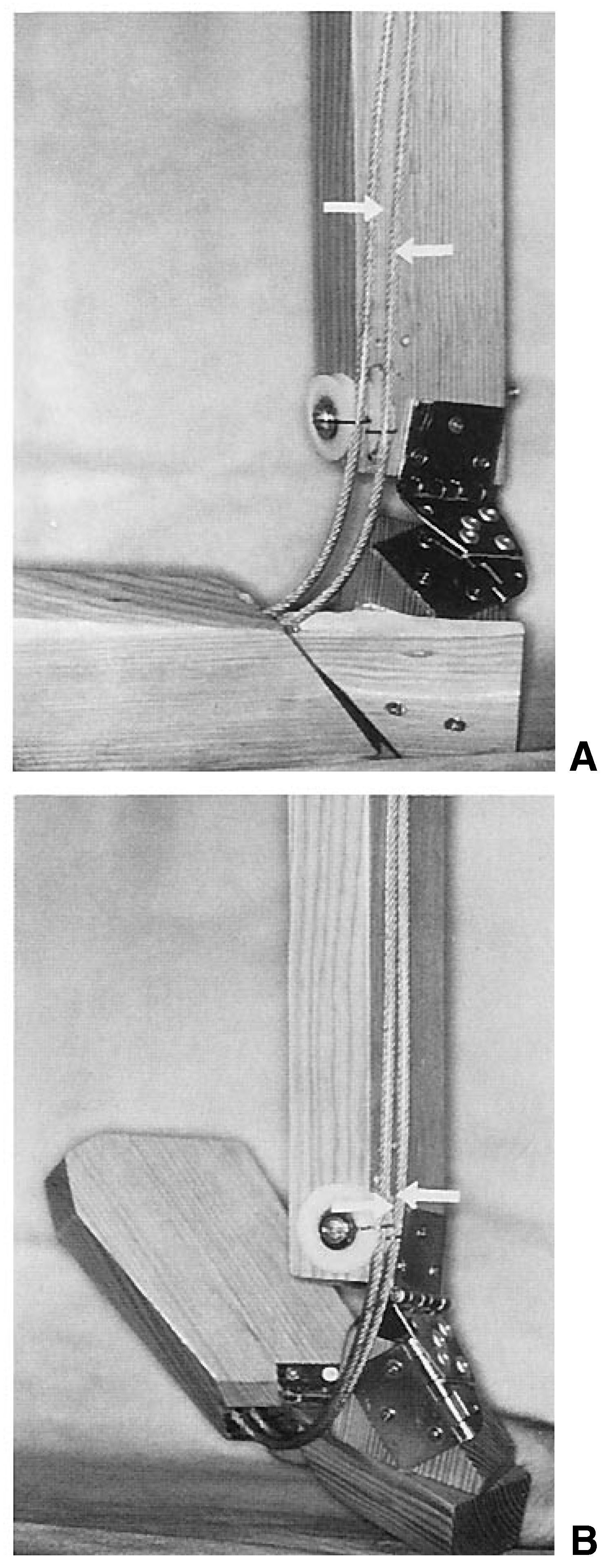
With the oblique midtarsal joint isolated and the cables side by side, a slight difference in the excursion between the two arrows can be noted during motion from plantarflexion and adduction to dorsiflexion and abduction (Fig. 4). The arrow attached to the medial cable moves further proximally during plantarflexion and adduction, and further distally during dorsiflexion and abduction, than the arrow attached to the lateral cable.
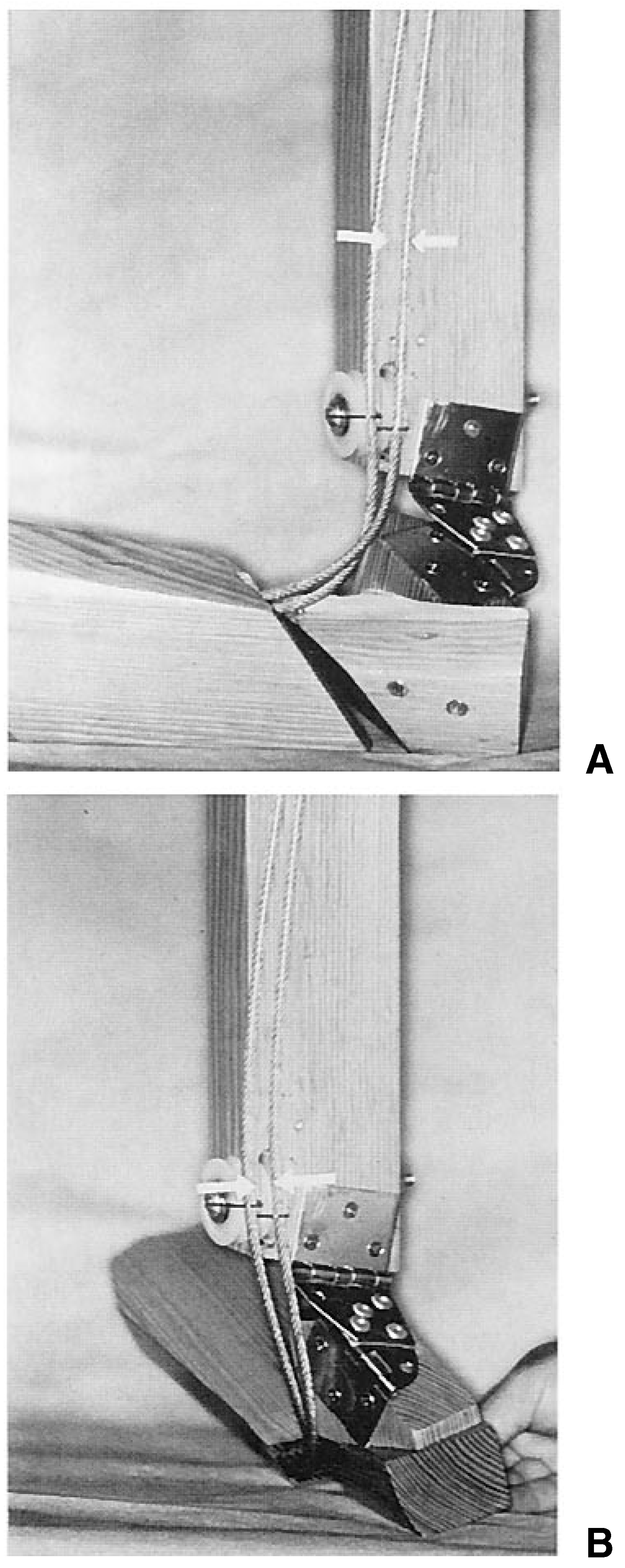
With the subtalar joint isolated and the cables side by side, a marked difference in the excursion of the two arrows can be noted during motion from inversion and adduction to eversion and abduction (Fig. 5). The arrow attached to the medial cable moves further proximally during inversion and adduction, and further distally during eversion and abduction, than the arrow attached to the lateral cable.
With the two cables side by side, motion from full supination to full pronation (ie, the combined motions produced at the ankle, subtalar, and oblique midtarsal joints) results in a significant difference in the amount of excursion between the two arrows (Fig. 6). The arrow attached to the medial cable moves further proximally during full supination, and further distally during full pronation, than the arrow attached to the lateral cable.
Discussion
The anatomy of the tendon of tibialis posterior, like all tendons, consists of a series of longitudinal, densely packed bundles of collages which move as a single, solid unit. [34,35] Figure 3 through 5 show that when the cables, which are meant to represent the medial and lateral fibers of the tendon of tibialis posterior, are side by side, the triplanar motions produced at each of the joints in the model result in unequal motion of the two cables. In each of these cases, the medial cable moves faster and through a greater excursion than the lateral cable, as shown by the misalignment of the arrows on the cables.
The amount of difference in the excursion of the two cables occurring across each joint is additive. Motion produced simultaneously across all three joints results in a greater excursion between the two cables than when motion is produced across any one of the isolated joints. This is shown by the marked difference in the excursion of the arrows on the cables in Figure 6 when compared with the amount of excursion the arrows undergo in any of the isolated situations presented in Figure 3 through 5. In a human limb, this would necessitate a significant amount of longitudinal motion between the individual fibers of the tendon of tibialis posterior. Because tendons function as a unit, any situation that requires longitudinal slippage between individual fibers is an unlikely and unnatural one.
In Figure 7, the cables representing the tendon of tibialis posterior have been twisted approximately 45°. In this arrangement, the medial cable passes posteriorly, and the lateral cable anteriorly, as they travel from proximal to distal. This is approximately the same amount and direction of torsion that has been previously described. [4] The presence of such a twist results in an equal excursion of the two cables throughout the triplanar motions of supination and pronation, as shown by the equal excursion of the arrows on the cables.
Figure 3.
The effects of motion around an isolated ankle joint axis. The cables, representing the medial and lateral fibers of the tendon of tibialis posterior, are side by side. An arrow has been fixed to each cable to show any unequal excursion between the two cables. Note that from plantarflexion (A) to dorsiflexion (B), there has been only a slight difference in the excursion of the two cables.
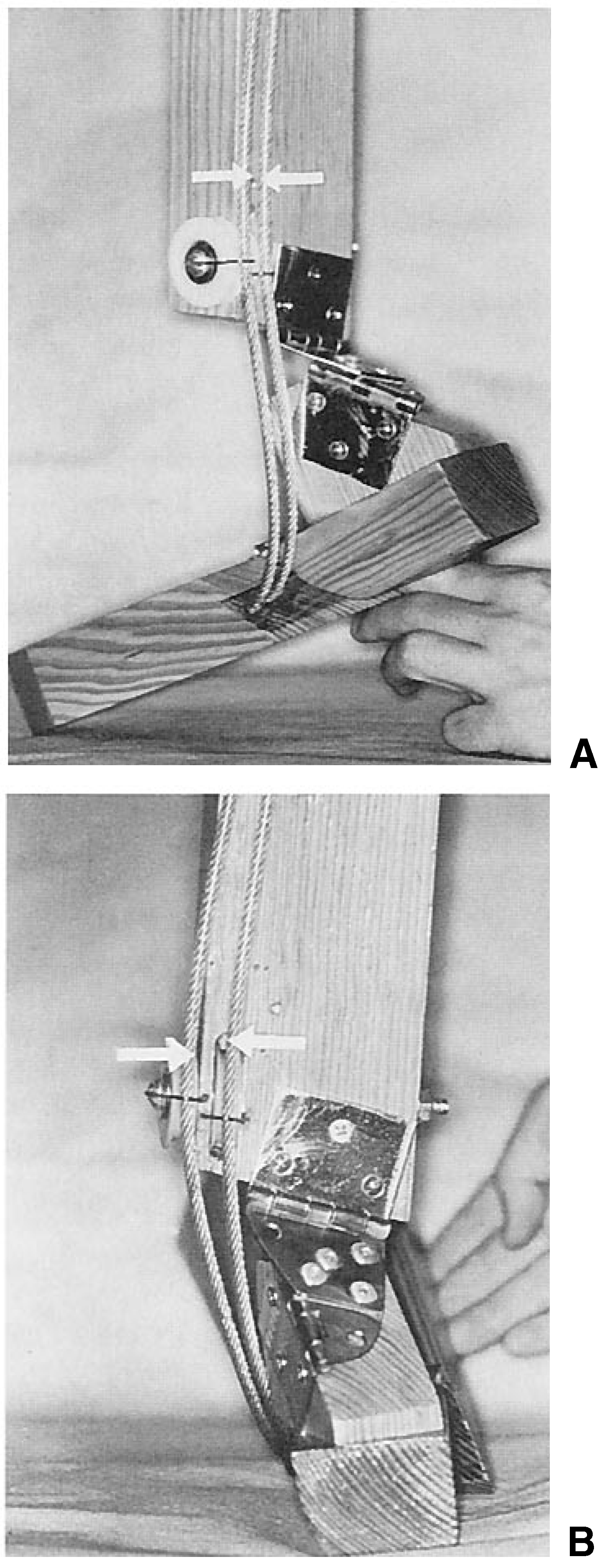
Figure 4.
The effects of motion around an isolated oblique midtarsal joint axis. The cables are side by side. Movement of this axis is one of plantarflexion and adduction (A) and dorsiflexion and abduction. (B). Note that such movement causes only a slight difference in the excursion of the two cables.
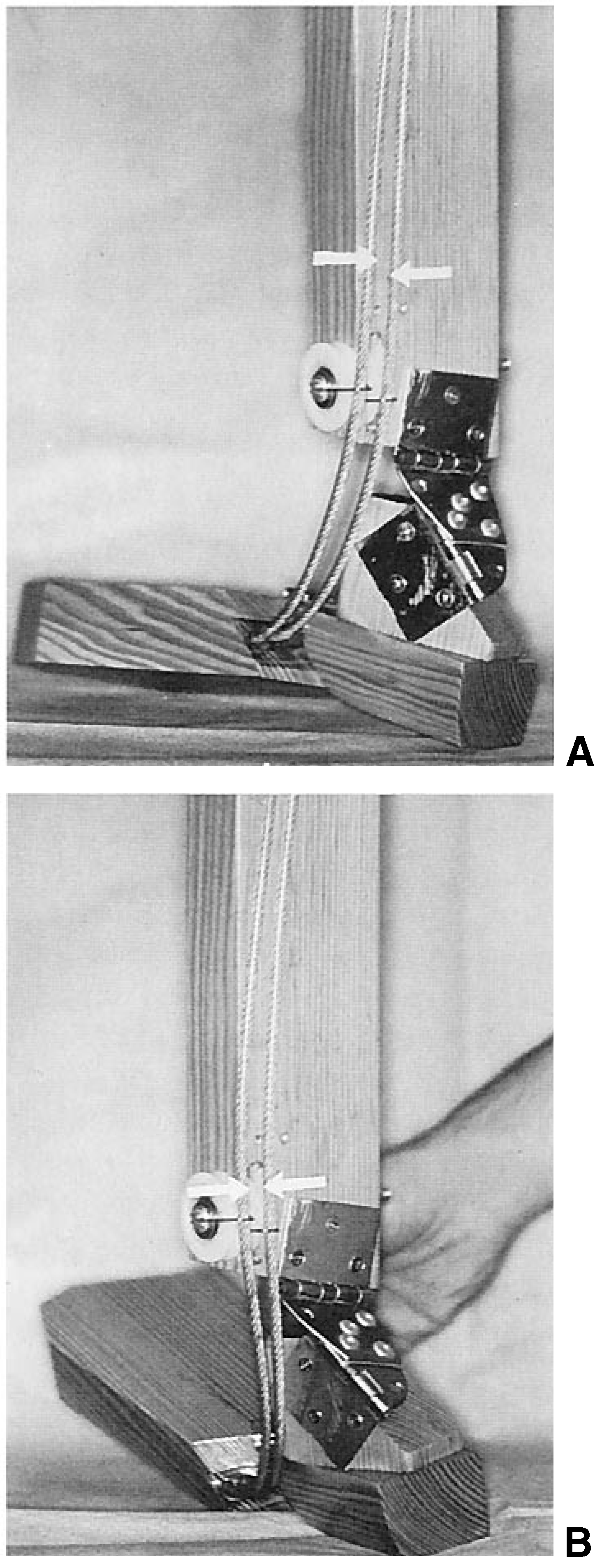
Figure 5.
The effects of motion around an isolated subtalar joint axis. The cables are side by side. Movement at the subtalar joint axis is inversion and adduction (A) and eversion and abduction (B). Note that such movement causes a marked difference in the excursion of the two cables.
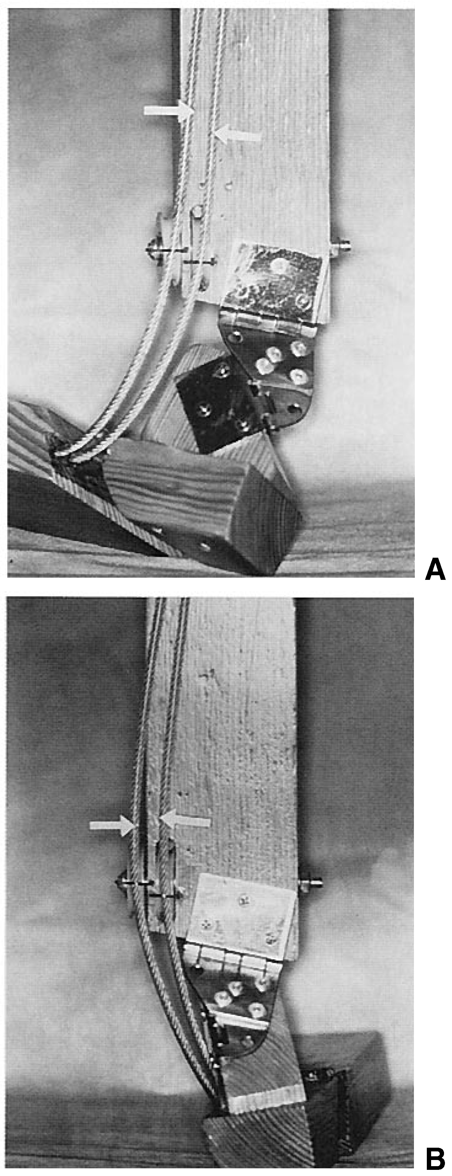
Figure 6.
The effects of the combined motions at all three joints. The cables are side by side. In this situation, movement from (A) supination (ie, plantarflexion of the ankle joint, plantarflexion and adduction of the oblique midtarsal joint, and inversion and adduction of the subtalar joint) to (B) pronation (ie, dorsiflexion of the ankle joint, dorsiflexion and abduction of the oblique midtarsal joint, and eversion and abduction of the subtalar joint) causes the greatest difference in the excursion of the two cables.
Therefore, the authors conclude that torsion of the tendon of tibialis posterior in a human limb equalizes the excursion of all fibers comprising the tendon, and eliminates the need for any longitudinal slippage between individual tendon fibers.
Figure 7.
The effect of twisting the wires approximately 45° is shown. The same model is used and the same motions are shown as in Figure 6. The medial cable now passes posteriorly, and the lateral cable passes anteriorly. This arrangement equalizes the excursion of the two cables, as shown by the alignment of the two arrows.
Conclusion
Torsion of the tendon of tibialis posterior has been shown to equalize the excursion of and eliminate the need for any longitudinal slippage between individual tendon fibers. A thorough discussion of the surgical significance of such a torsion is beyond the scope of this article but deserves further consideration. Since torsion is present within both the Achilles and posterior tibial tendons, it is probable that torsion also exists within some of the tendons of the other extrinsic muscles of the foot. This consideration warrants further investigation.
Acknowledgments
Lorin Miles and the Engineering Department at the California College of Podiatric Medicine for the wooden model; Chris Cushman for photographs; and Matthew Phelps for illustrations.
References
- INMAN, VT. “Application to Orthopedics,” in The Joints of the Ankle; Williams & Wilkins: Baltimore, 1976. [Google Scholar]
- SHEPHARD, E. Tarsal movements. J Bone Joint Surg 1951, 33B, 258. [Google Scholar] [CrossRef] [Scilit]
- HICKS, JH. The mechanics of the foot: part I. the joints. J Anat 1953, 87, 345. [Google Scholar]
- ROUKIS, TS; HURLESS, JS; PAGE, JC. Torsion of the tendon of tibialis posterior. JAPMA 1995, 85, 464. [Google Scholar] [CrossRef] [Scilit] [PubMed]
- ROOT, ML; ORIEN, WP; WEED, JH. “Motions of the Joints of the Foot,” Normal and Abnormal Functions of the Foot; Clinical Biomechanics Vol II, Clinical Biomechanics: Los Angeles, 1977. [Google Scholar]
- BARNETT, CH; NAPIER, JR. The axis of rotation at the ankle joint in man. J Anat 1952, 86, 1. [Google Scholar] [PubMed]
- PEPIN, WA; MARCHAND, ER. Some anatomical and physiological relationships in the foot. JAPA 1964, 54, 169. [Google Scholar]
- LUNDBERG, A; SVENSSON, OK; NEMETH, G; ET, AL. The axis of rotation of the ankle joint. J Bone Joint Surg 1989, 71B, 94. [Google Scholar] [CrossRef] [Scilit] [PubMed]
- Spoor, CW; Van Leeuwen, JL; Van der Meulen, WJ; et al. Eur J Morphol 1991, 29, 136.
- MORRIS, JL. A conceptual look at rearfoot motion. Clin Podiatr Med Surg 1994, 11, 297. [Google Scholar] [CrossRef] [Scilit]
- REIKERAS, O; HOISETH, A. Torsion of the leg determined by computed tomography. Acta Orthop Scand 1989, 60, 330. [Google Scholar] [CrossRef] [Scilit]
- CLEMENTZ, BG. Tibial torsion measured in normal adults. Acta Orthop Scand 1988, 59, 441. [Google Scholar] [CrossRef] [Scilit]
- BADELON, O; BENSAHEL, H; FOLINAIS, D; et al. Tibiofibural torsion from the fetal period until birth. J Pediatr Orthop 1989, 9, 169. [Google Scholar] [CrossRef] [Scilit] [PubMed]
- VALMASSY, R; STANTON, B. Tibial torsion: normal values in children. JAPMA 1989, 79, 432. [Google Scholar] [CrossRef] [Scilit]
- JAKOB, RP; HAERTEL, M; STUSSI, E. Tibial torsion calculated by computerized tomography and compared to other methods of measurement. J Bone Joint Surg 1980, 62B, 238. [Google Scholar] [CrossRef] [Scilit] [PubMed]
- COHEN-SOBEL, E; LEVITZ, SJ. Torsional development of the lower extremity: implications for in-toe and out-toe treatment. JAPMA 1991, 81, 344. [Google Scholar] [CrossRef] [Scilit]
- EKHOFF, DG; KRAMER, RC; WATKINS, JJ; ET, AL. Variations in tibial torsion. Clin Anat 1994, 7, 76. [Google Scholar] [CrossRef] [Scilit]
- HALL, MC. The normal movements at the subtalar joint. Can J Surg 1959, 2, 287. [Google Scholar] [PubMed]
- LAPIDUS, PW. Subtalar joint, its anatomy and mechanics. Bull Hosp J Dis 1955, 16, 179. [Google Scholar]
- JONES, RL. The functional significance of the declination of the axis of the subtalar joint. Anat Rec 1945, 93, 151. [Google Scholar] [CrossRef] [Scilit]
- MANTER, JT. Movements of the subtalar and transverse tarsal joints. Anat Rec 1941, 80, 397. [Google Scholar] [CrossRef] [Scilit]
- ROOT, ML; WEED, JH; SGARLATO, TE; ET, AL. Axis of motion of the subtalar joint: an anatomical study. JAPA 1966, 56, 149. [Google Scholar]
- PHILLIPS, RD; CHRISTECK, R; PHILLIPS, RL. Clinical measurement of the axis of the subtalar joint. JAPMA 1985, 75, 119. [Google Scholar] [CrossRef] [Scilit]
- KIRBY, KA. Methods for determination of positional variations in the subtalar joint axis. JAPMA 1987, 77, 228. [Google Scholar] [CrossRef] [Scilit]
- KIRBY, KA. Rotational equilibrium across the subtalar joint axis. JAPMA 1989, 79, 1. [Google Scholar] [CrossRef] [Scilit]
- PHILLIPS, RD; LIDTKE, RH. Clinical determination of the linear equation for the subtalar joint axis. JAPMA 1992, 82, 1. [Google Scholar] [CrossRef] [Scilit] [PubMed]
- ROMANES, GJ. “Muscles and Fasciae,” in Cunningham’s Textbook of Anatomy, 12th Ed; Romanes, GJ, Ed.; Oxford University Press: Oxford, England, 1981. [Google Scholar]
- O’RAHILLY, R. Gardner-Gray-O’Rahilly Anatomy, 5th Ed ed; WB Saunders: Philadelphia, 1986. [Google Scholar]
- DRAVES, DE. “Popliteal Fossa and the Leg,” in Anatomy of the Lower Extremity; Williams & Wilkins: Baltimore, 1986. [Google Scholar]
- SARRAFIAN, SK. Functional Anatomy of the Foot and Ankle. In Anatomy of the Foot and Ankle: Descriptive,Topographic, Functional, 2nd Ed ed; JB Lippincott: Philadelphia, 1993. [Google Scholar]
- KAYE, RA; JAHSS, MH. Foot fellows review tibialis posterior: a review of anatomy and biomechanics in relation to support of the medial longitudinal arch. Foot Ankle 1991, 11, 244. [Google Scholar] [CrossRef] [Scilit] [PubMed]
- MUELLER, TJ. Acquired flatfoot secondary to tibialis posterior dysfunction: biomechanical aspects. J Foot Surg 1991, 30, 2. [Google Scholar]
- KAPANDJI, I. “The Ankle,” in The Physiology of the Joints, Vol II, 2nd Ed ed; Churchill Livingstone: New York, 1970. [Google Scholar]
- JUNQUERIA, LC; CARNEIRO, J; KELLY, RO. “Connective Tissue,” in Basic ffistology, 7th Ed ed; Appleton & Lange: Norwalk, CT, 1992. [Google Scholar]
- DYKYJ, D; JULES, KT. The clinical anatomy of tendons. JAPMA 1991, 81, 358. [Google Scholar] [CrossRef] [Scilit] [PubMed]
© 1996 American Podiatric Medical Association