


Abstract
The authors present a quantitative analysis of the effect that first ray position has on motion of the first metatarsophalangeal joint. A goniometer was constructed to measure the degrees of first metatarsophalangeal joint dorsiflexion with the first ray in three positions: weightbearing resting position, dorsiflexed 4 mm from the weightbearing resting position, and dorsiflexed 8 mm from the weightbearing resting position. First metatarsophalangeal joint dorsiflexion decreased 19% as the first ray was moved from the weightbearing resting position to 4 mm dorsiflexed, 19.3% as the first ray was moved from 4 mm dorsiflexed to 8 mm dorsiflexed, and 34.7% as the first ray was moved from the weightbearing resting position to 8 mm dorsiflexed. The biomechanical significance of decreased first metatarsophalangeal joint dorsiflexion that results from first ray dorsiflexion is discussed, and proposed bases for the pathomechanics of hallux abducto valgus and hallux rigidus deformities are presented.
Sagittal plane motion of the first metatarsophalangeal joint is an important component of the normal function of the human foot during gait [1]. This motion has received the attention of several authors in reference to the development of pathology at the first metatarsophalangeal joint [1,2,3,4,5,6]. Functional and structural deformities such as functional hallux limitus [7,8,9], hallux abducto valgus [2,3,5,6,10], and hallux rigidus [5,6,11,12,13,14,15,16] have all been purported to develop secondary to lack of motion at the first metatarsophalangeal joint. Accordingly, numerous surgical procedures focus on the first metatarsophalangeal joint to correct these deformities [5,6,9,17,18,19]. However, current understanding of first metatarsophalangeal joint motion is based on a small number of studies [10,20,21,22,23,24,25]. Additionally, the wide variety of proposed etiologic factors responsible for determining motion of the first metatarsophalangeal joint suggests that an anatomically and biomechanically sound explanation has yet to be presented.
Review of the Literature
Originally, Hicks [26] concluded that first metatarsophalangeal joint motion was controlled solely by the windlass effect of the plantar aponeurosis. Hicks [26] believed that since the plantar aponeurosis is attached proximally to the calcaneus and distally to the proximal phalanx of the hallux, through its connection to the plantar pad, when the hallux is dorsiflexed, the plantar pad and plantar aponeurosis will move distally, shortening the distance between the calcaneus and head of the first metatarsal. Hicks [26] states, “The effect was as though a cable had been wound one-quarter of a turn on the drum of a windlass; the drum of the windlass being the head of the metatarsal, the handle which does the winding being the proximal phalanx, and the cable which is wound on to the drum being the plantar pad and the plantar aponeurosis.” As a result of this sequence of events, Hicks [26] concluded that, “The effective length of the cable was shortened [...] and the arch was thereby made shorter and higher.” He also proposed that this same mechanism would work in reverse. That is, as the arch of the foot lowered, the windlass would unwind and plantarflex the hallux at the first metatarsophalangeal joint, thereby decreasing the ability of the hallux to dorsiflex [26].
Durrant and Siepert [27] believed that certain strategically placed soft tissue structures determine the motion of the first metatarsophalangeal joint. They proposed that any soft tissue structure could individually restrict dorsiflexion of the hallux at the first metatarsophalangeal joint if it: 1) crosses, and lies below, the transverse axis of motion of the first metatarsophalangeal joint; 2) attaches to the plantar aspect of the proximal phalanx; 3) exerts a force that is parallel to the longitudinal axis of the first ray; and 4) is equally present on both sides of the longitudinal axis of the first ray. The flexor hallucis brevis and sesamoid apparatus, medial plantar fascial band, and joint capsule were all considered to meet the necessary criteria. A short flexor hallucis brevis; proximally located, elongated, and hypertrophic sesamoids; a congenitally short medial plantar fascial slip; and iatrogenically induced scarring of the joint capsule, were all considered capable of limiting motion of the first metatarsophalangeal joint [27].
Several authors have attempted to calculate the instant center of rotation of the first metatarsophalangeal joint [10,20,21,22]. An instant center of rotation is defined as the point in one object around which another object rotates [20]. Shereff et al [20] state that the instant center of rotation moves from the center of the head of the first metatarsal in a circular pattern from distal-dorsal to proximal-plantar, while Sammarco [22] states that the instant center of rotation moves from the center of the head of the first metatarsal in a continuous arc from proximal-plantar to distal-dorsal.
Rather than calculating the instant center of rotation during open kinetic chain motion, Hetherington et al [21] chose to study closed kinetic chain motion as this represents the actual function and motion of the first metatarsophalangeal joint during gait. They state that the instant center of rotation moves from distal-plantar, through the center of the head of the first metatarsal, to proximal-dorsal [21]. Shereff et al [10,20] have also described changes in the normal location and direction of the instant center of rotation of the first metatarsophalangeal joint in the presence of hallux abducto valgus and hallux rigidus deformities, and attributed these changes to anatomical variances and inherent properties of the first metatarsophalangeal joint.
Additionally, each of the following circumstances has been proposed as an etiologic factor directly responsible for decreasing motion at the first metatarsophalangeal joint: 1) heredity [28]; 2) primary or secondary osteoarthritis [1,29]; 3) osteochondritis dissecans [16,30,31,32]; 4) avascular necrosis [33]; 5) various arthritides (eg, rheumatoid, psoriatic, gouty and septic) [1,5]; 6) neuromuscular disorders resulting in either weakness of peroneus longus or spasticity of tibialis anterior [34]; 7) acute trauma [1]; 8) iatrogenic causes related to previous surgery of the first metatarsal or first metatarsophalangeal joint [5,6]; and 9) congenital fragmentation of the epiphysis in the base of the proximal phalanx of the hallux [30].
Rather than looking toward the first metatarsophalangeal joint for anatomical or functional variances, the authors believe that motion of the first metatarsophalangeal joint is governed principally by the position of the first ray. This concept is not new, as the position of the first ray has been inferred in the medical literature to affect the motion of the first metatarsophalangeal joint [35,36]. An excessively long or short first metatarsal, metatarsus primus elevatus, and immobilization or hypermobility of the first ray have all been purported to adversely affect the ability of the hallux to dorsiflex at the first metatarsophalangeal joint [1,5,12,29,37,38,39,40]. Although the medical literature supports the theory that the position of the first ray has an effect on motion of the first metatarsophalangeal joint, there exists no known quantitative documentation of this effect.
The purpose of this study was to determine if dorsiflexion of the first ray decreases first metatarsophalangeal joint dorsiflexion. The null hypothesis was that first ray dorsiflexion does not affect first metatarsophalangeal joint dorsiflexion. Additionally, the authors will present a theory as to how dorsiflexion of the first ray may result in pathologic changes at the first metatarsophalangeal joint.
The First Ray
Anatomically, the first ray consists of the collective articulations between the navicular, medial cuneiform, intermediate cuneiform, and base of the first metatarsal [1,41]. Motion of the first ray occurs at the first metatarsal-medial cuneiform joint, the medial cuneiform-intermediate cuneiform joint, and the medial cuneonavicular joint [1,35,41,42,43,44]. These individual joints essentially move in unison about a common axis of motion, and function as a single combined unit or ray.
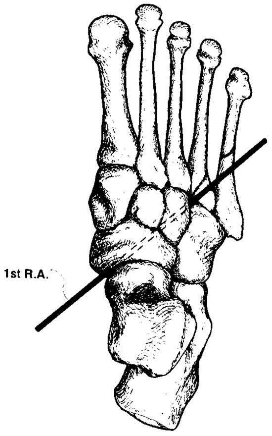
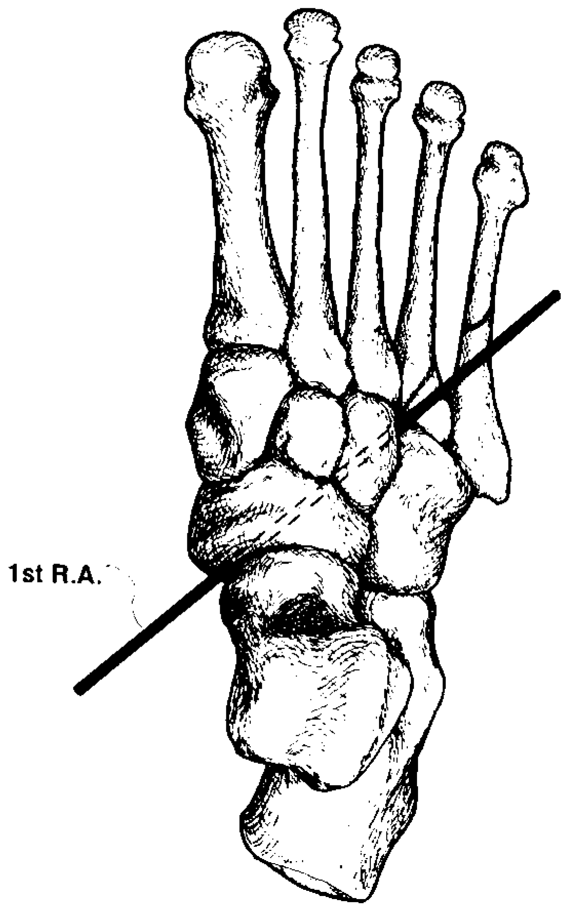
The axis of motion of the first ray passes obliquely, from the proximal-plantar-medial margin of the navicular tuberosity to the distal-dorsal-lateral aspect of the base of the third cuneiform [1]. The average axis of motion is angled approximately 45° from both the sagittal and frontal planes, and only slightly from the transverse plane (Figure 1) [1,41]. The actual amount of transverse plane motion is considered clinically insignificant, and although the first ray is said to exhibit triplanar motion, nearly all of the motion occurs within the frontal and sagittal planes [1,35,41,42,43,44]. Because of the orientation of its axis of motion, a proportionate amount of inversion occurs with dorsiflexion of the first ray, and a proportionate amount of eversion occurs with plantarflexion of the first ray [1,35,41,42,43,44].
Figure 1.
A view of the dorsal aspect of the right foot, showing the location and orientation of the axis of motion of the first ray (from ROOT ML, ORIEN WP, WEED JH: Normal and Abnormal Function of the Foot, Clinical Biomechanics, Vol II, Fig. 1-78, p 47, Clinical Biomechanics Corp, Los Angeles, 1977. Reprinted with permission).
Materials and Methods
Subjects in this study were required to meet the following conditions: 1) absence of digital deformities; 2) absence of hyperkeratotic lesions on the forefoot; 3) absence of either inversion or eversion of the calcaneus in resting calcaneal stance position; 4) presence of at least 10° of unrestricted ankle joint dorsiflexion with the knee joint fully extended; 5) presence of a total unrestricted motion of the functional subtalar joint of at least 20°; 6) presence of a total unrestricted range of motion of the longitudinal midtarsal joint of at least 15°; 7) presence of at least 8 mm of unrestricted dorsiflexion of the first ray; 8) presence of at least 20° of unrestricted dorsiflexion of the first metatarsophalangeal joint in resting calcaneal stance position; 9) a shoe size of 9.5; 10) negative history for any foot symptoms requiring medical attention, foot trauma, foot surgery, or clinical signs of hallux abducto valgus or hallux rigidus.
Ten male volunteer subjects met the above-defined conditions. Therefore, 20 feet (10 right, 10 left) were used in this study. The mean age of the subjects was 26 years, with a range from 23 to 30 years.
Subtalar joint, first ray, and first metatarsophalangeal joint ranges of motion were measured in the usual manners as described by Root et al [1]. Longitudinal midtarsal joint inversion was subjectively measured by manually stabilizing the subtalar joint in neutral position and then inverting the medial aspect of the forefoot relative to the stabilized rearfoot. The examiner then visually approximated the angle formed between the plantar aspect of the inverted forefoot and the plantar aspect of the rearfoot held in subtalar neutral position. In this manner, longitudinal midtarsal joint inversion could be estimated and pathology significant enough to restrict motion of the longitudinal midtarsal joint expressed.
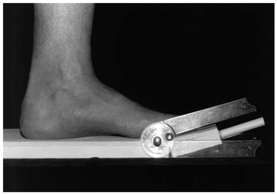
A weightbearing goniometer was constructed to measure closed kinetic chain dorsiflexion of the first metatarsophalangeal joint with the first ray in weightbearing resting position, dorsiflexed 4 mm from the weightbearing resting position, and dorsiflexed 8 mm from the weightbearing resting position. The proximal portion of the goniometer consisted of a segment of wood measuring 18 × 10 × 1 inches. The distal portion of the goniometer consisted of a segment of wood measuring 4 × 10 × 1 inches. A solid brass hinge was attached between the two segments of wood to allow sagittal plane motion of the distal segment on the proximal segment. A protractor was used to measure the degree of sagittal plane motion of the distal segment of the goniometer. The lower arm of the protractor was attached to the lower portion of the distal segment of the goniometer, and the upper arm of the protractor was attached to the upper portion of the distal segment of the goniometer (Figure 2).
Figure 2.
A view of the medial aspect of the goniometer used to measure first metatarsophalangeal joint dorsiflexion. Note the position and location of the upper and lower arms of the protractor in relation to the goniometer.
Subjects were instructed to stand in their angle and base of gait on the proximal segment of the goniometer with the center of their first metatarsophalangeal joint located at the level of the hinge (ie, between the proximal and distal segments of the goniometer) and with their hallux placed perpendicular to the axis of the hinge. This was considered to represent the weightbearing resting position of the first ray. The distal segment of the goniometer was then maximally dorsiflexed until resistance was felt by the examiner, but prior to visual plantarflexion of the first metatarsal. The authors considered this to represent the maximum degree of first metatarsophalangeal joint dorsiflexion. This position allowed an angle to be measured from the degree marker on the protractor.
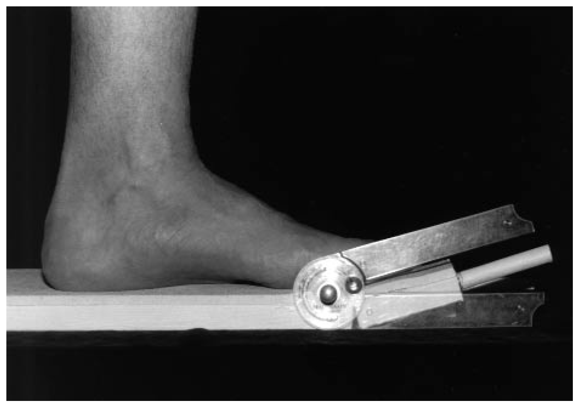
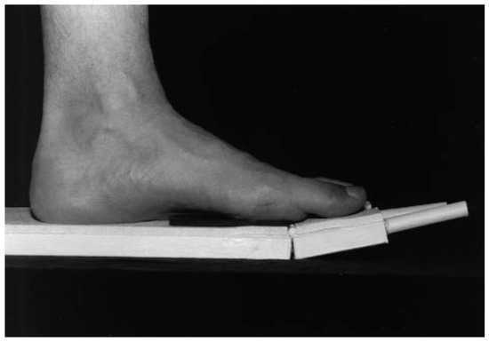
A 2 × 1-inch acrylic platform, 4 mm thick, was then placed directly below the head of the first metatarsal (Figure 3). With the first ray now dorsiflexed 4 mm from the weightbearing resting position, the distal segment was once again maximally dorsiflexed until resistance was felt by the examiner, and the degree of first metatarsophalangeal joint dorsiflexion was measured from the protractor. Another 4-mm thick acrylic platform was then placed on top of the first 4-mm platform. With the first ray now dorsiflexed 8 mm from the weightbearing resting position, the distal segment of the goniometer was maximally dorsiflexed until resistance was felt by the examiner, and the degree of first metatarsophalangeal joint dorsiflexion was once again measured from the protractor.
Figure 3.
A view of the medial aspect of the goniometer and the medial aspect of the left foot, while measuring the degree of first metatarsophalangeal joint dorsiflexion. The distal segment of the goniometer is maximally dorsiflexed and a 4-mm thick, 2 × 1-inch acylic platform was placed directly below the head of the first metatarsal.
The degrees of first metatarsophalangeal joint dorsiflexion were recorded to the nearest degree, with the first ray in weightbearing resting position, dorsiflexed 4 mm and 8 mm. After a period of acclimation to the use of the goniometer, this entire procedure was repeated three times by one examiner. Thus, three values of first metatarsophalangeal joint dorsiflexion were obtained for each of the three positions of the first ray. The mean of these values was used during statistical analysis.
The Student’s t-test was performed to compare the mean degrees of first metatarsophalangeal joint dorsiflexion with the first ray in weightbearing resting position, dorsiflexed 4 mm and 8 mm, between the right and left feet. The Student’s t-test was performed to compare the mean degrees of first metatarsophalangeal joint dorsiflexion with the first ray in weightbearing resting position versus dorsiflexed 4 mm, dorsiflexed 4 mm versus dorsiflexed 8 mm, and weightbearing resting position versus dorsiflexed 8 mm, for all 20 feet.
All statistical analysis was performed using the Stat View 4.02® (Abacus Concepts, Inc, Berkeley, CA) program, and the graph was produced on a personal computer using Cricketgraph 1.3® (Cricket Software, Malvern, PA).
Results
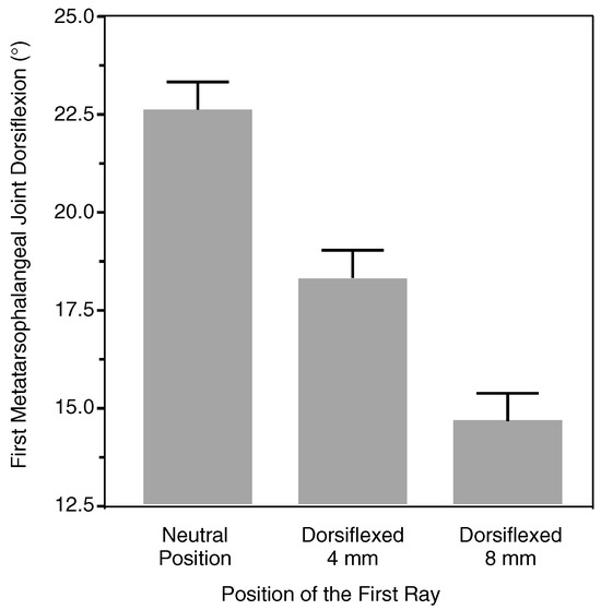
The results of this part of the study are summarized in Table 1. These results document the proportionate decrease of first metatarsophalangeal joint dorsiflexion that occurs with dorsiflexion of the first ray. The mean degrees of first metatarsophalangeal joint dorsiflexion available with the first ray in weightbearing resting position, 4 mm dorsiflexed, and 8 mm dorsiflexed were calculated and plotted (Figure 4). The data presented in Figure 4 graphically represent the proportionate decrease in first metatarsophalangeal joint dorsiflexion as the first ray proceeds from the weightbearing resting position to a dorsiflexed position.
Table 1.
Degrees of First Metatarsophalangeal Joint Dorsiflexion with the First Ray in Weightbearing Resting Position, and Dorsiflexed 4 mm and 8 mm.
Figure 4.
Degrees of first metatarsophalangeal joint dorsiflexion with the first ray in weightbearing resting position, dorsiflexed 4 mm from the weightbearing resting position, and dorsiflexed 8 mm from the weightbearing resting position. Values indicate mean; N = 20 for each measurement.
Mean first metatarsophalangeal joint dorsiflexion was 22.7° ± 0.4° (range 20° to 26°) with the first ray in weightbearing resting position, 18.4° ± 0.5° (range 15° to 23°) with the first ray dorsiflexed 4 mm, and 14.8° ± 0.6° (range 10° to 20°) with the first ray dorsiflexed 8 mm. The amount of dorsiflexion at the first metatarsophalangeal joint decreased 19% when the first ray was moved from the weightbearing resting position to the dorsiflexed 4-mm position, 19.3% when the first ray was moved from the dorsiflexed 4-mm position to the dorsiflexed 8-mm position, and 34.7% when the first ray was moved from the weightbearing resting position to the dorsiflexed 8-mm position.
Comparison, between the right and left feet, of the difference in the mean degrees of first metatarsophalangeal joint dorsiflexion with the first ray in weightbearing resting position (p = 0.3293), dorsiflexed 4 mm (p = 0.3382), and dorsiflexed 8 mm (p = 0.7558), was not significant. Comparison, for all 20 feet, of the difference in the mean degrees of first metatarsophalangeal joint dorsiflexion with the first ray in weightbearing resting position versus dorsiflexed 4 mm (p ≤ 0.0001), dorsiflexed 4 mm versus dorsiflexed 8 mm (p ≤ 0.0001), and weightbearing resting position versus dorsiflexed 8 mm (p ≤ 0.0001), were significant.
Discussion
The null hypothesis stated that first ray dorsiflexion does not affect first metatarsophalangeal joint dorsiflexion. The results of this study, however, support the theory that motion of the first metatarsophalangeal joint is influenced by the position of the first ray [35,36]. The authors’ data are the only sources now available that show that there is a quantitative decrease in first metatarsophalangeal joint dorsiflexion as the first ray dorsiflexes around its axis of motion (Table 1). Furthermore, the data show that there was a proportionate and predictable decrease in first metatarsophalangeal joint dorsiflexion as the first ray dorsiflexed from the weightbearing resting position (Figure 4). Increasing the dorsiflexion of the first ray by twice the amount of millimeters decreased dorsiflexion of the first metatarsophalangeal joint by twice the degrees. Based on these findings, the authors rejected the null hypothesis.
Although the data were obtained with as much accuracy and reproducibility as the goniometer would permit, the use of a tensionometer or digital goniometer could have resulted in more precise measurements and greater reproducibility. Likewise, more stringent research protocol and a greater patient population could have further delineated statistical significance. However, it is the authors’ contention that the basic findings of this study would not have been greatly altered with more precise measuring devices, improved protocol, or an increased patient population.
Theorem
The authors propose that the decrease of first metatarsophalangeal joint dorsiflexion that results from dorsiflexion of the first ray is the predominant factor behind the development of hallux abducto valgus and hallux rigidus deformities. The final deformity is theorized to depend on the actual amount of first ray dorsiflexion that occurs during gait, rather than representing anatomical variances or inherent properties of the first metatarsophalangeal joint. It should be noted that in the following discussion, any sagittal or frontal plane motion of the first ray is considered relative to the second metatarsal. Fracture or other trauma of the first metatarsophalangeal joint is beyond the scope of this discussion.
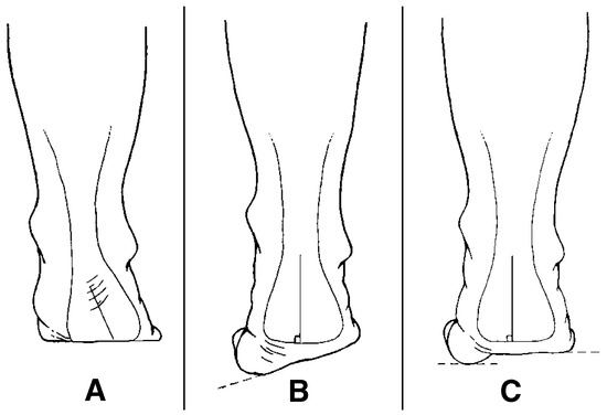
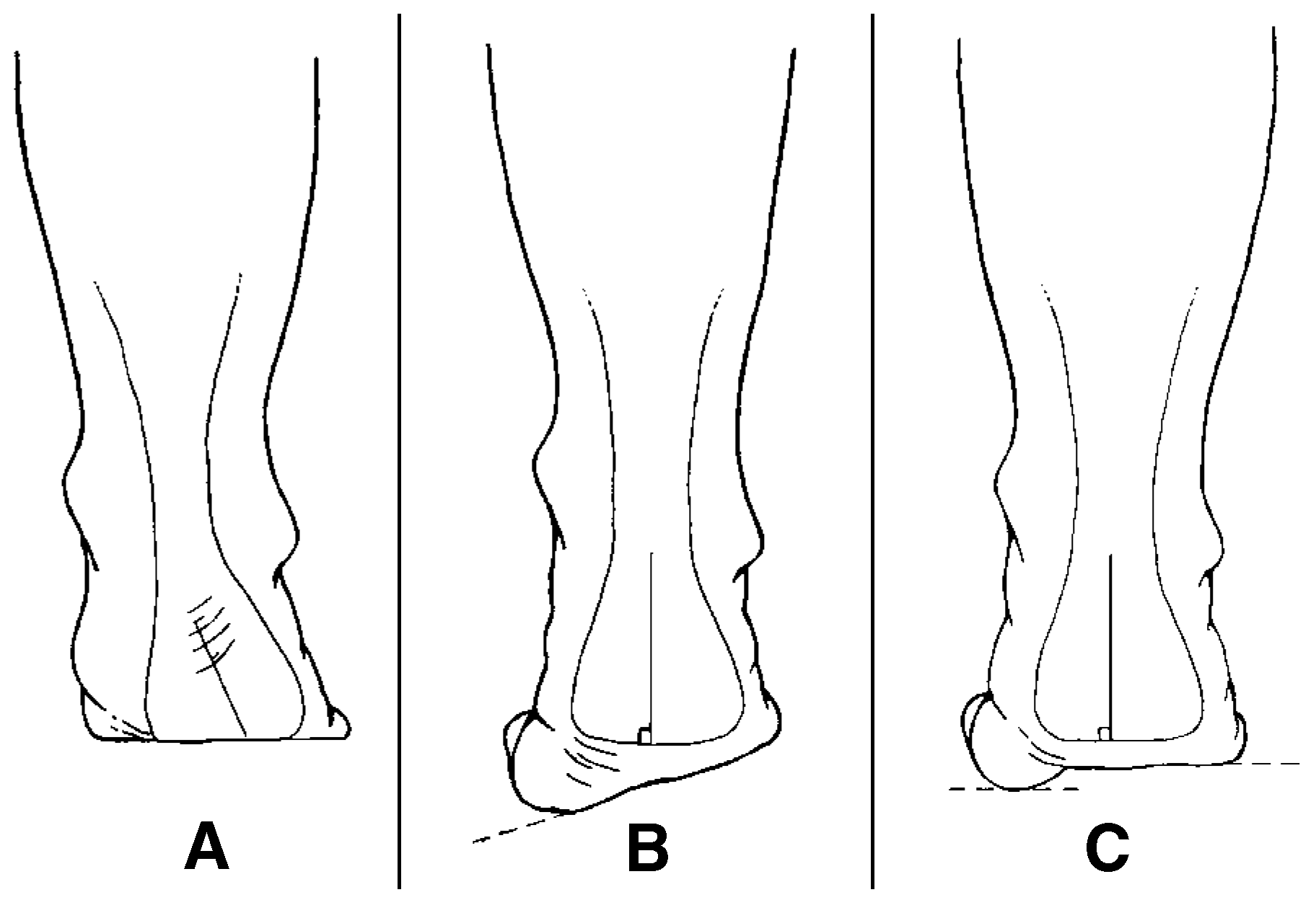
Any structural or functional deformity that increases ground reactive force under the head of the first metatarsal during the midstance period of the gait cycle can potentially dorsiflex the first ray. Three possibilities are: 1) any deformity that causes eversion of the calcaneus; 2) a flexible forefoot valgus or everted forefoot-to-rearfoot deformity; and 3) a congenital plantarflexed first metatarsal (Figure 5). Each of these situations increases ground reactive force under the head of the first metatarsal during the midstance period of the gait cycle by driving the medial aspect of the forefoot into the ground. Any joint that possesses an axis of motion that permits movement of the medial aspect of the forefoot within the frontal plane (ie, inversion) and the sagittal plane (ie, dorsiflexion) will attempt to compensate for the excessive ground reactive force created by these deformities. Two joints possess axes of motion that permit such movements: 1) inversion of the medial aspect of the forefoot can occur at the longitudinal midtarsal joint [41,45]; and 2) dorsiflexion and inversion of the medial aspect of the forefoot can occur at the first ray. [1,35,41,42,43,44].
Figure 5.
A graphic representation of the three foot types commonly resulting in a dorsiflexed first ray: (A) eversion of the calcaneus; (B) flexible forefoot valgus; and (C) congenital plantarflexed first metatarsal.
The theory of proximal stability states that no motion can occur around an axis of motion until the segment proximal to that axis of motion is stable. [1] The longitudinal midtarsal joint is considered capable of approximately 4° to 8° of closed kinetic chain inversion. [1,41] In response to the increased ground reactive force under the head of the first metatarsal in the deformities previously stated, the longitudinal midtarsal joint attempts to compensate by inverting the medial aspect of the forefoot around its axis of motion. Based on the theory of proximal stability, the authors propose that only after the longitudinal midtarsal joint has reached its end range of motion in the direction of inversion and further compensation is required, will the first ray begin to dorsiflex and invert the medial aspect of the forefoot around its own axis of motion.
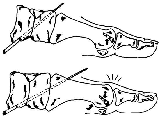
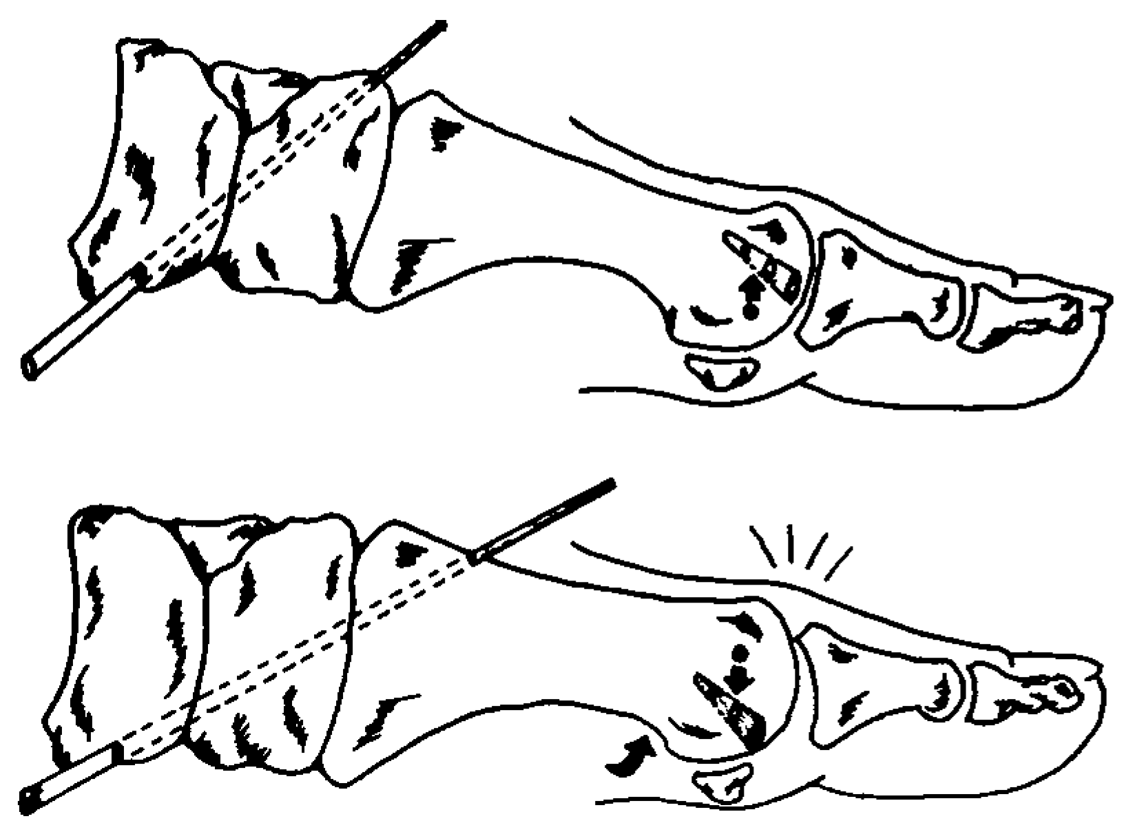
As presented earlier, when the first metatarsal plantarflexes and the hallux dorsiflexes at the first metatarsophalangeal joint during closed kinetic chain, the instant center of rotation of the first metatarsophalangeal joint proceeds from distal-plantar, through the center of the head, to proximal-dorsal. [21] The authors theorize that if this sequence is accepted as correct, then the opposite also must occur. That is, when the first metatarsal dorsiflexes and the hallux attempts to dorsiflex at the first metatarsophalangeal joint during closed kinetic chain, the instant center of rotation of the first metatarsophalangeal joint must proceed from proximal-dorsal, through the center of the head, to distal-plantar (Figure 6). The authors suggest that this position would decrease first metatarsophalangeal joint dorsiflexion by creating compression between the articular surfaces of the base of the proximal phalanx of the hallux and head of the first metatarsal. Since functional hallux limitus is, by definition, a decrease of first metatarsophalangeal joint dorsiflexion, whenever the first ray is dorsiflexed, a functional hallux limitus will be present. [7] The authors propose that the pathology that follows this event depends on how much the first ray dorsiflexes before it reaches its end range of motion.
Figure 6.
A graphic representation of the location of the axis of rotation of the first metatarsophalangeal joint with the first ray plantarflexed (top) and dorsiflexed (bottom).
It is important to remember that as a result of orientation of its axis of motion, as the first ray dorsiflexes it also inverts a proportionate amount. [1,35,41,42,43,44] Therefore, individuals who possess a large amount of dorsiflexion of the first ray will invert the first ray a large amount, and individuals who possess a small amount of dorsiflexion of the first ray will invert the first ray a small amount. The authors propose that as the first ray dorsiflexes and inverts, it would concomitantly dorsiflex and invert the first metatarsophalangeal joints’ transverse and vertical axes of motion. In closed kinetic chain motion, as a result of this dorsiflexion and inversion of the transverse and vertical axes, pure sagittal plane motion of the first metatarsophalangeal joint would decrease, while transverse plane motion would increase.
Additionally, the authors propose that as a result of first metatarsophalangeal joint inversion, the plantar-lateral aspect of the hallux would contact the ground with greater pressure than the plantar-medial aspect. Over time, as the first metatarsophalangeal joint continued to function in this position, subluxation or dislocation of the hallux on the head of the first metatarsal within both the transverse plane (ie, abduction) and the frontal plane (ie, eversion) would occur. Therefore, the authors propose that an individual with a large first ray range of motion would progress from a functional hallux limitus into a hallux abducto valgus deformity. Conversely, individuals who possess a small end range of dorsiflexion at the first ray would invert the first ray only a small amount. Accordingly, the transverse and vertical axes of motion of the first metatarsophalangeal joint would be expected to dorsiflex and invert only a small amount.
As the authors’ data show, only 4 mm of first ray dorsiflexion is required to decrease first metatarsophalangeal joint dorsiflexion by 19%. The authors propose that over time, this relentless blockage of sagittal plane motion and continuous articular surface compression would deform the first metatarsophalangeal joint and progress from a functional hallux limitus into a hallux rigidus deformity.
With the previous discussion in mind, the authors propose that the initial stage in the development of pathology of the first metatarsophalangeal joint is dorsiflexion of the first ray and concomitant decrease in first metatarsophalangeal joint dorsiflexion (ie, functional hallux limitus). The authors suggest that the deformity that follows this event depends on the actual amount the first ray dorsiflexes. Accordingly, the authors propose that hallux abducto valgus deformities are the end result of a large range of first ray dorsiflexion, and that hallux rigidus deformities are the end result of a small range of first ray dorsiflexion. The authors believe that the changes that Shereff et al [20] described for the location and direction of the instant center of rotation in the presence of hallux abducto valgus and hallux rigidus deformities, would primarily depend on how far the first ray dorsiflexes before it reaches its end range of motion, rather than represent anatomical variances of the first metatarsophalangeal joint.
Since motion of the first metatarsophalangeal joint is influenced by the position of the first ray, attention should be given to the first ray when considering the choice and prognosis of surgical procedures dealing with pathology of the first metatarsophalangeal joint. In addition, there is a need to dispense functional foot orthoses to control abnormal biomechanical foot function following surgical correction of hallux abducto valgus and hallux rigidus deformities. These considerations warrant further investigation.
Conclusion
A goniometer was constructed to measure degree of first metatarsophalangeal joint dorsiflexion with the first ray in weightbearing resting position, dorsiflexed 4 mm and 8 mm. The data revealed a quantitative and proportionate decrease of first metatarsophalangeal joint dorsiflexion as the first ray was dorsiflexed away from its weightbearing resting position. The authors proposed that hallux abducto valgus and hallux rigidus deformities are both the end result of dorsiflexion of the first ray. The final deformity was suggested to depend on the actual amount the first ray dorsiflexes and inverts, rather than representing anatomical variances or inherent properties of the first metatarsophalangeal joint. Attention to the first ray during conservative and surgical management of first metatarsophalangeal joint deformities was alluded to and deserves further consideration.
Acknowledgments
Sonjia Johnson, DPM, for illustrations.
References
- ROOT ML, ORIEN WP, WEED JH: Normal and Abnormal Function of the Foot, Clinical Biomechanics, Vol II, Clinical Biomechanics Corp, Los Angeles, 1977.
- DONICK II, BERLIN SJ, BLOCK LD, ET AL: An approach for hallux valgus surgery: fifteen year review. part I. J Foot Surg 19: 113, 1980.
- BRAHM SM: Shape of the first metatarsal head in hallux rigidus and hallux valgus. JAPMA 78: 300, 1988.
- GIRDLESTONE GR, SPOONER HJ: A new operation of hallux valgus and hallux rigidus. J Bone Joint Surg 19: 30, 1937.
- MCGLAMRY ED, BANKS AS, DOWNEY MS: Comprehensive Textbook of Foot Surgery, Vol II, Williams & Wilkins, Baltimore, 1992.
- GERBERT J: Textbook of Bunion Surgery, 2nd Ed, Futura Publishing, New York, 1991.
- DANANBERG HJ: Functional hallux limitus and its relationship to gait efficiency. JAPMA 76: 648, 1986.
- DANANBERG HJ: Gait style as an etiology to chronic postural pain: part I. functional hallux limitus. JAPMA 83: 433, 1993.
- COHN I, KANAT IO: Functional limitation of motion of the first metatarsophalangeal joint. J Foot Surg 23: 477, 1984.
- SHEREFF MJ: Pathology, anatomy and biomechanics of hallux valgus. Orthopaedics 13: 939, 1990.
- JACK EA: The aetiology of hallux rigidus. Br J Surg 27: 492, 1940.
- BINGOLD AC, COLLINS DH: Hallux rigidus. J Bone Joint Surg 32B: 214, 1950.
- KESSEL L, BONNEY G: Hallux rigidus in the adolescent. J Bone Joint Surg 40B: 668, 1958.
- KASHUK KB: Hallux rigidus, hallux limitus, and other functionally limiting disorders of the great toe joint: background, treatment, and case studies. J Foot Surg 14: 45, 1975.
- DRAGO JJ, OLOFF L, JACOBS AM: A comprehensive review of hallux limitus. J Foot Surg 23: 213, 1984.
- MCMASTER MJ: The pathogenesis of hallux rigidus. J Bone Joint Surg 60B: 82, 1978.
- MANN RA, COUGHLIN MJ: Surgery of the Foot and Ankle, 6th Ed, Vol I, CV Mosby, St Louis, 1992.
- KELIKIAN H: Hallux Valgus: Allied Deformities of the Forefoot and Metatarsalgia, WB Saunders, Philadelphia, 1965.
- HETHERINGTON VJ: Hallux Valgus and Forefoot Surgery, Churchill Livingstone, New York, 1994.
- SHEREFF MJ, BEJJANI FJ, KUMMER FJ: Kinematics of the first metatarsophalangeal joint. J Bone Joint Surg 68A:392, 1986.
- HETHERINGTON VJ, CARNETT J, PATTERSON BA: Motion of the first metatarsophalangeal joint. J Foot Surg 28: 13, 1989.
- SAMMARCO JG: “Biomechanics of the Foot,” in Basic Biomechanics of the Skeletal System, ed by VH Frankel, M Nordin, Lea & Febiger, Philadelphia, 1980.
- MYGIND HB: Some views on the treatment of hallux valgus. Acta Orthop Scand 23: 152, 1953.
- BOJSEN-MOLLER F, LAMOREUX L: Significance of free dorsiflexion of the toes in walking. Acta Orthop Scand 50: 471, 1979.
- HETHERINGTON VJ, JOHNSON RE, ALBRITTON JS: Necessary dorsiflexion of the first metatarsophalangeal joint during gait. J Foot Surg 29: 218, 1990.
- HICKS JH: The mechanics of the foot: part II. the plantar aponeurosis and the arch. J Anat 88: 25, 1954.
- DURRANT MN, SIEPERT KK: Role of soft tissue structures as an etiology of hallux limitus. JAPMA 83: 173, 1993.
- HARDY RH, CLAPHAM JCR: Hallux valgus: predisposing anatomical causes. Lancet i: 118, 1952.
- NILSONNE H: Hallux rigidus and its treatment. Acta Orthop Scand 1: 295, 1930.
- LYRITIS G: Developmental disorders of the proximal epiphysis of the hallux. Skeletal Radiol 10: 250, 1983.
- CAMASTA CA, PITTS TE, COREY SV: Bilateral osteochondritis dissecans of the first metatarsophalangeal joint. JAPMA 84: 297, 1994.
- GOODFELLOW J: Aetiology of hallux rigidus. Proc R Soc Med 59: 821, 1965.
- STEINHAUSER W: Osteochodrose der basalen epiphyse der grundphalanx: grosszehe und hallux rigidus. Beit Ges Arbeit Orthop 6: 177, 1959.
- LAPIDUS PW: “Dorsal bunion”: its mechanics and operative correction. J Bone Joint Surg 22: 627, 1940.
- EBISUI JM: The first ray axis and the first metatarsophalangeal joint: an anatomical and pathomechanical study. JAPA 58: 160, 1968.
- KELSO SF, RICHIE DH JR, COHEN IR, ET AL: Direction and range of motion of the first ray. JAPA 72: 600, 1982.
- PLASTER HM: Hallux valgus as related to the first metatarsal length pattern. J Nat Assoc Chir 44: 17, 1954.
- PLASTER HM: Hallux valgus as related to the first metatarsal length pattern. J Nat Assoc Chir 47: 176, 1957.
- PHILLIPS AJ, MCGLAMRY ED: “Hallux Limitus: Technique,” in Reconstructive Surgery of the Foot and Leg ’89, ed by ED McGlamry, Podiatry Institute Publishing, Tucker, GA, 1989.
- LAMBRINUDI C: Metatarsus primus elevatus. Proc R Soc Med 31: 1273, 1938.
- HICKS JH: The mechanics of the foot: part I. the joints. J Anat 87: 345, 1953.
- D’AMICO JC, SCHUSTER RO: Motion of the first ray: clarification through investigation. JAPA 69: 17, 1979.
- GRODE SE, MCCARTHY DJ: The anatomical implications of hallux abducto valgus: a cryomicrotomy study. JAPA 70: 539, 1980.
- OLDENBROOK LL, SMITH CE: Metatarsal head motion secondary to rearfoot pronation and supination: an anatomical investigation. JAPA 69: 24, 1979.
- MANTER JT: Movements of the subtalar and transverse tarsal joints. Anat Record 80: 397, 1941.

Disclaimer/Publisher’s Note: The statements, opinions and data contained in all publications are solely those of the individual author(s) and contributor(s) and not of MDPI and/or the editor(s). MDPI and/or the editor(s) disclaim responsibility for any injury to people or property resulting from any ideas, methods, instructions or products referred to in the content. |
© 1996 American Podiatric Medical Association