


Abstract
Background/Objectives: Intractable plantar keratosis may remain symptomatic despite conservative care, and comparative data on local excision techniques are scarce. This study retrospectively evaluated total elliptical excision and core excision, two surgical methods used in patients whose symptoms persisted despite conservative treatment. Methods: The records of 80 adult patients with histopathologically confirmed plantar keratosis who were treated surgically at a single center between April 2023 and August 2025 were reviewed. All patients had received conservative treatment before surgery. The primary outcome was symptomatic keratosis that reappeared at the operative site, was confirmed clinically, and required reoperation. Pain at the last visit, rated on a visual analog scale (VAS), served as the secondary outcome. The groups were compared with respect to patient characteristics, reoperation for recurrence, and final pain scores. Results: Reoperation for symptomatic recurrence was performed in 17 patients (21.3%): 3 of 37 (8.1%) in the total elliptical excision group and 14 of 43 (32.6%) in the core excision group (p = 0.012). Final VAS scores were 1.3 ± 0.8 and 1.5 ± 0.9, respectively, with no statistically significant difference (p = 0.41). Mean follow-up was 17.7 ± 9.6 and 18.4 ± 8.6 months (p = 0.638). Conclusions: In this retrospective cohort, symptomatic recurrence requiring reoperation occurred less often after total elliptical excision than after core excision. However, the non-randomized choice of technique, baseline differences between the groups, and differing postoperative protocols preclude firm conclusions about the independent superiority of either technique.
1. Introduction
Repetitive pressure and shear forces acting on the sole of the foot can produce well-demarcated, tender, hyperkeratotic lesions. These lesions, termed intractable plantar keratosis (IPK), usually develop in areas where plantar loading is concentrated. Load applied to the lesion during walking causes pain and may impair footwear tolerance, activities of daily living, and functional capacity [1,2].
Plantar keratosis should be regarded not merely as epidermal thickening but as the clinical consequence of chronic mechanical stress and altered plantar pressure distribution. Foot deformities, plantar prominence of the metatarsal heads, hallux valgus, hammer toe deformity, atrophy of the plantar fat pad, and ill-fitting footwear may all contribute to increased local pressure [1,2,3]. Histopathological examination may show epidermal hyperplasia, compact hyperkeratosis, and a central keratin core. The pressure that this central core exerts on the surrounding tissues during weight bearing is one of the possible mechanisms of pain [4,5].
First-line treatment is conservative and aims to reduce local pressure and relieve symptoms. Available measures include regular debridement, footwear modification, metatarsal pads, cushioning insoles, pressure-relieving orthoses, and topical keratolytic agents such as salicylic acid [6]. Nevertheless, symptoms may persist or lesions may recur when the underlying mechanical loading is not adequately addressed. Surgical treatment may be considered in selected patients whose pain and functional limitation persist despite conservative treatment [5,7,8].
Surgical options reported for IPK include core excision, total elliptical excision, plantar condylectomy, metatarsal osteotomy, and other offloading procedures [9,10]. For local soft-tissue procedures, one of the fundamental questions is how wide the excision should be. Core excision focuses on removing the central keratotic portion, whereas total elliptical excision aims to remove the lesion in full thickness together with a narrow skin margin; when necessary, subcutaneous tissue that appears macroscopically fibrotic may also be included in the excision [11]. The effect of these differences on clinical recurrence should be assessed with directly comparative data.
A substantial part of the existing surgical literature consists of small case series [10,12]. The scarcity of clinical data comparing total elliptical excision with core excision makes it difficult to select the appropriate technique. We therefore compared the two techniques in plantar keratosis treated surgically after conservative care had failed, focusing on symptomatic recurrence requiring reoperation and on pain scores at the last visit. We hypothesized that total elliptical excision would be associated with a lower rate of recurrence requiring reoperation.
2. Materials and Methods
2.1. Design and Eligibility
We carried out a retrospective cohort comparison at one center, going through the records of adults operated on between April 2023 and August 2025 for plantar keratosis refractory to conservative treatment. The hospital’s ethics committee for scientific research gave its approval (protocol no. 2026-137; 24 September 2026), and the work complied with the Declaration of Helsinki.
All patients received conservative treatment before surgery, including orthotic insoles, topical retinoic acid, footwear modification, topical salicylic acid, and advice to rest. Surgical excision was reserved for patients with persistent severe pain that interfered with daily activities despite these measures. Preoperative pain severity was assessed clinically; standardized pain scores were not recorded.
Preoperative evaluation included clinical examination of the foot and radiographic assessment. Instrumented plantar pressure measurements, pedobarography, and formal gait analysis were not performed.
Patients were eligible if they had a clinical diagnosis of plantar keratosis, symptoms persisting despite conservative treatment before surgery, treatment by total elliptical excision or core excision, histopathological confirmation of the diagnosis, medical records adequate for evaluation, and at least 12 months of follow-up data. All lesions were located on the plantar surface.
Patients with another diagnosis, such as plantar wart, epidermal cyst, foreign body reaction, or malignant skin lesion; dorsal keratotic lesions; patients who underwent concomitant metatarsal osteotomy, plantar condylectomy, or another offloading bone procedure; those with neuropathic diabetic foot ulcers; those younger than 18 years; and those with less than 12 months of follow-up were excluded. The final cohort comprised 80 patients, of whom 37 had total elliptical excision and 43 had core excision.
2.2. Surgical Techniques and Postoperative Care
The choice of technique was not randomized and was determined by the preoperative clinical preference of the operating surgeon. The procedures were performed using the standard approaches described below. No tourniquet was used in either technique. Local anesthesia was achieved with 3 mL of 2% prilocaine.
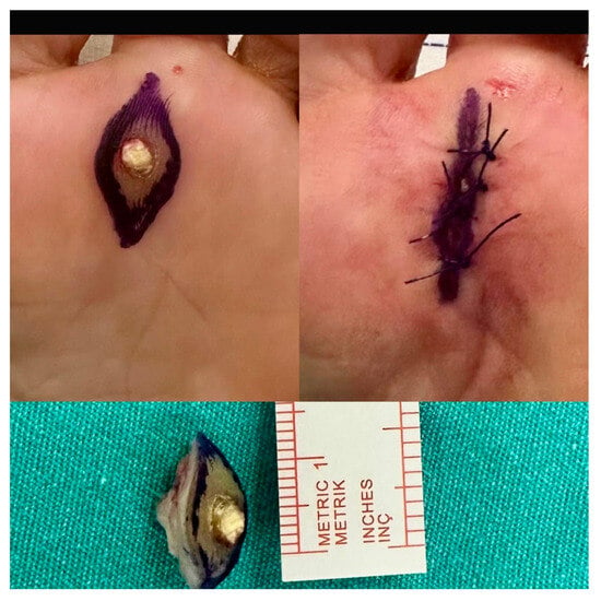
Total elliptical excision: A full-thickness elliptical incision was planned to encompass the entire lesion with a narrow margin of healthy skin. The hyperkeratotic lesion was removed with a No. 15 scalpel blade. When necessary, subcutaneous tissue that appeared macroscopically fibrotic was also included in the excision. After hemostasis, the wound was closed primarily with non-absorbable sutures (Figure 1). Patients were advised not to bear weight until the sutures were removed. The sutures were removed at approximately two weeks, and gradual full weight bearing was allowed once the wound had healed.
Figure 1.
Total elliptical excision. Top left: elliptical marking around the lesion. Top right: primary closure with interrupted non-absorbable sutures. Bottom: excised specimen containing the central keratin core, with a measuring ruler.
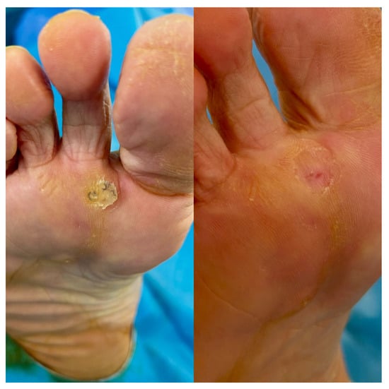
Core excision: The painful central keratotic core was removed in a limited fashion with a No. 15 scalpel blade, preserving the surrounding plantar skin and healthy soft tissue as much as possible. The defect was left open to heal by secondary intention (Figure 2). Regular dressings were applied, and early weight bearing was allowed as tolerated.
Figure 2.
Core excision. Left: preoperative appearance of the plantar lesion with a central keratotic core. Right: appearance after healing by secondary intention.
All excised specimens were sent for histopathological examination. Following wound healing and completion of dressing care, topical silicone gel was initiated to minimize hypertrophic scar formation. All patients continued using orthotic insoles and appropriate footwear modifications after resuming weight bearing. Because the method of wound closure and the timing of weight bearing differed between the groups, the postoperative protocols were not identical.
2.3. Outcome Measures and Recorded Variables
The primary outcome was defined as symptomatic recurrence requiring reoperation. This outcome required that symptomatic keratosis reappearing at the operative site be confirmed by clinical examination and treated by repeat surgery. Histopathological examination was performed in all reoperated cases.
The secondary outcome was the pain score at the final follow-up. Pain was scored between 0 and 10 by visual analog scale (VAS). Preoperative VAS values had not been reported, so the change in pain score was not calculated.
The electronic records provided age, sex, side (right, left, or bilateral), diabetes, smoking, number of lesions (single or multiple), anatomical region (toe, forefoot, or hindfoot), duration of follow-up, and the outcomes. Dimensions were determined from the longest diameter of the excised specimen and the diameter perpendicular to it, as stated in the pathology reports. These values were not regarded as standardized preoperative clinical measurements of the lesion. Because conservative treatment had been given to all patients, it was not used as a variable for comparison between the groups.
2.4. Statistical Analysis
IBM SPSS Statistics 29.0 was used throughout. Means with standard deviations describe continuous data, and counts with percentages describe categorical data. Our check for normality was the Kolmogorov–Smirnov test. When normality held, group comparisons of continuous data relied on the independent-samples t test; when it did not, we turned to the Mann–Whitney U test, also our choice for VAS scores. For categorical data the Pearson chi-square test was applied or, where cells were sparse, the Fisher exact test. For the primary outcome, the comparison across the two groups was made by the two-sided Fisher exact test.
Sample size was dictated by the eligible records available during the study period. Multivariable analysis was not performed because there were only 17 reoperations for recurrence and a model containing numerous covariates could yield unstable estimates. The results were interpreted as unadjusted comparisons. Significance required a two-sided p under 0.05.
3. Results
3.1. Patient Characteristics
Of the 80 patients in the study, 37 (46.3%) had undergone total elliptical excision and 43 (53.8%) core excision. All lesions were plantar. Mean age was 49.2 ± 16.9 and 42.3 ± 19.2 years, respectively (p = 0.092). The two groups were comparable in age, share of male patients, diabetes, and smoking, with no significant differences. Among patients treated by total elliptical excision, multiple lesions occurred more often (32.4% against 11.6%; p = 0.030). Table 1 lists baseline features and outcomes.
Table 1.
The two surgical groups compared: baseline features and outcomes.
3.2. Follow-Up and Clinical Outcomes
Mean follow-up was 17.7 ± 9.6 months in the total elliptical excision group and 18.4 ± 8.6 months in the core excision group (p = 0.638). Reoperation for symptomatic recurrence was performed in 17 patients (21.3%). The rate was 8.1% (3/37) in the total elliptical excision group and 32.6% (14/43) in the core excision group. By the Fisher exact test this gap was significant (p = 0.012); core excision carried a rate 24.5 percentage points higher.
Final VAS scores in the total elliptical excision and core excision groups were 1.3 ± 0.8 and 1.5 ± 0.9, respectively (p = 0.41). This comparison reflects pain levels at the final follow-up; it does not demonstrate a reduction in pain relative to the preoperative state or the equivalence of the two methods.
3.3. Variables Associated with Reoperation for Recurrence
In univariable analyses, no statistically significant association was found between symptomatic recurrence requiring reoperation and age (p = 0.601), sex (p = 0.727), side (p = 0.560), diabetes (p = 0.145), smoking (p = 0.528), multiple lesions (p = 0.940), or anatomical region (p = 0.579). The absence of statistically significant associations does not exclude a potential effect of these variables on symptomatic recurrence requiring reoperation.
Within each surgical group, the dimensions of the pathological excision specimen did not differ significantly between patients who were and were not reoperated for recurrence (total elliptical excision, p = 0.228; core excision, p = 0.843). Because these measurements reflect the tissue removed, no firm conclusion can be drawn as to whether the size of the original lesion predicts recurrence.
4. Discussion
Symptomatic recurrence requiring reoperation was less frequent after total elliptical excision than after core excision in our cohort (8.1% vs. 32.6%). Pain scores at the last visit were comparable and did not differ significantly. However, the comparisons are unadjusted; because of the non-randomized choice of technique, the baseline characteristics, and the differing postoperative care, the difference cannot be attributed solely to the extent of excision.
Repetitive mechanical loading and local pressure distribution are central to how IPK develops [1,2,3,5]. Removing the lesion may therefore not, by itself, correct the underlying loading conditions. Total elliptical excision removes the entire hyperkeratotic lesion and, when necessary, macroscopically fibrotic tissue, which may contribute to more durable local control than core excision. Due to the fact that residual tissue and the histological margins of the excision were not assessed, and the amount of fibrosis was not quantified in this study, this explanation remains hypothetical. However, ongoing mechanical stress may contribute to recurrent symptoms after either method.
Much of the research on IPK surgery has focused on metatarsal osteotomy, condylectomy, and other bone-based offloading procedures [9,13,14,15]. Comparative evidence on local excision methods is more limited. The scoping review by Reilly et al. also highlighted the limitations of the evidence in this field and the need for stronger clinical studies [10]. Although approaches such as lesion excision with a rotation flap have been evaluated in case series, those results cannot be generalized directly to the present two techniques because of the different surgical and care protocols [12]. The contribution of our study is that it presents comparative results for two local excision approaches performed at the same center.
The postoperative protocols are particularly important when interpreting the observed difference. In the total elliptical excision group, primary closure and non-weight bearing until suture removal were used, whereas in the core excision group open wound care and early weight bearing as tolerated were used. The study therefore compares not only different extents of excision but also the different care approaches applied with them. The contribution of early loading or of the mode of healing to the outcomes cannot be separated with the available data. This difference limits any claim of technical superiority and shows the need to standardize postoperative care in future comparisons.
The groups differed in the frequency of multiple lesions. Although multiple lesions were more common in the total elliptical excision group, no significant association was found between multiple lesions and recurrence requiring reoperation in the cohort as a whole. This result does not, however, show that multiple lesions have no effect on recurrence. In a cohort limited to seventeen events, the sensitivity of subgroup comparisons is low. The failure to demonstrate an association for age, sex, and diabetes should be interpreted with the same caution. Because no multivariable adjustment was made, a contribution of measured and unmeasured confounders to the observed difference between techniques cannot be excluded.
Smoking was not significantly associated with symptomatic recurrence requiring reoperation in this cohort. However, the small number of reoperation events limits the interpretation of this finding. Furthermore, because all patients had established IPK, this study could not evaluate the association between smoking and the development of these lesions.
The definition of the outcome also affects the assessment. Symptomatic recurrence requiring reoperation is a clinically important outcome that can be traced in the records. However, it does not capture recurrences in patients who declined repeat surgery or were managed non-surgically. The decision to reoperate may be influenced by patient preference and the surgeon’s decision threshold as much as by the severity of symptoms. The reported rates should therefore not be presented as the rate of all clinical recurrences. In future studies, assessing clinical recurrence, reoperation, and patient satisfaction as separate outcomes will provide more comprehensive information.
The absence of a significant difference in final VAS scores indicates that low pain levels were observed in both groups at the end of follow-up. However, because preoperative scores were not reported, the change in pain could not be calculated. Moreover, the absence of a significant difference is not evidence of statistical equivalence. In patients who underwent repeat surgery, the timing of the VAS measurement relative to the revision is also important; pain measured after revision may not reflect the effect of the first operation alone.
Mercier et al. used lesion surface area to assess the response to injection treatment [8]. Bhingradia et al. reported an elliptical excision approach for large plantar corns [11]. The size data in our study were obtained from pathology reports. A total elliptical excision specimen may include a margin of healthy skin and surrounding tissue, whereas a core excision specimen represents only the central portion. These measurements therefore do not reflect the same anatomical extent in the two methods. The lack of a significant association in the within-group comparisons does not exclude a prognostic role for preoperative lesion size; the available data assess only the size of the excised specimen.
Although a lower rate of repeat surgery is clinically valuable, the overall success of treatment cannot be determined by this outcome alone. Time to wound healing, infection, wound dehiscence, painful scarring, and return to weight bearing and daily activities are also important. Smoking has been reported to impair wound healing [16]; however, wound healing and recurrence of keratosis are different outcomes. Because systematic data on complications and functional outcomes are not presented in this study, the superiority of either method in terms of safety or overall clinical benefit has not been demonstrated.
The strengths of the study are that all lesions were plantar, the diagnosis was confirmed histopathologically, and two local surgical approaches were compared directly at the same center. Nevertheless, because the underlying mechanical loading was not assessed, the biomechanical explanation of the surgical results is limited. Conservative treatment and, where necessary, continued pressure-relieving measures remain important in the management of IPK [5,6].
5. Limitations
Because of the retrospective, non-randomized design, selection bias and unmeasured confounders cannot be excluded. Because surgeon preference determined the choice of technique, the effects of surgeon experience and of the technique are difficult to separate. The groups differed in the frequency of multiple lesions, and no multivariable adjustment was made. With so few reoperation events, the subgroup results are hard to interpret. The results should therefore not be regarded as an independent or causal effect of the technique.
Postoperative wound care and weight-bearing protocols differed. Because the primary outcome required reoperation, recurrences managed without surgery were not captured, and overall clinical recurrence may be underestimated. A mean follow-up of approximately 18 months may not be sufficient to detect later recurrences. The time from initial surgery to clinical recurrence was not available; therefore, the mean time to recurrence could not be determined.
Plantar pressure measurement, pedobarography, and gait analysis were not available. Preoperative VAS values, validated functional scales, patient satisfaction, and systematic complication outcomes were not reported. The dimensions obtained from the pathology specimen may not represent the whole clinical lesion, and the extent measured differs between the techniques. The single-center design also limits generalizability.
6. Conclusions
In this retrospective cohort, total elliptical excision was associated with a lower rate of symptomatic recurrence requiring reoperation than core excision. Final pain scores did not differ significantly between groups. These findings warrant confirmation in prospective studies with standardized postoperative care.
Author Contributions
V.E.Ç. and M.M.A. conceived the study. V.E.Ç. designed it, ran the analyses and drafted the text. Operations were performed by V.E.Ç., Ü.G. and M.M.A. Data were collected by Ü.G., B.I., K.E.G. and A.T.D. assessed the dermatological and histopathological findings. M.M.A. supervised the work. All authors have read and agreed to the published version of the manuscript.
Funding
This research received no external funding.
Institutional Review Board Statement
The Scientific Research Ethics Committee of Kocaeli City Hospital approved the protocol (code 2026-137; 24 September 2026), and its conduct followed the Helsinki Declaration.
Informed Consent Statement
Individual patient consent was not required, since this retrospective study drew only on anonymized information already held in medical records. No identifying features appear in the clinical photographs.
Data Availability Statement
The corresponding author will share anonymized patient-level data when a reasonable request is made. To protect patient confidentiality, the dataset was not placed in a public repository.
Conflicts of Interest
The authors declare no conflicts of interest.
References
- Freeman, D.B. Corns and Calluses Resulting from Mechanical Hyperkeratosis. Am. Fam. Physician 2002, 65, 2277–2280. [Google Scholar]
- Spink, M.J.; Menz, H.B.; Lord, S.R. Distribution and Correlates of Plantar Hyperkeratotic Lesions in Older People. J. Foot Ankle Res. 2009, 2, 8. [Google Scholar] [CrossRef] [Scilit]
- Menz, H.B.; Morris, M.E. Clinical Determinants of Plantar Forces and Pressures during Walking in Older People. Gait Posture 2006, 24, 229–236. [Google Scholar] [CrossRef] [Scilit]
- Yu, G.V.; Judge, M.S. Hyperkeratotic Disorders of the Foot. In McGlamry’s Comprehensive Textbook of Foot and Ankle Surgery; Lippincott Williams & Wilkins: Philadelphia, PA, USA, 2002; pp. 667–713. [Google Scholar]
- Mann, R.A. Intractable Plantar Keratosis. Instr. Course Lect. 1984, 33, 287–301. [Google Scholar]
- Singh, D.; Bentley, G.; Trevino, S.G. Callosities, Corns, and Calluses. BMJ 1996, 312, 1403–1406. [Google Scholar] [CrossRef]
- Cangialosi, C.P. Plantar Keratoses. Why Treatment Often Fails and What to Do about It. Postgrad. Med. 1984, 75, 128–129. [Google Scholar] [CrossRef] [Scilit]
- Mercier, M.-P.; Blanchette, V.; Cantin, V.; Brousseau-Foley, M. Effectiveness of Saline Water and Lidocaine Injection Treatment of Intractable Plantar Keratoma: A Randomised Feasibility Study. J. Foot Ankle Res. 2021, 14, 30. [Google Scholar] [CrossRef] [Scilit]
- Leventen, E.O.; Pearson, S.W. Distal Metatarsal Osteotomy for Intractable Plantar Keratoses. Foot Ankle 1990, 10, 247–251. [Google Scholar] [CrossRef] [Scilit]
- Reilly, I.N.; Longhurst, B.; Vlahovic, T.C. Surgical Excision of Intractable Plantar Keratoses (Corns) of the Foot: A Scoping Review. J. Am. Podiatr. Med. Assoc. 2023, 113, 22-044. [Google Scholar] [CrossRef] [Scilit]
- Bhingradia, Y.M.; Thorat, D.D.; Pundir, A. Elliptical Excision as a Surgical Approach for Large Plantar Corns. J. Cutan. Aesthet. Surg. 2026, 19, 369–371. [Google Scholar] [CrossRef] [Scilit]
- Saipoor, A.; Maher, A.; Hogg, L. A Retrospective Audit of Lesion Excision and Rotation Skin Flap for the Treatment of Intractable Plantar Keratosis. Foot 2018, 34, 23–27. [Google Scholar] [CrossRef] [Scilit]
- Idusuyi, O.B.; Kitaoka, H.B.; Patzer, G.L. Oblique Metatarsal Osteotomy for Intractable Plantar Keratosis: 10-Year Follow-Up. Foot Ankle Int. 1998, 19, 351–355. [Google Scholar] [CrossRef] [Scilit]
- Berkun, R.N.; DeVincentis, A.; Goller, W.L. The Tilt-Up Osteotomy for Correction of Intractable Plantar Keratoses. J. Foot Surg. 1984, 23, 52–55. [Google Scholar]
- Pontious, J.; Lane, G.D.; Moritz, J.C.; Martin, W. Lesser Metatarsal V-Osteotomy for Chronic Intractable Plantar Keratosis. Retrospective Analysis of 40 Procedures. J. Am. Podiatr. Med. Assoc. 1998, 88, 323–331. [Google Scholar] [CrossRef] [Scilit]
- McDaniel, J.C.; Browning, K.K. Smoking, Chronic Wound Healing, and Implications for Evidence-Based Practice. J. Wound Ostomy Cont. Nurs. 2014, 41, 415–423. [Google Scholar] [CrossRef] [Scilit]

Disclaimer/Publisher’s Note: The statements, opinions and data contained in all publications are solely those of the individual author(s) and contributor(s) and not of MDPI and/or the editor(s). MDPI and/or the editor(s) disclaim responsibility for any injury to people or property resulting from any ideas, methods, instructions or products referred to in the content. |
© 2026 by the authors. Published by MDPI on behalf of the American Podiatric Medical Association. Licensee MDPI, Basel, Switzerland. This article is an open access article distributed under the terms and conditions of the Creative Commons Attribution (CC BY) license.