


Abstract
Background: Hallux valgus (HV) is a three-dimensional deformity, and preoperative assessment of sesamoid position and first metatarsal rotation is important for surgical planning. Weight-bearing computed tomography (WBCT) enables three-dimensional assessment but was unavailable at our institution during the study period. Therefore, this study was a retrospective, exploratory, descriptive radiographic study of a modified weight-bearing metatarso-sesamoid (MS) projection, rather than a diagnostic-accuracy, comparative, or feasibility study. Methods: Between 2022 and 2024, 18 patients (16 female, 2 male; mean age 46 years, range 25–75 years) with 22 feet scheduled for HV surgery were included. Preoperative weight-bearing AP, lateral, and modified MS radiographs were evaluated. The alpha angle, sesamoid rotation angle (SRA), AOFAS tibial sesamoid grade, and the pronation/subluxation (P/S) pattern were recorded on the modified MS view. Because the alpha angle and P/S classification were originally defined on axial CT, we applied them to plain radiography as an exploratory approach. We did not validate them against a CT reference standard. Intraobserver and interobserver measurement agreement on the same existing radiographs was assessed through repeated blinded measurements by the primary observer after four weeks and through independent evaluations by a second blinded observer. We quantified reliability using ICC(2,1), unweighted kappa, and linearly weighted kappa, with 95% CIs. Technical acquisition performance was recorded. Results: On AP radiographs, IMA1–2 showed 2 mild, 15 moderate, and 5 severe deformities; HVA showed 1 normal, 11 mild, and 10 moderate cases; and Agrawal lateral sesamoid (LS) severity showed 1 mild, 20 moderate, and 1 severe case. On the modified MS view, the alpha angle ranged from 6° to 30° (mean ± SD 15.6 ± 6.6°), and the SRA from 6° to 45° (18.2 ± 8.3°). Using the CT-derived thresholds as descriptive reference labels only, alpha values were at or below 15.8° in 13 feet and above 15.8° in 9 feet, and SRA values were within the 7–18° reference range in 11 feet. AOFAS tibial sesamoid grade was 0 in 6 feet, 1 in 11, and 2 in 5. P/S patterns were P−/S− in 9 feet, P−/S+ in 4, P+/S− in 4, and P+/S+ in 5. Intraobserver ICCs were 0.996 (95% CI 0.991–0.998) for the alpha angle and 0.998 (0.994–0.999) for the SRA; interobserver ICCs were 0.988 (0.972–0.994) and 0.975 (0.947–0.981), respectively. Interobserver agreement was substantial for the P/S pattern (κ = 0.746, 95% CI 0.476–0.937) and excellent for the AOFAS grade (linear weighted κ = 0.878, 95% CI 0.663–1.000); intraobserver agreement was κ = 1.000 for both. The modified MS view provided a diagnostic image on the first acquisition in 15 of 18 patients; 3 patients required a single repeat acquisition, which was adequate in every case, and no procedure-related discomfort or intolerance was recorded. Acquisition time and radiation exposure were not recorded. Conclusions: Weight-bearing CT remains the reference standard for three-dimensional assessment. In this small cohort study, the modified MS projection could be obtained in every patient and showed high intraobserver and interobserver measurement agreement when the same existing radiographs were assessed. These findings describe measurement agreement on the same existing radiographs and the acquisition experience. Because no WBCT reference standard was available, the study cannot determine which projection reflects the underlying anatomy and provides no evidence of diagnostic accuracy, superiority over AP radiography, or clinical benefit. Standardized prospective studies with direct WBCT validation are required.
1. Introduction
Hallux valgus (HV) is a three-dimensional deformity characterized by lateral deviation of the first metatarsophalangeal joint, metatarsal varus, pronation, and sesamoid subluxation [1,2]. The primary lesion is insufficiency of the medial stabilizers, resulting in medial displacement of the metatarsal head and lateral sesamoid dislocation. Subsequent disruption of the metatarsal-sesamoid ligaments induces metatarsal pronation, while the bowstring effect of displaced hallucis tendons further aggravates the deformity [3,4].
The surgical goal is not only to correct the deformity but also to restore the sesamoids to their anatomical position. Inadequate sesamoid reduction is a major risk factor for recurrence, regardless of the surgical technique used [5,6,7]. Dayton et al. emphasized that metatarsophalangeal joint incongruity in HV is associated with metatarsal pronation and sesamoid subluxation, and that surgeons should consider these factors during correction [8].
Various methods have been developed to assess HV severity on weight-bearing dorsoplantar (AP) radiographs. Sesamoid subluxation from the first metatarsal head generally indicates HV [9,10]. Hardy and Clapham examined the relationship between medial (tibial) sesamoid position and HV and classified HV into seven stages; however, identification is difficult because the sesamoids overlap with the first metatarsal head [11]. Agrawal et al. evaluated the lateral sesamoid position relative to the first metatarsal head and its association with HV severity using radiographic measurements (IMA1–2 and HVA) on weight-bearing AP radiographs [12].
In recent years, weight-bearing computed tomography (WBCT) has enabled three-dimensional evaluation of the deformity, and numerous foot studies have used this modality [13,14,15,16]. Because WBCT was unavailable at our institution during the study period, we obtained a modified weight-bearing metatarso-sesamoid (MS) radiograph as an exploratory alternative to assess the relationship between the sesamoid complex and the first metatarsal.
We developed and validated the alpha angle and P/S classification using axial CT images [1]. We have not validated their transfer to a modified plain radiographic projection, and no WBCT reference standard was available in the present series. Consequently, this study was designed as a retrospective, exploratory, descriptive radiographic investigation. Its objectives were limited to (i) describing the measurements and classifications obtained from the modified MS projection in a consecutive surgical HV cohort, (ii) quantifying intraobserver and interobserver measurement agreement for those measurements on the same available images, and (iii) reporting the technical acquisition experience. The study does not establish which imaging modality reflects true anatomy, nor does it define diagnostic accuracy, comparative effectiveness, feasibility, or clinical outcome endpoints.
2. Materials and Methods
2.1. Study Population and Design
The hospital ethics committee approved our study, and we conducted it in accordance with the 1964 Declaration of Helsinki. This retrospective, exploratory, descriptive radiographic study included foot radiographs from 18 patients (16 female, 2 male) with a mean age of 46 years (range 25–75 years), totaling 22 feet (12 right, 10 left), who were scheduled for surgical treatment of hallux valgus at our clinic between 2022 and 2024. Four patients contributed both feet; we identified these paired observations and did not treat them as statistically independent. Patients younger than 18 years and those diagnosed with inflammatory arthritis (e.g., gout, rheumatoid arthritis) or hallux rigidus were excluded.
We obtained weight-bearing AP, lateral, and modified MS foot radiographs in the same X-ray room using identical equipment. For the AP view, the patient stood with both feet on the image detector and knees fully extended, with the X-ray beam centered on the 3rd tarsometatarsal joint and angled 10 degrees posteriorly.
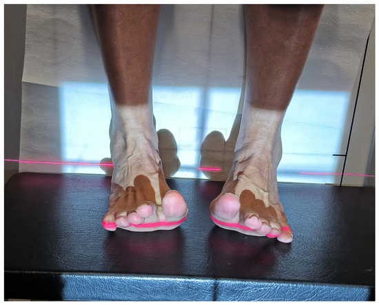
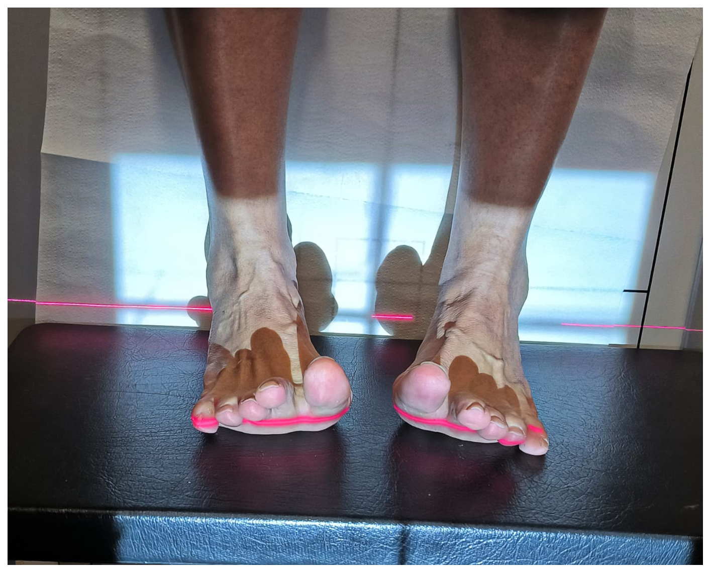
The modified MS radiograph was obtained with the patient in a weight-bearing standing position. With the big toe dorsiflexed 30–40 degrees, the beam was directed at an oblique angle of 20°, with a source-to-cassette distance of 100 cm, focusing on the first metatarsal head and sesamoid complex (Figure 1). The examining physician targeted the 30–40° dorsiflexion angle using visual and clinical assessment rather than a goniometer or a dedicated positioning device (orthoposer). We reviewed all radiographs at the time of acquisition, and repeated images that were inadequately positioned or of insufficient quality. Because neither hallux dorsiflexion nor whole-foot rotation was instrumented, positioning may have influenced the projected metatarsal-sesamoid relationship; we address this explicitly in Section 5.
Figure 1.
Clinical image of a modified metatarso-sesamoid (MS) radiograph taken while standing with the big toe in a dorsiflexion position. Both feet in this illustrative photograph appear supinated; this positioning may alter the projected metatarso-sesamoid relationship compared with that obtained using a dedicated orthoposer and is discussed as a limitation.
The first author initially evaluated all radiographs using the Fonet PACS Viewer digital imaging system. To assess intraobserver and interobserver measurement agreement, the same observer repeated the full set of modified-MS measurements and classifications after a minimum interval of four weeks, with the images re-randomized and blinded to the initial values; a second observer independently assessed the same images, blinded to the initial measurements and all clinical data. We therefore formally quantified measurement agreement in the present analysis (Section 2.4).
2.2. Measurement of Ima1–2, Hva, and Lateral Sesamoid (Ls) Position on AP Radiographs
On AP radiographs, the intermetatarsal angle (IMA1–2) and hallux valgus angle (HVA) were measured. IMA1–2 was calculated as the angle between the long axes of the first and second metatarsals, following the method described by Miller [17]. HVA was defined as the angle between the shaft axis of the first metatarsal and the long axis of the proximal phalanx of the great toe. IMA values were classified as normal (<9°), mild (9–13°), moderate (14–18°), and severe (>18°); HVA values were classified as normal (<15°), mild (15–30°), moderate (30–40°), and severe (>40°) [18].
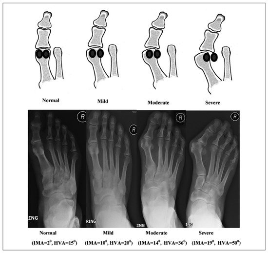
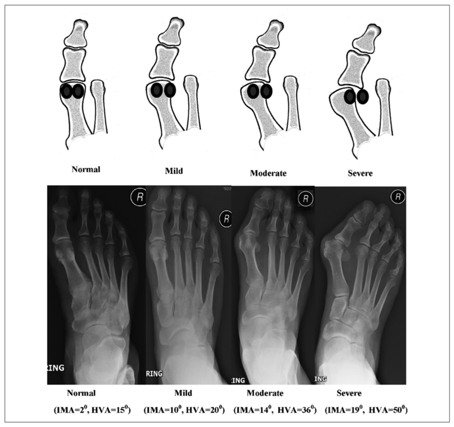
Deformity on weight-bearing AP radiographs was graded using the classification proposed by Agrawal et al., which is based on the relationship between the lateral border of the first metatarsal head and the lateral sesamoid position [12]. In this classification, the lateral sesamoid normally lies medial to the lateral cortical border of the first metatarsal head; in mild deformity, it is lateralized by up to 50%; in moderate deformity, more than 50% extends beyond the cortical border while contact is maintained; and in severe deformity, a distinct gap forms with complete lateralization of the sesamoid bone (Figure 2, Table 1).
Figure 2.
Sesamoid classification from Agrawal et al. [12].
Table 1.
Severity grading based on the position of the lateral sesamoid (LS) [12].
2.3. Assessment of Sesamoid Subluxation and Metatarsal Pronation on the Modified MS View
On the modified MS radiograph, the most prominent point of the first metatarsal crista served as the reference. A line was drawn between the lateral and medial sulci, and a perpendicular line from the crista apex was added. We transposed this line to the widest point of the tibial sesamoid using the Talbot method to determine its position [9]. Sesamoid subluxation was defined as the proportion of the medial sesamoid displaced laterally (Figure 3). Hallux dorsiflexion may induce a bowstring effect through the flexor hallucis brevis, which can alter the apparent position of the sesamoid complex; this is acknowledged as a potential source of measurement bias for all modified MS measurements reported below.
Figure 3.
Measurement of the subluxation ratio of the tibial (medial) sesamoid (left foot) according to the Talbot method in modified metatarso-sesamoid (MS) radiography [9].
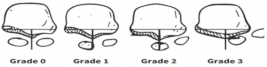
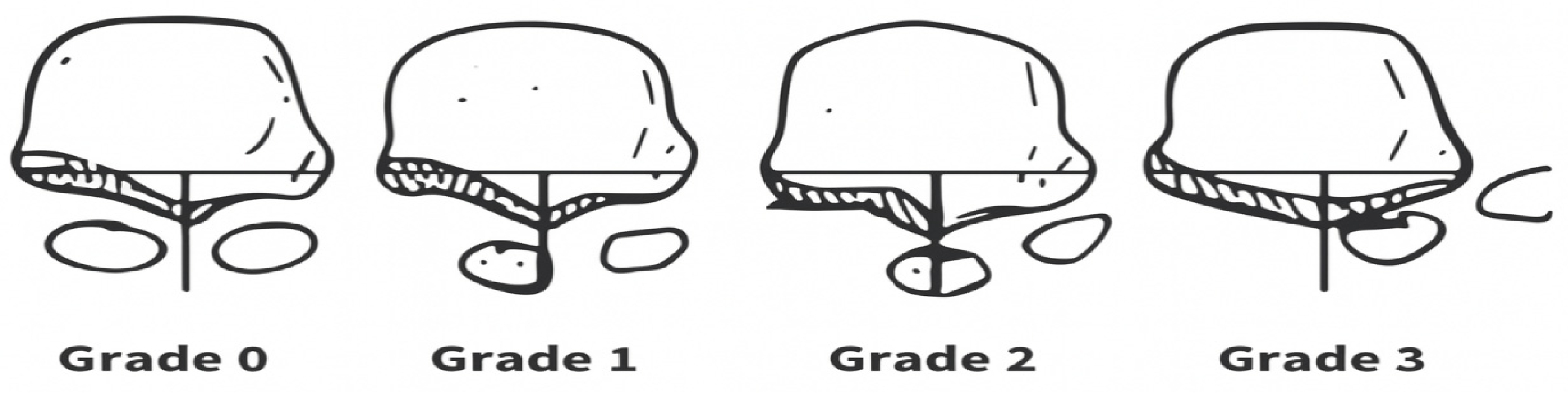
Smith et al. [19] classified tibial sesamoid position on weight-bearing tangential radiographs into four grades relative to the intermetatarsal ridge, per the American Orthopedic Foot and Ankle Society (AOFAS) (Figure 4).
Figure 4.
Smith et al. described an AOFAS staging classification for tibial sesamoid position on weight-bearing tangential radiographs [19].
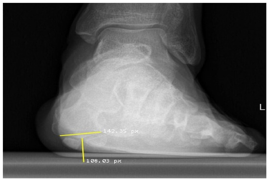
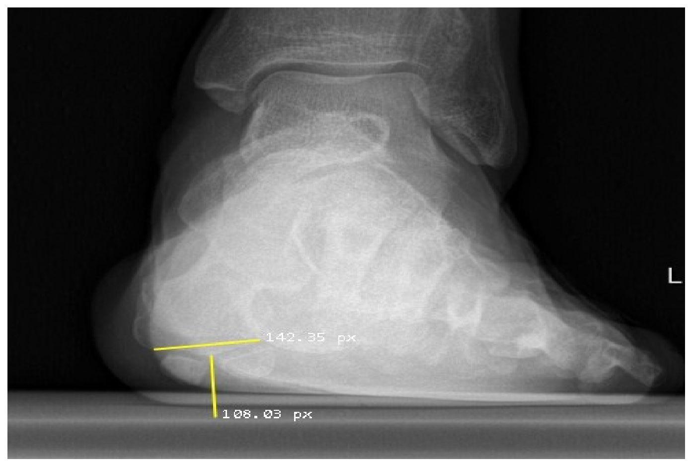
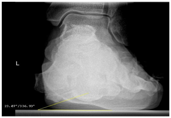
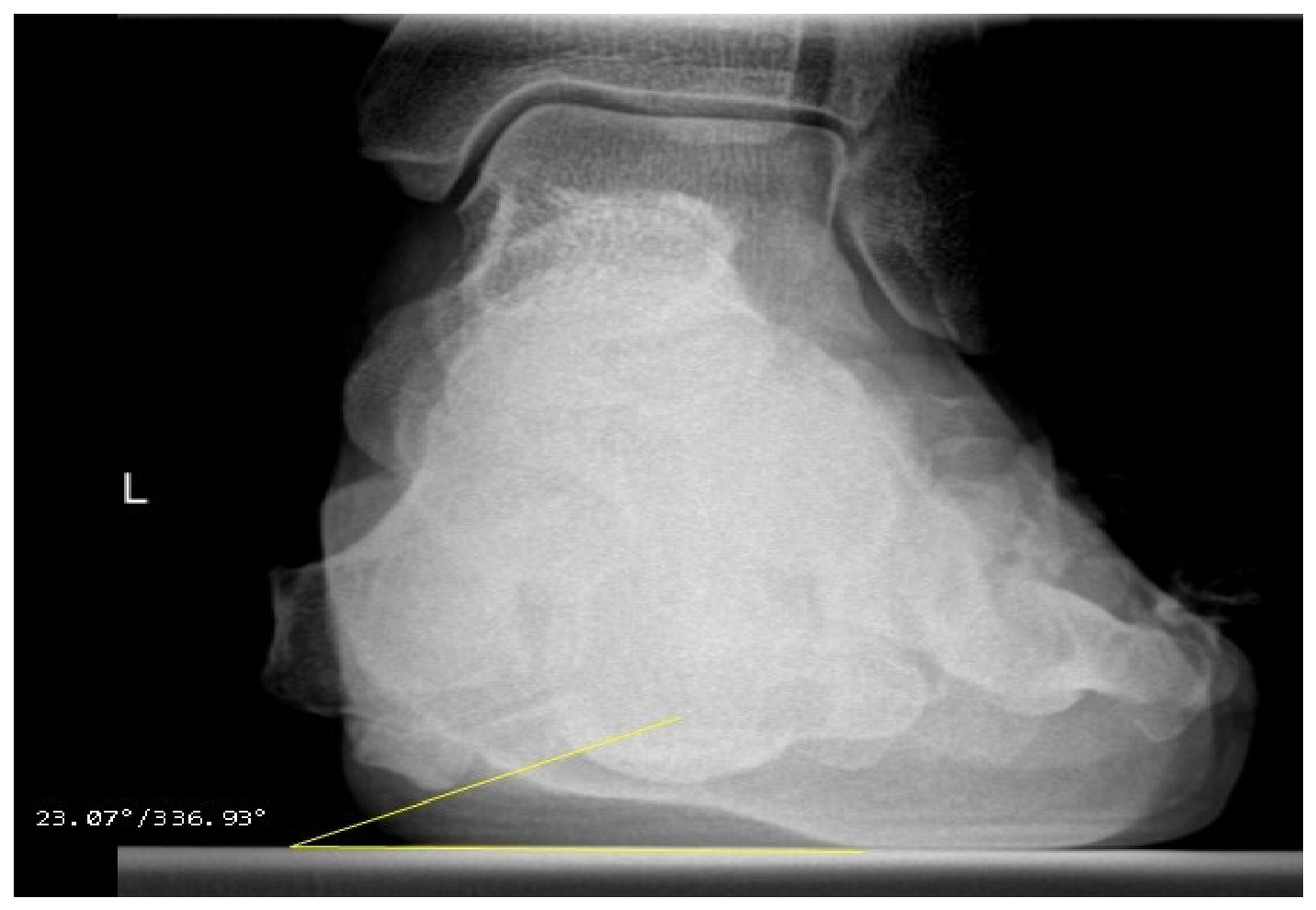
The sesamoid rotation angle (SRA) was measured as the angle between the line connecting the most inferior points of the medial and lateral sesamoids and the horizontal axis on the modified MS radiograph, following the method described by Kuwano et al. [10], an SRA of 7° to 18° was considered normal and served as the reference in our study (Figure 5).
Figure 5.
Measurement of SRA (sesamoid rotation angle) on modified metatarso-sesamoid radiography (23.07°) (left foot).
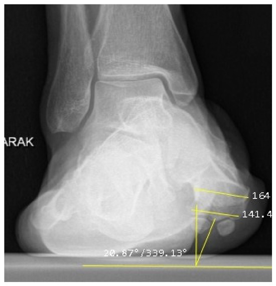
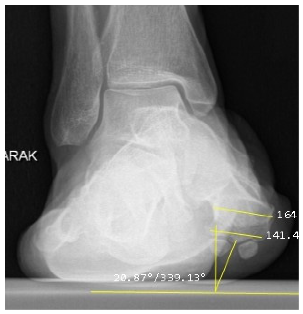
Kim et al. defined the alpha angle on axial CT sections to assess first metatarsal pronation in HV [1]. The reference range was accepted as 0–15.8 degrees. The angle is measured between the line passing through the midpoint of the parallel lines between the metatarsal head and the sulcus and the line drawn perpendicular to the ground (Figure 6). Using the same method, tibial sesamoid subluxation was also assessed, and four types of HV deformity were defined: no pronation, no sesamoid subluxation (P−, S−); no pronation, sesamoid subluxation (P−, S+); pronation, no sesamoid subluxation (P+, S−); and both pronation and sesamoid subluxation (P+, S+).
Figure 6.
Measurement of alpha angle as defined by Kim et al. [1] on modified metatarso-sesamoid radiograph (20.87°).
Critical methodological clarification. Because the alpha angle and the P/S classification were originally defined and validated on axial CT rather than on plain radiography, their application to the modified MS view in this study was strictly exploratory. In this manuscript, we use the 0–15.8° alpha-angle threshold, the 7–18° SRA reference range, and the P/S labels only as descriptive reference categories to compare our numerical results with the published CT-based literature. No foot was classified as anatomically normal or abnormal on this basis; no measurement was interpreted as demonstrating true metatarsal pronation or true sesamoid subluxation; and these labels should not be regarded as validated diagnostic criteria until they have been confirmed against a CT reference standard.
We described weight-bearing AP radiographs using the staging system (LS severity grade) described by Agrawal [12]. On the modified MS radiographs obtained in our study, we recorded and tabulated the first metatarsal rotation label, sesamoid subluxation label, SRA measurement, and AOFAS staging descriptively (Section 3).
2.4. Observer Measurement Agreement Assessment
Continuous variables (alpha angle, SRA) were assessed using two-way random-effects, absolute-agreement, single-measure intraclass correlation coefficients (ICC(2,1)) calculated from paired observer measurements. Categorical variables (P/S pattern, AOFAS tibial sesamoid grade) were assessed using Cohen’s unweighted kappa and linearly weighted kappa, respectively. Because four patients contributed bilateral feet, we obtained 95% confidence intervals for all agreement statistics by nonparametric bootstrap resampling at the patient level (10,000 resamples), preserving within-patient pairing, rather than using analytic methods that assume independent foot-level observations. We analyzed observer measurement agreement only for the modified-MS variables; we obtained the AP variables from routine clinical radiographs and did not remeasure them. These analyses quantify how consistently images can be measured and classified; they do not validate the projection against a CT reference standard.
2.5. Statistical Analysis
Descriptive statistics (mean ± standard deviation, minimum–maximum, case count, and percentage) were calculated using SPSS version 27.0 (IBM Corp., Armonk, NY, USA); the ICC and kappa estimates and their bootstrap confidence intervals were calculated in Python 3 using NumPy and scikit-learn (Section 2.4). No inferential hypothesis testing was performed in this study. In particular, we removed the chi-square test and all p-values reported in the previous version. This decision was made for three reasons: (i) the AP LS severity categories were severely imbalanced (1 mild, 20 moderate, and 1 severe case), so that expected cell frequencies were far below the assumptions of the chi-square test and the categories with n = 1 cannot support any comparison; (ii) four patients contributed bilateral feet, so the foot-level observations are not statistically independent; and (iii) p-values are not descriptive statistics, and labeling an analysis as exploratory does not make an invalid test interpretable. Consequently, between-category numerical differences are reported as observed values only, and no statement is made anywhere in this manuscript regarding the presence or absence of a statistically significant association or difference.
3. Results
3.1. Cohort Characteristics and Standard AP Radiographic Findings
Radiographs of 22 feet (12 right, 10 left) from 18 patients were reviewed. Four patients contributed bilateral feet; therefore, foot-level observations are not fully statistically independent, and we report the results descriptively at the foot level below. For observer-agreement statistics, we performed bootstrap resampling at the patient level, resampling paired feet from the same patient together.
According to IMA1–2 values, no foot was classified as normal: 2 feet (9.1%) showed mild deformity, 15 (68.2%) showed moderate deformity, and 5 (22.7%) showed severe deformity (mean ± SD: 15.5° ± 2.8° (range: 10–21°). For HVA, 1 foot (4.5%) was classified as normal, 11 (50%) as mild, and 10 (45.5%) as moderate; no foot showed severe deformity (mean ± SD: 28.6° ± 6.4° (range: 14–38°). Thus, the series, particularly for IMA1–2, clustered in the moderate- and severe-deformity groups.
According to the severity grading based on lateral sesamoid (LS) position described by Agrawal et al. [12], among 22 feet, 1 was mild, 20 were moderate, and 1 was severe. No foot with a normal LS position was identified. This distribution describes the appearance of the lateral sesamoid on the AP projection of this surgical cohort. Because no reference standard was available, this report is descriptive and not interpreted as a quantification of true anatomical displacement.
3.2. Modified Metatarso-Sesamoid View Findings
Weight-bearing MS radiographs were categorized using the P/S scheme defined by Kim et al. [1]. The distribution on a per-foot basis was as follows: P−, S− in 9 feet; P−, S+ in 4 feet; P+, S− in 4 feet; and P+, S+ in 5 feet. This small series therefore included all four label combinations.
The alpha angle measured on the modified MS radiographs ranged from 6° to 30° (mean ± SD, 15.6 ± 6.6°). Using the CT-derived 0–15.8° range as a descriptive reference, values were at or below 15.8° in 13 feet and above 15.8° in 9 feet.
The SRA values measured on the modified MS radiographs ranged from 6° to 45° (mean ± SD 18.2 ± 8.3°). Relative to the 7–18° reference range reported by Kuwano [10], 11 feet fell within this range, 10 exceeded 18°, and 1 measured 6°. The highest value, 45°, was recorded in the left foot of patient 3. These figures describe the angular relationship projected in this view; they are not interpreted as true sesamoid rotation measurements, and high values may partly reflect hallux position or whole-foot rotation.
When the tibial sesamoid position was assessed using the AOFAS classification [19], grades 0 to 2 were observed: grade 0 in 6 feet, grade 1 in 11 feet, and grade 2 in 5 feet (mean grade 1.0 ± 0.7). No inferential comparison between the AOFAS grades and the alpha-angle or SRA values was performed.
Table 2 presents the joint distribution of the modified-MS P/S labels and the AP LS severity grades. No test of association was performed. The counts are provided solely to describe the composition of this cohort, and the very small and unequal sizes of the mild (n = 1) and severe (n = 1) AP LS categories make any formal test of association between these two classifications uninterpretable.
Table 2.
Distribution of P/S patterns on weight-bearing modified metatarso-sesamoid (MS) radiographs according to Agrawal AP lateral sesamoid (LS) severity (counts; no statistical test performed).
Because two of the four AP LS severity categories contain a single foot, group-wise means and standard deviations for the modified-MS variables were not calculated; such estimates would be unstable and could be misread as evidence of a between-group effect. The descriptive statistics of the modified-MS variables for the whole cohort are therefore reported in the text above and summarized in Table 3.
Table 3.
Descriptive summary of the modified metatarso-sesamoid measurements for the whole cohort (n = 22 feet). We did not perform hypothesis testing; no p-values are reported. (Note to Table 3: values are reported for the whole cohort only. The 0–15.8° and 7–18° intervals are CT-derived descriptive reference ranges and do not constitute validated diagnostic thresholds on plain radiography).
3.3. Observer Measurement Agreement for the Modified MS Measurements
Table 4 presents agreement statistics for repeat primary-observer readings and independent second-observer readings. Intraobserver agreement for the continuous measurements was essentially perfect (alpha angle ICC 0.996, 95% CI 0.991–0.998; SRA ICC 0.998, 95% CI 0.994–0.999), and interobserver agreement was excellent (alpha angle ICC 0.988, 95% CI 0.972–0.994; SRA ICC 0.975, 95% CI 0.947–0.981). For the categorical assessments, intraobserver agreement was perfect (kappa 1.000 for both the P/S pattern and the AOFAS grade). Interobserver agreement was substantial for the P/S pattern (kappa 0.746, 95% CI 0.476–0.937) and excellent for the AOFAS grade (linear weighted kappa 0.878, 95% CI 0.663–1.000). The confidence intervals for the categorical statistics are wide, reflecting the small sample size and the low frequency of some categories rather than instability in the continuous measurements.
Table 4.
Intraobserver and interobserver measurement agreement for the modified metatarso-sesamoid measurements on the same existing radiographs (n = 22 feet, 18 patients). We obtained 95% confidence intervals using patient-level bootstrap resampling (10,000 resamples).
3.4. Technical Acquisition Experience
Of the 18 patients, 15 (83.3%) provided a diagnostic modified MS radiograph on the first acquisition. In the remaining three patients (16.7%), we judged the first image at the time of acquisition to be inadequately positioned or of insufficient quality, and we performed a single repeat acquisition; in all three cases, the repeat image was adequate for measurement. No patient reported procedure-related discomfort, and no examination was abandoned because of intolerance. We did not record acquisition time or radiation exposure and therefore cannot report them. These counts describe our technical experience with the projection; they are not presented as a formal feasibility outcome because this retrospective design had no predefined feasibility endpoint, no a priori sample size, and no dosimetric measurement.
4. Discussion
Hallux valgus is a multifaceted deformity characterized by varus and pronation of the first metatarsal bone, sesamoid subluxation, and valgus of the first proximal phalanx [1]. Kay proposed in 1989 that great toe pronation develops secondary to first metatarsal rotation and adductor tendon tension [20]. Eustace et al. first described metatarsal rotational deformity in 1993, using morphological markers at the base of the first metatarsal to estimate rotation on plain radiographs [21]. Although initially considered confined to the AP plane, recent studies have shown this deformity to have a three-dimensional pathogenesis [14].
The primary observation of this exploratory study is descriptive: in a cohort whose AP radiographs were heavily clustered in a single severity category, the modified MS projection yielded a heterogeneous distribution of alpha-angle values, SRA values, AOFAS grades, and P/S labels. In our study of 22 feet, IMA1–2 classified 15 feet as moderate and 5 as severe (2 mild, no normal feet); HVA classified 11 feet as mild, 10 as moderate, and 1 as normal; and AP LS grading according to Agrawal et al. [12] clustered 20 of 22 feet in the moderate stage (1 mild, 1 severe). On the same feet, the modified MS projection produced the four P/S label combinations in 9, 4, 4, and 5 feet, respectively. Our data demonstrate this difference between the two radiographic projections. However, they do not establish which appearance corresponds to the underlying three-dimensional anatomy, because no WBCT reference standard was available.
Our AP findings, showing lateral sesamoid displacement in 21 of 22 feet, are consistent with previous reports by Agrawal et al. [12] and Kim SH et al. [22], who identified sesamoid displacement on AP views as a characteristic of surgical HV. Agrawal et al. demonstrated that LS position correlates with conventional planar severity and provides a framework for corrective osteotomies [12,23].
In the modified MS view, 8 of the 20 feet graded as moderate on the AP projection carried the P−/S− label. The isolated sesamoid subluxation (P−/S+) and isolated pronation (P+, S−) labels were recorded in four feet each, and the combined (P+, S+) label in five feet. We did not formally test the association between the AP LS grade and these labels for the methodological reasons set out in Section 2.5.
Talbot [9] and Saltzman [24] provide relevant context: using their own designs and reference standards, they reported that overlapping projections on AP radiographs can misclassify sesamoid position and may not reflect spatial alignment when the first ray is rotated. In 1996, Saltzman et al. used tangential weight-bearing radiographs to measure coronal plane rotation of the first metatarsal [24], and Talbot reported that restoring subluxated sesamoids to their normal position is critical for surgical success in HV [9]. We cite these studies as background; the present series neither replicates their designs nor independently demonstrates misclassification in our cohort, and our unvalidated labels cannot support such a claim.
Regarding first metatarsal rotation, our modified MS measurements using the alpha angle described by Kim et al. [1] recorded values within the CT-derived 0–15.8° reference range in 13 feet and above it in 9 feet. This distribution is consistent with the CT-based findings of Kim et al. [1] and Kim and Young [14] that first metatarsal rotation is variable rather than an obligatory feature of every moderate-to-severe HV deformity; however, because we obtained our measurements on an unvalidated plain-radiographic projection, they support that concept only descriptively.
Kuwano et al. developed a weight-bearing tangential radiographic method in 2002 [10], proposed SRA measurements to assess sesamoid rotational position, and defined 7–18° as the reference range. In our series, SRA values ranged from 6° to 45°, with 11 feet within the reference range and 10 above 18°. Elevated values, such as the 45° recorded in the left foot of patient 3, may reflect pronounced projected rotation, positioning effects, or both, and cannot be attributed to a specific anatomical cause without CT. This individual variation aligns with the literature on indirect morphological markers on AP radiographs, such as the rounded lateral metatarsal head edge described by Okuda et al. [25] and Yamaguchi et al. [26]. In 2007, Okuda et al. reported that a rounded lateral border is strongly associated with HV and that its persistence after surgery increases recurrence risk; they later showed that metatarsal supination corrected this abnormal head shape [7]. Yamaguchi et al. [26] reported that a round lateral border on AP radiographs indicates pronation, whereas the metatarsal condyles become more visible with outward rotation. These findings, derived from the cited authors’ own designs, support the concept emphasized by Wagner [6,27] that surgical derotation should be customized to the individual deformity rather than applied uniformly based on AP angular severity alone.
Hardy and Clapham examined the relationship between medial (tibial) sesamoid position and HV and classified it into seven stages; however, identification is difficult because of overlap with the first metatarsal head [11]. Smith et al. classified tibial sesamoid position on weight-bearing tangential radiographs into four grades relative to the intermetatarsal ridge [19]. Our evaluation using the AOFAS tibial sesamoid grading [19] on modified MS radiographs recorded grade 0 in 6 feet, grade 1 in 11 feet, and grade 2 in 5 feet, so that grades 1 or 2 were recorded in 16 of 22 feet. This distribution is consistent with the high prevalence of sesamoid displacement expected in a surgical HV cohort and with the findings of Katsui et al. [28] and Lewis et al. [29] regarding disruption of joint congruency in progressive HV. Complete reduction in the sesamoids beneath the first metatarsal crista is widely recognized as important for restoring joint mechanics and preventing postoperative recurrence [5,7,29]. Although tangential sesamoid imaging has been advocated by Yıldırım et al. [30] to overcome AP superimposition, caution is required because passive hallux dorsiflexion during positioning may create a bowstring effect via the flexor hallucis brevis tendon, potentially retracting the sesamoids or altering their projected alignment relative to the metatarsal head [1,30].
In recent years, weight-bearing CT has become the most reliable method for revealing the triplanar nature of the deformity in preoperative HV planning [1,14]. Kim et al. defined four CT-based types using the alpha angle and tibial sesamoid subluxation, demonstrating that HV has a three-dimensional component [1]. Those CT-based results, not our plain-radiographic labels, support that conclusion.
Observer measurement agreement. We have directly addressed the central methodological concern raised in the previous review. The modified-MS measurements showed high observer measurement agreement on the same existing radiographs: intraobserver ICCs of 0.996 (alpha angle) and 0.998 (SRA), and interobserver ICCs of 0.988 and 0.975 indicate that the two observers extracted essentially the same numerical information from the same images. Categorical agreement was substantial for the P/S pattern (κ = 0.746) and excellent for the AOFAS grade (weighted κ = 0.878). We interpret this result narrowly. Observer measurement agreement and validity are distinct properties: demonstrating that a measurement can be obtained consistently does not demonstrate that it measures what it is assumed to measure. Consistent measurement of a projection whose relationship to three-dimensional anatomy has not been validated does not make that measurement anatomically correct, and the wide confidence intervals around the categorical statistics reflect the small sample.
Interpretative Considerations and Limitations
Weight-bearing computed tomography remains the established reference standard for three-dimensional volumetric assessment of first-ray alignment, as detailed by Welck et al. [13] and Bouredoucen [15,16], who reported that WBCT reveals the three-dimensional nature of hallux valgus and enhances diagnostic accuracy and surgical planning [15,16]. These conclusions are based on the authors’ own methods.
The present study does not extend those conclusions. It demonstrates only that (i) a modified MS projection could be obtained in all 18 patients, and (ii) the resulting measurements and classifications showed intraobserver and interobserver agreement when the same existing radiographs were assessed. Combining the Agrawal LS classification, AOFAS tibial sesamoid position, SRA, and the CT-derived P/S scheme on this projection may provide additional descriptive information, but we did not assess whether it changes surgical decision-making or improves outcomes, and these data do not support that conclusion.
Several limitations must be stated explicitly. First, the sample size was small (22 feet from 18 patients); four patients contributed bilateral feet, and there was no asymptomatic control cohort. Second, two observers assessed measurement agreement only for the modified-MS variables; the AP variables were not re-measured, no third reader was involved, and the confidence intervals for the categorical statistics were wide because of the small sample. We established observer agreement on the same existing radiographs, but we did not assess accuracy, acquisition reproducibility after patient repositioning, or validity against a CT reference standard. Third, great toe dorsiflexion (30–40°) was targeted clinically without a positioning jig (orthoposer) or goniometer, and visual evaluation showed subtle foot supination, which may have influenced the projected metatarsal-sesamoid relationship; the reported P/S labels and SRA values may therefore partly reflect position-dependent projection effects rather than anatomical differences, and the observer-agreement analysis does not address this limitation, because both observers assessed the same positioning. Fourth, the authors did not record acquisition time or radiation exposure, so no dosimetric or formal feasibility conclusion can be drawn. Finally, the study design was retrospective and cross-sectional; thus, the study could not establish correlations between preoperative radiographic classifications and postoperative correction, clinical satisfaction, or recurrence rates. Prospective, multicenter studies incorporating larger cohorts and direct validation against WBCT are necessary before this modified radiographic view can be recommended as a standardized clinical tool.
5. Conclusions
In this retrospective exploratory descriptive study of 22 feet from 18 patients, a modified weight-bearing metatarso-sesamoid projection could be obtained for analysis in every patient, showed high intraobserver and interobserver measurement agreement for alpha-angle, SRA, AOFAS-grade, and P/S-label assessments when the same existing radiographs were assessed, and required a single repeat acquisition in 3 of 18 patients with no recorded intolerance. These findings describe measurement agreement on existing radiographs and the acquisition experience; they do not establish reproducibility of image acquisition after patient repositioning.
WBCT remains the reference standard for three-dimensional assessment of the metatarsal-sesamoid relationship. Because no CT reference standard, clinical or postoperative outcome data, or assessment of surgical decision-making were available, our results do not demonstrate that the modified MS view reflects true anatomy, that it is superior or equivalent to AP radiography, that it corrects AP misclassification, or that it improves surgical planning. Standardized prospective studies with direct WBCT validation are required before recommending this projection for routine clinical use.
Author Contributions
Conceptualization, M.A.Ş.; methodology, M.A.Ş. and M.U.; formal analysis, M.U.; observer measurement agreement assessment and second-observer measurements, F.D. and K.D.; investigation, E.C.B.; data curation, M.U. and M.A.Ş.; writing—original draft preparation, M.A.Ş.; writing—review and editing, M.A.Ş. and M.U.; visualization, F.D.; supervision, M.A.Ş. and M.U.; project administration, E.C.B. and K.D. All authors have read and agreed to the published version of the manuscript.
Funding
This research received no external funding.
Institutional Review Board Statement
The study was conducted in accordance with the Declaration of Helsinki and approved by the Ethics Committee of Health Science University Gazi Yaşargil Training and Research Hospital (protocol code 750, approval date 21 November 2025).
Informed Consent Statement
Informed consent was obtained from all subjects involved in the study.
Data Availability Statement
The data supporting the findings of this study are available from the corresponding author upon reasonable request. The data are not publicly available due to patient privacy and institutional restrictions.
Conflicts of Interest
The authors declare no conflicts of interest.
References
- Kim, Y.; Kim, J.S.; Young, K.W.; Naraghi, R.; Cho, H.K.; Lee, S.Y. A new measure of tibial sesamoid position in hallux valgus in relation to the coronal rotation of the first metatarsal in CT scans. Foot Ankle Int. 2015, 36, 944–952. [Google Scholar] [CrossRef] [Scilit] [PubMed]
- Doğan, A.; Üzümcügil, O.; Akman, Y.E. Halluks valgus. TOTBİD Derg. 2007, 6, 88–94. [Google Scholar]
- Perera, A.M.; Mason, L.; Stephens, M.M. The pathogenesis of hallux valgus. J. Bone Jt. Surg. Am. 2011, 93, 1650–1661. [Google Scholar] [CrossRef] [Scilit] [PubMed]
- Mortier, J.P.; Bernard, J.L.; Maestro, M. Axial rotation of the first metatarsal head in a normal population and hallux valgus patients. Orthop. Traumatol. Surg. Res. 2012, 98, 677–683. [Google Scholar] [CrossRef] [Scilit] [PubMed]
- Chen, J.Y.; Rikhraj, K.; Gatot, C.; Lee, J.Y.Y.; Rikhraj, I.S. Tibial sesamoid position influence on functional outcome and satisfaction after hallux valgus surgery. Foot Ankle Int. 2016, 37, 1178–1182. [Google Scholar] [CrossRef] [Scilit] [PubMed]
- Wagner, E.; Wagner, P. Metatarsal pronation in hallux valgus deformity: A review. J. Am. Acad. Orthop. Surg. Glob. Res. Rev. 2020, 4, e20.00091. [Google Scholar] [CrossRef] [Scilit] [PubMed]
- Okuda, R.; Yasuda, T.; Jotoku, T. Supination stress of the great toe for assessing intraoperative correction of hallux valgus. J. Orthop. Sci. 2012, 17, 129–135. [Google Scholar] [CrossRef] [Scilit] [PubMed]
- Dayton, F.P.; Kauwe, B.M.; Hirschi, B.J. Relationship of frontal plane rotation of first metatarsal to proximal articular set angle and hallux alignment in patients undergoing tarsometatarsal arthrodesis for hallux abducto valgus: A case series and critical review of the literature. J. Foot Ankle Surg. 2013, 52, 348–354. [Google Scholar] [CrossRef] [Scilit] [PubMed]
- Talbot, K.D.; Saltzman, C.L. Assessing sesamoid subluxation: How good is the AP radiograph? Foot Ankle Int. 1998, 19, 547–554. [Google Scholar] [CrossRef] [Scilit] [PubMed]
- Kuwano, T.; Nagamine, R.; Sakaki, K.; Urabe, K.; Iwamoto, Y. New radiographic analysis of sesamoid rotation in hallux valgus: Comparison with conventional evaluation methods. Foot Ankle Int. 2002, 23, 811–817. [Google Scholar] [CrossRef] [Scilit] [PubMed]
- Hardy, R.H.; Clapham, J.C. Observations on hallux valgus. J. Bone Jt. Surg. Br. 1951, 33B, 376–391. [Google Scholar] [CrossRef] [Scilit] [PubMed]
- Agrawal, Y.; Desai, A.; Mehta, J. Lateral sesamoid position in hallux valgus: Correlation with the conventional radiological assessment. Foot Ankle Surg. 2011, 17, 308–311. [Google Scholar] [CrossRef] [Scilit] [PubMed]
- Welck, M.; Singh, D.; Cullen, N.; Goldberg, A. Evaluation of the 1st metatarso-sesamoid joint using standing CT—The Stanmore classification. Foot Ankle Surg. 2018, 24, 314–319. [Google Scholar] [CrossRef] [Scilit] [PubMed]
- Kim, J.S.; Young, K.W. Sesamoid position in hallux valgus in relation to the coronal rotation of the first metatarsal. Foot Ankle Clin. 2018, 23, 219–230. [Google Scholar] [CrossRef] [Scilit] [PubMed]
- Bouredoucen, H. Hallux valgus: Advances in weightbearing CT imaging and three-dimensional assessment of first ray deformity. Skelet. Radiol. 2026, 55, 2713–2731. [Google Scholar] [CrossRef] [Scilit] [PubMed]
- Bouredoucen, H. Surgical management and postoperative imaging assessment of hallux valgus. Skelet. Radiol. 2026, 55, 3031–3059. [Google Scholar] [CrossRef] [Scilit] [PubMed]
- Miller, J.W. Distal first metatarsal displacement osteotomy: Its place in the scheme of bunion surgery. J. Bone Jt. Surg. Am. 1974, 56, 923–931. [Google Scholar]
- Campbell, J. Hallux valgus adult and juvenile. In Orthopaedic Knowledge Update—Foot and Ankle; Richardson, G., Ed.; AAOS: Rosemont, IL, USA, 2003; Volume 3, pp. 3–15. [Google Scholar]
- Smith, R.W.; Reynolds, J.C.; Stewart, M.J. Hallux valgus assessment: Report of research committee of the American Orthopaedic Foot and Ankle Society. Foot Ankle 1984, 5, 92–103. [Google Scholar] [CrossRef] [Scilit] [PubMed]
- Kay, D.B.; Njus, G.; Parrish, W.; Theken, R. Basilar crescentic osteotomy: A three-dimensional computer simulation. Orthop. Clin. N. Am. 1989, 20, 571–582. [Google Scholar]
- Eustace, S.; O’Byrne, J.; Stack, J.; Stephens, M.M. Radiographic features that enable assessment of first metatarsal rotation: The role of pronation in hallux valgus. Skelet. Radiol. 1993, 22, 153–156. [Google Scholar] [CrossRef] [Scilit] [PubMed]
- Kim, S.H.; Kim, Y.H.; Cha, J.Y.; Lee, Y.K. Correlations of sesamoid bone subluxation with the radiologic measures of hallux valgus and its clinical implications. Medicina 2023, 59, 876. [Google Scholar] [CrossRef] [Scilit] [PubMed]
- Xie, Z.M.; Wang, Z.M.; Zhai, S.M.; Wu, J.M.; Zhang, L.M.; Gao, Y.M.; Wu, H.M. Comparison of osteotomy schemes for the repair and treatment of moderate-to-severe hallux valgus deformities. Medicine 2025, 104, e41251. [Google Scholar] [CrossRef] [Scilit] [PubMed]
- Saltzman, C.L.; Brandser, E.A.; Anderson, C.M.; Berbaum, K.S.; Brown, T.D. Coronal plane rotation of the first metatarsal. Foot Ankle Int. 1996, 17, 157–161. [Google Scholar] [CrossRef] [Scilit] [PubMed]
- Okuda, R.; Kinoshita, M.; Yasuda, T. The shape of the lateral edge of the first metatarsal head as a risk factor for recurrence of hallux valgus. J. Bone Jt. Surg. Am. 2007, 89, 2163–2172. [Google Scholar] [CrossRef] [Scilit] [PubMed]
- Yamaguchi, S.; Sasho, T.; Endo, J.; Yamamoto, Y.; Akagi, R.; Sato, Y.; Takahashi, K. Shape of the lateral edge of the first metatarsal head changes depending on the rotation and inclination of the first metatarsal: A study using digitally reconstructed radiographs. J. Orthop. Sci. 2015, 20, 868–874. [Google Scholar] [CrossRef] [Scilit] [PubMed]
- Wagner, P.; Wagner, E. Proximal rotational metatarsal osteotomy for hallux valgus (PROMO): Short-term prospective case series with a novel technique and topic review. Foot Ankle Orthop. 2018, 3, 2473011418790071. [Google Scholar]
- Katsui, R.; Samoto, N.; Taniguchi, A.; Akahane, M.; Isomoto, S.; Sugimoto, K.; Tanaka, Y. Relationship between displacement and degenerative changes of the sesamoids in hallux valgus. Foot Ankle Int. 2016, 37, 1303–1309. [Google Scholar] [CrossRef] [Scilit] [PubMed]
- Lewis, T.L.; Ferreira, G.F.; Nunes, G.A.; Lam, P.; Ray, R. Impact of sesamoid coverage on clinical foot function following fourth-generation percutaneous hallux valgus surgery. Foot Ankle Orthop. 2024, 9, 2473011241230560. [Google Scholar] [CrossRef] [Scilit] [PubMed]
- Yildirim, Y.; C¸aBukoglu, C.; Erol, B.; Esemenli, T. Effect of metatarsophalangeal joint position on the reliability of the tangential sesamoid view in determining sesamoid position. Foot Ankle Int. 2005, 26, 247–250. [Google Scholar] [CrossRef] [Scilit] [PubMed]

Disclaimer/Publisher’s Note: The statements, opinions and data contained in all publications are solely those of the individual author(s) and contributor(s) and not of MDPI and/or the editor(s). MDPI and/or the editor(s) disclaim responsibility for any injury to people or property resulting from any ideas, methods, instructions or products referred to in the content. |
© 2026 by the authors. Published by MDPI on behalf of the American Podiatric Medical Association. Licensee MDPI, Basel, Switzerland. This article is an open access article distributed under the terms and conditions of the Creative Commons Attribution (CC BY) license.