


Abstract
Background: The symptoms of tarsal tunnel syndrome (TTS) have traditionally been considered the result of entrapment of the tibial nerve due to the laciniate ligament or compression of the nerve’s distal branches as they course beneath the abductor hallucis (AH) muscle. Recent research shows that TTS may be due to compression of the tibial nerve proximal to these structures, at the level of the high ankle (termed high TTS). This study appears to be among the first to investigate the incidence of compression at either or both of these sites in persons with clinical findings of TTS. Methods: The medical records of 91 adult patients (56 women and 35 men) who presented to a podiatric medicine practice with clinical findings of TTS and underwent electrodiagnostic (EDX) testing were retrospectively reviewed for the prevalence of nerve compression at the level of the high ankle (ie, high TTS) or the laciniate ligament/AH muscle in the foot. Results: Of the 91 patients with clinical findings of TTS, EDX testing found that 59 had nerve compression at the laciniate ligament/AH muscle, 69 had nerve compression at the level of the high ankle, 51 had compression in both areas, and 13 did not have any entrapment. Of the 69 patients found to have an entrapment at the high ankle, 41 were women and 28 were men, with the compression occurring bilaterally in 44 of the 69 patients. Conclusions: In patients presenting with clinical findings of TTS, EDX testing found that compression most often occurred at the level of the high ankle, beneath the fascia of the leg, followed by entrapment at the traditionally recognized site, beneath the laciniate ligament/AH muscle, with a significant number of patients having compression at both sites. A small number of patients had no EDX evidence of nerve compression.
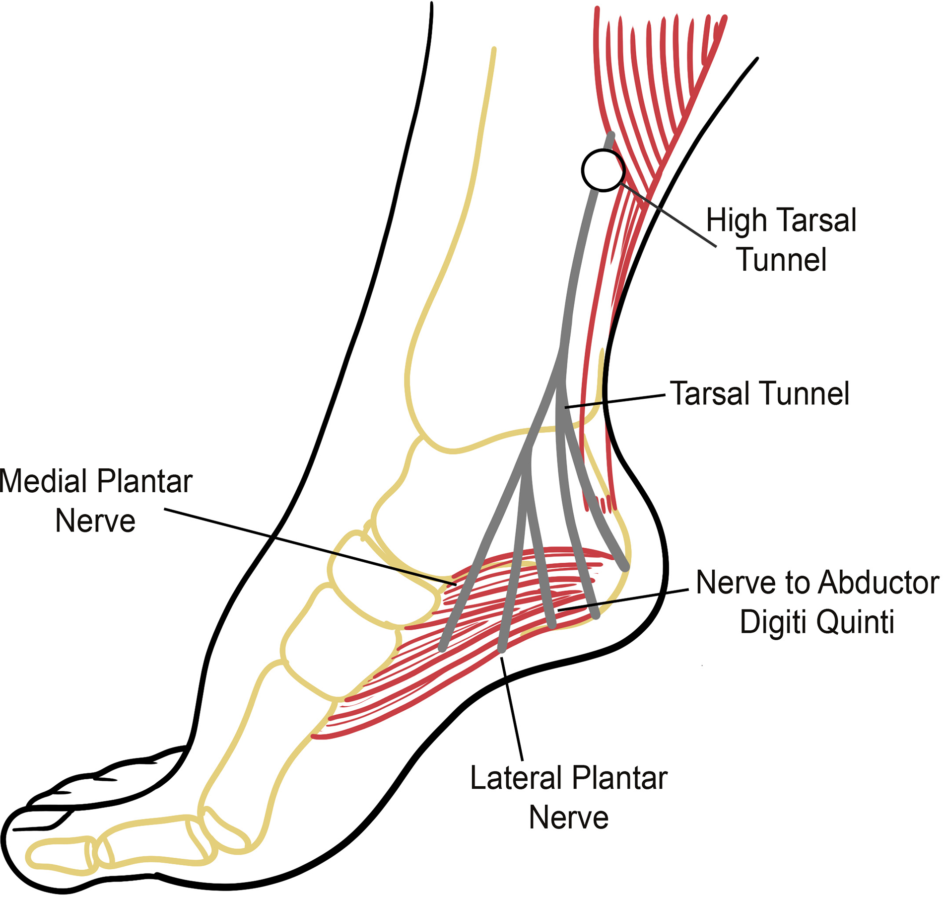
In 1962, Keck [1] defined the term tarsal tunnel syndrome (TTS) as a compression of the tibial nerve as it courses beneath the laciniate ligament. Two years later, McGill [2] noted that a further component of TTS is entrapment of the tibial nerve or its distal branches by the abductor hallucis (AH) muscle and its fascia. In 1995, an additional site of entrapment at the level of the high ankle was recognized by Baxter [3]. This site of compression has come to be further recognized by additional authors [4,5,6,7] and has been referred to as Segura’s area [8]. Entrapment of the tibial nerve at this site in the presence of clinical findings of TTS has been referred to as high TTS (HTTS) (Fig. 1) [3,4,6]. Finally, clinical findings of TTS can also result from tibial nerve entrapment at the soleal sling, which has been referred to as proximal TTS [9,10,11].
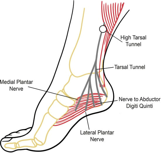
Figure 1.
Locations of possible tibial nerve compression: the high ankle in high tarsal tunnel syndrome, the tarsal tunnel, and under the abductor hallucis muscle.
The symptoms of TTS were first described in 1918 by Malaise [12] and since then have been well-established [13,14,15,16]. These include burning, numbness, paresthesia, or other pain-inducing sensations; a cold or swollen sensation; or a feeling like sandpaper or gravel on the bottom of the foot or in the toes. In long-standing cases, the muscles of the toes may atrophy and weaken, leading to digital deformities [14,16,17].
Clinical diagnosis of patients who have symptoms of TTS focuses on their history and physical examination. The physician’s examination may include checking for the presence of a Tinel’s sign, evaluating the sole of the foot for areas of sensory loss, evaluating the strength of the toes or their ability to spread apart, performing diagnostic nerve blocks, or using a provocative maneuver test. Provocative tests attempt to elicit paresthesia, pain, or other sensations by stimulating the tibial nerve, and such tests include the dorsiflexion-eversion test, plantarflexion-inversion test, and triple compression stress test [13,18,19,20,21,22,23].
Electrodiagnostic (EDX) testing has been established as useful in diagnosing TTS, although in 2005 Patel et al [12] found that the scientific support for EDX testing in TTS is low. More recently, in 2010, a more precise technique referred to as inching, originally developed to identify nerve compression in the upper extremity [24,25], has been used in the diagnosis of TTS by Segura [5]. This technique allows for localization of the tibial nerve’s compression site or sites [6,26,27]. This retrospective medical record evaluation investigated the site or sites of tibial nerve compression, determined by EDX testing, in patients presenting with clinical findings of TTS.
Materials and Methods
The medical records of 91 patients who presented to a podiatric medicine practice with clinical findings of TTS were retrospectively reviewed for the prevalence of nerve compression at the level of the high ankle (ie, HTTS) or the laciniate ligament/AH muscle in the foot. Patients were examined by a podiatric physician (M.S.N.) who was a fellow of the Association of Extremity Nerve Surgeons. If a clinical suspicion of TTS was present, they were referred for EDX testing.
The study group ranged in age from 28 to 85 with a mean of 57.8. The patients comprised 56 women and 35 men. All patients had adequate circulation to their feet, as determined by physical examination, and none had any open wounds, infections, masses, anatomical variants, complex regional pain syndrome, or history of prior TTS surgery or trauma to the extremity.
A clinical neurophysiologist (R.P.S.) performed EDX testing on the lower extremities of each patient. This evaluation consisted of conventional EDX testing, which was completed first and involved motor conduction studies on the tibial and common peroneal nerves and sensory conduction studies on the sural, superficial peroneal, and medial and lateral plantar nerves. The evaluation included analysis of latencies, amplitudes, and distances, and these were compared with the contralateral limb. A lower limit of 40 m/sec was deemed normal for intersegmental conduction velocities [26]. Needle electromyography was also performed to evaluate for proximal pathology, such as radiculopathy.
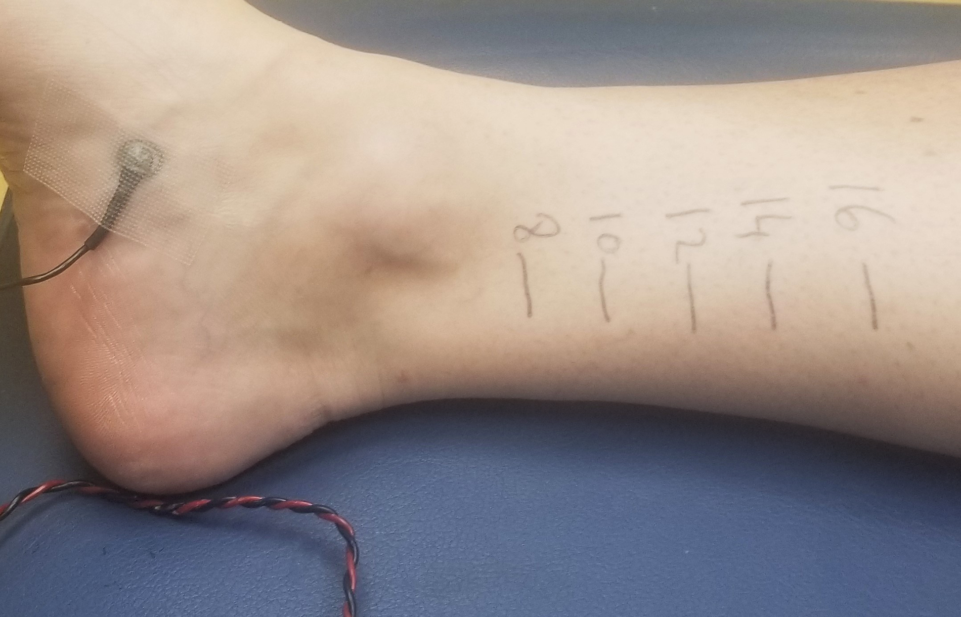
Following conventional EDX testing, the tibial nerve was evaluated using the inching technique. Readings were made from the AH muscle with the tibial nerve stimulated at intervals of 2 cm, as prior research had demonstrated this length to yield acceptable results [5]. Measuring from the AH muscle in the foot proximally, the tibial nerve was stimulated along its course at 8, 10, 12, 14, and 16 cm (Fig. 2). This area is above the laciniate ligament and corresponds to the level of the high ankle [27].
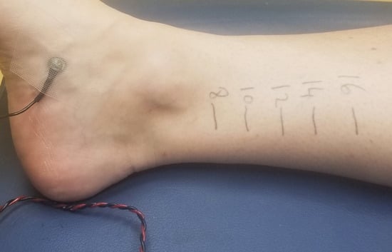
Figure 2.
Anatomical sites (labeled with black marker) where the tibial nerve was stimulated on a patient’s lower leg.
The patients’ medical information was in existence prior to the study’s start date, and patient data used in this investigation were anonymized, with no identifying information present. All procedures were performed in accordance with the ethical standards of the 1964 Declaration of Helsinki and its amendments or comparable ethical standards. An institutional board review was not required because the study was not considered human subject research, as the study’s protocol (ie, anonymized records) precluded the identification of any persons.
Results
Of the 91 patients determined to have clinical findings of TTS and sent for EDX testing, 59 (64.8%) had nerve compression at the laciniate ligament/AH muscle in the foot, 69 (75.8%) had nerve compression at the level of the high ankle, 51 (56%) had compression in both areas, and 13 (14.3%) were found to have no EDX evidence of tibial nerve entrapment or any other entrapment, such as radiculopathy. Of the 69 patients found to have an entrapment at the high ankle, 41 were women and 28 were men, accounting for 73% and 80% of women and men, respectively, with the compression occurring bilaterally in 44 of the 69 patients (with the remaining 25 instances occurring unilaterally).
Analyzing this data, 85.7% of patients determined to have a clinical suspicion of TTS were confirmed with EDX testing to have a tibial nerve compression; 64.8% had a compression of the nerve at the laciniate ligament/AH muscle; 75.8% had compression at the level of the high ankle, between 10 and 16 cm from the AH muscle; and 56% had compression in both anatomical sites. Of those patients who had EDX findings of TTS, 75.6% had a compression of the tibial nerve at the high ankle, 88.5% had a compression of the nerve at the laciniate ligament/AH muscle, and 65.4% had compression at both anatomical sites.
Discussion
Although compression of the tibial nerve at the level of the high ankle was first recognized approximately 30 years ago [3], research on it is sparse, and the authors were unable to find another study investigating both this compression (HTTS) and entrapment under the laciniate ligament/AH muscle in a given individual. Interestingly, this study found that compression of the tibial nerve occurred more often at the high ankle (HTTS) than at the laciniate ligament/AH muscle (69 versus 59 patients). This finding is contrary to the traditional understanding of TTS, which has been that the primary compression site is in the foot, at the laciniate ligament/AH muscle.
Research has shown that the incidence of failure of TTS surgery varies, with rates ranging from 4% to 56% [28]. Although a myriad of reasons may explain why surgical decompression for TTS fails, including the presence of a space-occupying mass, the development of adhesive neuritis or scar tissue, or an insufficient release [14,28,29], Raikin and Minnich [29] noted that failure can occur when HTTS is overlooked. The present study provides support for Raikin and Minnich’s observation, with HTTS occurring in conjunction with entrapment beneath the laciniate ligament/AH muscle 65.4% of the time and alone, without entrapment at the laciniate ligament/AH muscle, 23.1% of the time. These findings suggest that a significant number of failed TTS surgeries (or those in which patients obtain only partial improvement) may be due to HTTS being overlooked.
The finding that compression occurs at the high ankle more than at the conventional site (laciniate ligament/AH muscle) suggests that a new paradigm in TTS may need to be considered. Perhaps physicians need to examine the tibial nerve at the ankle, checking for such findings as a Tinel’s sign or provocative sign, and electrodiagnosticians need to employ an inching technique along the tibial nerve when evaluating a patient for TTS.
As this study found HTTS occurring independently of nerve entrapment at the laciniate ligament/AH muscle, perhaps some patients who require surgery for TTS could avoid surgery at this area of the foot and require only a small incision at the ankle. Barrett [30] found that one of the more common complications of TTS surgery in the foot is postoperative wound dehiscence. Avoiding surgery at this area in the foot could eliminate this complication for some persons with TTS and also lessen postoperative pain and disability.
The 14.3% of patients who did not show any compression with EDX testing suggests another etiology for their problems, such as a musculoskeletal issue. Alternatively, the EDX testing may have produced a false-negative outcome. In this regard, conventional EDX testing has been noted in the scientific literature to not always identify TTS [24,30,31,32,33].
This study was limited by geographic location, age range, and sample size. Future research should consider a larger sample size, broader distribution of patient demographics, patient biomechanics, and the influence of other medical issues, such as diabetes.
Conclusions
This study found that compression of the tibial nerve in patients presenting with TTS occurs most often at the level of the high ankle, 10 to 16 cm from the AH muscle, not at the laciniate ligament/AH muscle in the foot. Importantly, the study suggests that HTTS has been overlooked by much of the medical and scientific community with regard to patients with clinical findings of TTS.
We believe a new approach should be taken by clinicians and neurophysiologists involved in the treatment of patients with clinical findings of TTS. Both practitioners should consider the possibility of HTTS, examining the high ankle for signs of compression in their clinical examination and during EDX testing, employing an inching technique. Finally, this research finds that surgical decompression for TTS appears to have a significant risk of failure or incomplete result in patients in whom the presence of HTTS has been overlooked.
Financial Disclosure: None reported.
Conflict of Interest: None reported.
References
- Keck C: The tarsal-tunnel syndrome. J Bone Joint Surg Am 44: 180, 1962.
- McGill DM: Tarsal tunnel syndrome. Proc R Soc Med 57: 1125, 1964.
- Baxter DE: “Functional nerve disorders,” in The Foot and Ankle in Sport, p 9, CV Mosby, St. Louis, 1995.
- Kennedy JG, Baxter DE: Nerve disorders in dancers. Clin Sports Med 27: 329, 2008.
- Segura RP: Incidence of tarsal tunnel syndrome in a diabetic population: redefining topographic and electrophysiologic correlates. Presented at the 13th World Congress on Pain, August 29–September 3, 2010, Montreal, QC, Canada.
- Segura RP, Nirenberg MS: Prevalence of obesity in high tarsal tunnel syndrome: a cross-sectional study. JAPMA 113: 22, 2023.
- Baxter D, Schon LC: “Assessment and treatment of the elite athlete: helpful hints and pertinent pearls,” in Baxter’s The Foot and Ankle in Sport, 2nd Ed, edited by DA Porter, LC Schon, p 3, CV Mosby, St. Louis, 2008.
- Parker R: Parker pass & MIS tarsal tunnel trifecta update. Paper presented at: Association of Extremity Nerve Surgeons Annual Symposium, November 4-6, 2022, Fort Worth, TX.
- Williams EH, Rosson GD, Hagan RR, et al: Soleal sling syndrome (proximal tibial nerve compression): results of surgical decompression. Plast Reconstr Surg 129: 454, 2012.
- Rader AJ, Cancelliere P, Kelly T: Diagnostic ultrasound of the soleal sling. JAPMA 106: 147, 2016.
- Maguire A, Garcia V, Nasser E: When lightning strikes twice: tarsal tunnel syndrome in the presence of soleal sling syndrome. FASTRAC 4: 100351, 2024.
- Patel AT, Gaines K, Malamut R, et al: Usefulness of electrodiagnostic techniques in the evaluation of suspected tarsal tunnel syndrome: an evidence‐based review. Muscle Nerve 32: 236, 2005.
- Abouelela AA, Zohiery AK: The triple compression stress test for diagnosis of tarsal tunnel syndrome. Foot (Edinb) 22: 146, 2012.
- Ahmad M, Tsang K, Mackenney PJ, et al: Tarsal tunnel syndrome: a literature review. Foot Ankle Surg 18: 149, 2012.
- Gould JS: Tarsal tunnel syndrome. Foot Ankle Clin 16: 275, 2011.
- McSweeney SC, Cichero M: Tarsal tunnel syndrome—a narrative literature review. Foot (Edinb) 25: 244, 2015.
- Haq II, Banerjee AA, Arshad Z, et al: The management of tarsal tunnel syndrome: a scoping review. J Clin Orthop Trauma 54: 102489, 2024.
- Oh SJ: Neuropathies of the foot. Clin Neurophysiol 118: 954, 2007.
- Yunoki M: Analysis of surgical cases of tarsal tunnel syndrome in our department: case series and literature review. Asian J Neurosurg 15: 59, 2020.
- Hendrix CL, Jolly GP, Garbalosa JC, et al: Entrapment neuropathy: the etiology of intractable chronic heel pain syndrome. J Foot Ankle Surg 37: 273, 1998.
- Kinoshita M, Okuda R, Morikawa J, et al: The dorsiflexion-eversion test for diagnosis of tarsal tunnel syndrome. J Bone Joint Surg Am 83: 1835, 2001.
- Franson J, Baravarian B: Tarsal tunnel syndrome: a compression neuropathy involving four distinct tunnels. Clin Podiatr Med Surg 23: 597, 2006.
- Dellon AL: Partial joint denervation II: knee and ankle. Plast Reconstr Surg 123: 208, 2009.
- Kimura J: A method for determining median nerve conduction velocity across the carpal tunnel. J Neurol Sci 38: 1, 1978.
- Miller RG: The cubital tunnel syndrome: diagnosis and precise localization. Ann Neurol 6: 56, 1979.
- Oexeman S, Arroyo CI, Rodriguez-Collazo ER, et al: Redefining the topography of the tibial nerve: updated treatment guide for tibial nerve entrapments: an addendum to surgical treatment protocol for peripheral nerve dysfunction of the lower extremity: a systematic approach. Clin Podiatr Med Surg 38: e7, 2021.
- Segura RP, Nirenberg MS: High tibial nerve entrapment: a common component of tarsal tunnel syndrome. JAPMA 115: 22, 2025.
- Yalcinkaya M, Ozer UE, Yalcin MB, et al: Neurolysis for failed tarsal tunnel surgery. J Foot Ankle Surg 53: 794, 2014.
- Raikin SM, Minnich JM: Failed tarsal tunnel syndrome surgery. Foot Ankle Clin 8: 159, 2003.
- Barrett SL: Practical Pain Management for the Lower Extremity Surgeon, Data Trace Publishing Company, Brooklandville, MD, 2015.
- Dellon AL: The four medial ankle tunnels: a critical review of perceptions of tarsal tunnel syndrome and neuropathy. Neurosurg Clin N Am 19: 629, 2008.
- De Prado M, Cuervas-Mons M, Golanó P, et al: The tarsal tunnel syndrome. FussSprungg 13: 227, 2015.
- Oishi C, Ichikawa Y, Chiba A, et al: O-2-31. The usefulness of electrophysiological examinations in the diagnosis of tarsal tunnel syndrome. Clin Neurophysiol 129: e34, 2018.
© 2025 American Podiatric Medical Association