


Abstract
This case involves a 35-year-old female who underwent a custom talonavicular replacement with subtalar fusion for spontaneous osteonecrosis of the navicular and talus. Preoperatively, she had an American Orthopedic Foot and Ankle Society (AOFAS) score of 42, a talocalcaneal height of 64.6 mm, and a talar declination angle of 23.6°. Imaging showed severe erosive changes in the talonavicular and anterior subtalar joints, with marrow edema throughout the navicular and anterior talus. The procedure involved removing the talus and navicular and replacing them with a titanium, nitride-coated talonavicular implant, secured by two 5.5-mm screws. One year postoperatively, her AOFAS score improved to 65, with a talar declination of 15.9° and a talocalcaneal height of 63.8 mm. She had an ankle range of motion with 12° dorsiflexion and 40° plantarflexion. Radiographs showed proper implant seating and no radiolucency. The patient expressed satisfaction with the outcome. This article aims to contribute to the literature on custom implants for severe deformities and pathologies on patients who wish to maintain motion within vital joints. In conclusion, it appears a custom-made talonavicular implant may be a viable solution for spontaneous osteonecrosis of the talus and navicular.
The navicular bone is crucial for the medial column, contributing to both the medial longitudinal and transverse arches. The medial tuberosity serves as an attachment point for the posterior tibialis tendon, aiding in support for the medial longitudinal arch. The bone’s blood supply is by the dorsalis pedis, supplying the dorsal lateral one third of the navicular, and the medial plantar artery, supplying the plantar aspect. The dorsalis pedis and medial plantar artery form an anastomosis to provide a blood supply to the medial tuberosity. Unfortunately, the central zone lacks a strong blood supply, coinciding with the area of bone that receives maximum shear force [1]. The talus, with a limited vascular support, bears a significant amount of weight during various activities. The restricted blood supply is due to the talus having five articular facets, covering 60% to 70% of its surface with cartilage. As a result, the majority of its blood supply comes from extra-osseous sources rather than the periosteum [2]. The posterior tibial artery is the primary contributor of blood to the talus, responsible for more than 47% of the vascularity supplied by its branches [3]. Both bones may have a disruption in the blood supply leading to avascular necrosis (AVN) [4]. Avascular necrosis is most commonly a result of trauma, disrupting a bone with delicate blood supply [5]. Other reported causes of AVN may include alcoholism, steroid use, dyslipidemia, or idiopathic [6] The closest staging system for this case report describes ankle avascular necrosis by the Ficat and Arlet classification: stage I being normal, stage II having cystic lesions within a normal contour of talus, stage III having subchondral collapse, and stage IV showing joint-space narrowing with secondary distal tibial changes [7]. Isolated cases of osteonecrosis or structural abnormalities to the talus or navicular have been documented. Avascular necrosis or structural abnormalities to the navicular is linked to conditions such as Kohler’s and Müller-Weiss; AVN to the talus is associated with Diaz’s disease.
Vaishya [8] was the first to describe a case of spontaneous osteonecrosis of the navicular and talus (SONNT) in 2016. This condition is characterized by symptomatic osteonecrosis in both the talus and navicular simultaneously occurring in the adult population. Reasons for SONNT include AVN, where a decrease in blood supply affects the aerobic metabolism, leading to ischemic necrosis. Another possible reason for SONNT is a developmental issue [8,9]. Vaishya’s case involved a 24-year-old male with bilateral SONNT. The patient presented with a gradual increase in bilateral hindfoot pain, a planovalgus deformity, and limitations in supination and pronation. Radiographs revealed fragmentation of the navicular and sclerosis with partial collapse of the talus and navicular. Their patient underwent left foot talonavicular-cuneiform arthrodesis with core decompression of the talar body, overall achieving successful results. Vaishya suggests their patient’s SONNT may be secondary to abnormal embryologic development. This was based off the bilateral presentation and involvement of the preaxial regions (second ray, talus, navicular, and cuneiform) [8].
A condition with a similar presentation to SONNT is Müller-Weiss disease. This pathology often observed in middle-aged females who are experiencing chronic midfoot and hindfoot pain. Radiographs typically reveal compression on the lateral aspect of the navicular bone [10]. Currently the cause of Müller-Weiss remains unclear, with various overlapping theories. Maceira [11] a suggests Müller-Weiss is a consequence from a delay in navicular ossification. This delay causes the bone to be inadequate in structurally handling a lateral shift in compressive forces. The shift in forces may be a result of first ray brachymetatarsia, primary varus of the subtalar joint, or undiagnosed clubfoot. These compressive forces cause the navicular to flatten in the dorsal lateral aspect and eventually result in the formation of a sagittal plane cleft dividing the navicular into two parts. The bony changes in the navicular also lead to the lateral shift of the talar head and a hindfoot varus deformity. Although many articles describe the condition due to osteonecrosis or avascular necrosis of the navicular bone, histopathologic studies have yet to provide conclusive evidence of osteonecrosis with the disease most likely secondary to a developmental and biomechanical issue [11]. It may be important to distinguish between SONNT and Müller-Weiss disease due to a potential source of SONNT being AVN, whereas Müller-Weiss disease is secondary to compressive forces. The source of AVN may impact healing at an arthrodesis site.
A more recent trend in treating AVN of the talus is a custom three-dimensional implant. Harnrrongroj and Vanadurognwan [12] were among the first in describing the use of a talar implant device, specifically the first generation talar body implant, to address talar body AVN. Their aim was to maintain anatomic motion through the tibiotalar joint and restore ankle height. The first-generation featured a talar body prosthesis with a peg inserted into the talar neck and head. Although early clinical results were promising, issues with loosening arose at the fixation site. The second-generation implant omitted the peg and was not anchored to the talar head. Again, initial outcomes were positive, but follow-up revealed talar head collapse in some patients. The standard third-generation implant replaces the entire of the talus and is designed to restore normal ankle joint [13]. Now with further advances in technology, more specific custom implants may be tailored to the patient’s pathology, including a custom implant replacing the talus and navicular.
The joint-sparing implants may be beneficial in maintaining function to the joint while maintaining the structure of the foot and filling bone void. We report a case of a 35-year-old female with SONNT who was treated with a talonavicular implant with subtalar fusion.
Case Report
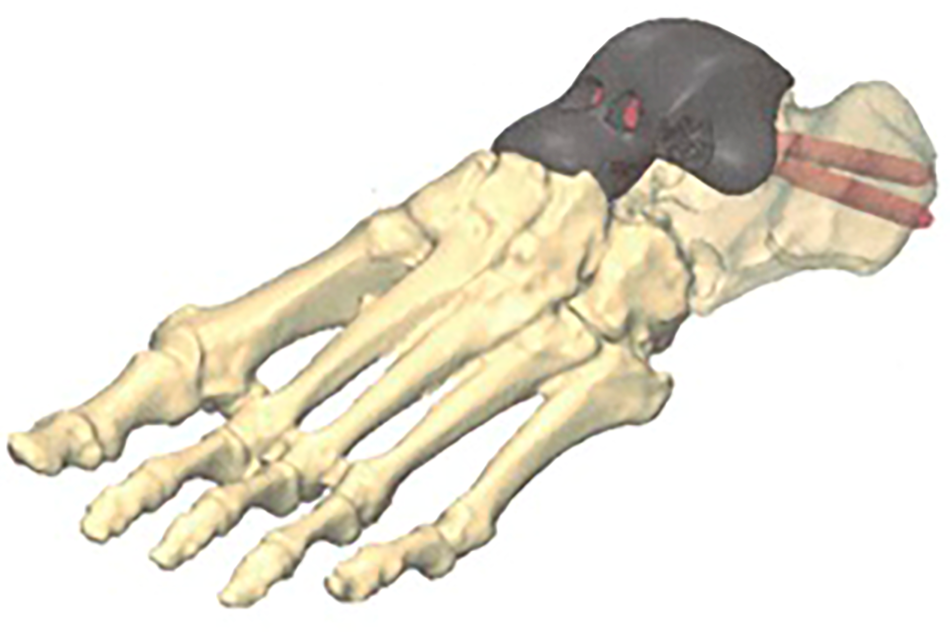
A 35-year-old female presented with left foot pain persisting for more than a year, which began shortly after having a newborn child. She reports a history of trauma to the left ankle (a sprain 3 years prior) that resolved shortly after the incident. The patient reports a history of controlled gestational diabetes, which had resolved following pregnancy, and denied other forms of neuropathy, diabetes mellitus, hyperlipidemias, inflammatory arthritis, metabolic disorders, connective tissue disorders, or autoimmune disorders. She denied use of alcohol and tobacco products. Physical examination revealed a planovalgus deformity in the left foot, with noticeable pain and guarding during supination and pronation and pain to the subtalar, talonavicular, and calcaneocuboid joints. Radiographs displayed extensive erosive changes in the talar head and proximal navicular, along with arthritis in the subtalar joint and calcaneocuboid (Fig. 1). Magnetic resonance imaging (MRI) indicated collapse with severe erosive changes in the talonavicular and middle facet of the anterior subtalar joint, with underlying marrow edema throughout the navicular and anterior half of the talus (Fig. 2). We attempted conservative treatments, including anti-inflammatories, medications, corticosteroid injections, physical therapy, and changes in shoe gear but achieved minimal improvement. Due to the severe pain with failed conservative treatment, the patient opted for a custom talonavicular implant with subtalar fusion. The custom implant design was based on a computed tomography scan of the contralateral limb 2 months before surgery. Made of titanium, the implant featured a mirror-polished finish at the surfaces facing the naviculocuneiform joint and tibiotalar joint. There were two 6.2-mm holes originating from the talar neck of the implant, directed from dorsal anterior to plantar posterior, to accommodate the threads of two 5.5-mm screws and achieve appropriate apposition of the screw heads (Fig. 3). Also, there was a porous surface along the subtalar surface to aid in fusion at the subtalar joint (Fig. 2).
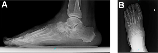
Figure 1.
Lateral (A) and anteroposterior (B) radiographs of the right foot demonstrating erosive changes to the talonavicular joint with surrounding arthritis to the subtalar and calcaneocuboid joint.
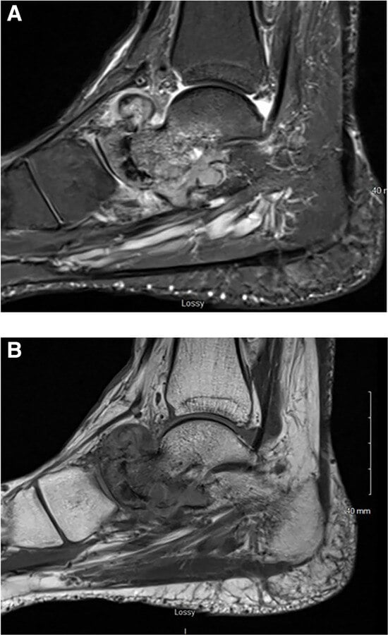
Figure 2.
A, T1- and B, T2-weighted magnetic resonance images demonstrating severe erosive change of the talonavicular articulation, middle facet of the subtalar joint, and of the calcaneocuboid articulation. This shows the collapse of the talonavicular joint with superior osteophyte formation.
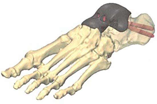
Figure 3.
Preoperative three-dimensional rendering of the talonavicular implant.
During the surgery, an anterior linear incision was made overlaying the ankle joint. This incision was centered between the tibialis anterior tendon and the extensor hallucis longus, extending distally over the talonavicular joint. Subperiosteal dissection exposed the talonavicular and naviculocuneiform joints. The posterior tibial tendon was identified and resected from its insertion on the navicular tuberosity. The navicular was then cut into multiple pieces with a sagittal saw and removed, followed by the talus. We denuded the articular surface for the subtalar joint on the calcaneus and fenestrated it using a 2.0-mm drill bit. Subsequently, we inserted the trial sizer implant and then the custom implant, demonstrating good apposition and ankle joint range of motion (Fig. 4). After carefully placing the implant and maintaining the foot in a neutral position at 90°, we secured the posterior tibial tendon to the medial aspect of the implant using designated suture holes with size two fiberwire. Temporary fixation was achieved by driving two 0.062 Kirschner wires through the predrilled holes in the dorsal aspect of the implant into the subtalar joint. After fixation, we drilled and inserted two 5.5 x 75-mm partially threaded cannulated screws through the prosthesis, across the subtalar joint, and into the calcaneus. The navicular bone specimen was sent to pathology, and the report indicated necrosis with no signs of infection. Postoperative protocol involved the patient remaining nonweightbearing in a fiberglass cast until the second week. Sutures were then removed at the second week, and the patient was transitioned into a controlled ankle motion (CAM) boot with continued nonweightbearing to the left lower extremity. During this time, the patient was encouraged to take off the CAM boot and work nonweightbearing ankle range of motion (ROM) via dorsiflexion and plantarflexion. At the sixth week, the patient transitioned into being weightbearing as tolerated to the right lower extremity in an over-the-shoe walking brace (TayCo; South Bend, Indiana) with physical therapy.
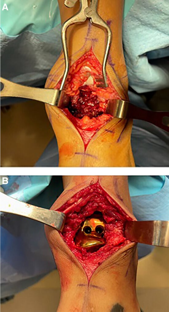
Figure 4.
Intraoperative photograph showing the anterior incision approach after (A) talonavicular removal and (B) implant insertion.
Before the surgery, the patient reported an American Orthopedic Foot and Ankle Society (AOFAS) score of 42, a talocalcaneal height of 64.6 mm, and a talar declination angle of 23.6°. After the operation at the 1-year mark, the patient reported an increased AOFAS score of 65, with an ankle dorsiflexion measuring 12° and plantarflexion at 40°. Additionally, there was an observed improvement in the talar declination to 15.9°, with minimal changes to the talocalcaneal height, now measured at 63.8 mm (Table 1). Serial radiographs were obtained of the patient following surgery. Radiographs at the 1-year mark revealed no signs of radiolucency and proper seating of the implant within the tibiotalar joint. Also, a slight amount of dorsal displacement of the medial collumn at the medial cueniform occurred at the the 1-year mark (Fig. 5C).

Table 1.
Preoperative and Postoperative Results for AOFAS, ROM, and Radiographic Measurements
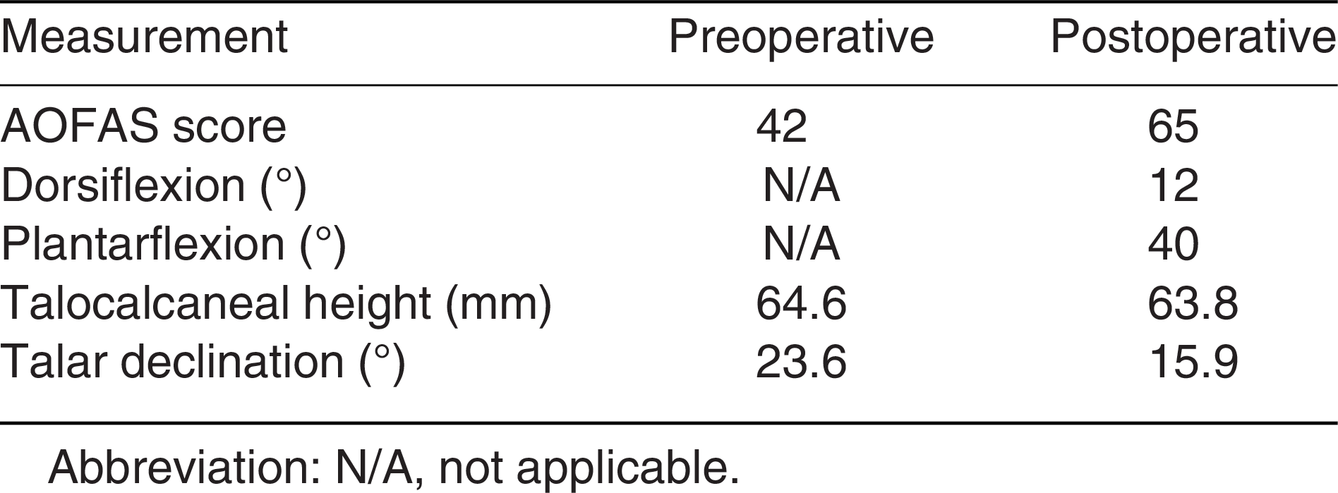

Figure 5.
Lateral postoperative radiographs taken at 1-month (A), 6 months (B), and 1 year (C).

Discussion
A recent uptake in literature demonstrates growth of popularity of the use of custom three-dimensional implants [14,15]. Due to the complexity of the foot and ankle, the use of custom implants are helpful when replacing multiple segments of bone or joints [16]. Currently there is limited research on the outcomes for a talonavicular implant. To our knowledge, the only publication is a case study by Giannini [17]. They reported a 30-month follow-up for a custom talonavicular replacement with subtalar arthrodesis for a patient with post-traumatic arthritis. They demonstrated an increase in the AOFAS score from 36 to 81 and a postoperative ankle range of motion with dorsiflexion of 15° and 4° of plantarflexion [17].
Our patient preoperatively reported an AOFAS score of 42, talocalcaneal height of 64.6 mm, and talar declination angle of 23.6°. One year postoperatively, the patient demonstrated an improved AOFAS score of 65 and talar declination of 15.9°. The talocalcaneal height remained almost unchanged at 63.8 mm. Radiographs at the 1-year mark revealed no signs of radiolucency and proper seating of the implant within the tibiotalar joint. Additionally, the patient had an ankle range of motion with 12° of dorsiflexion and 40° of plantarflexion. In the normal gait, the required ankle range of motion at the loading response phase is a mean of 5° and a mean of 12° at the terminal stance phase. Based on those results, our patient demonstrated enough ankle ROM for normal gait [18]. When comparing our study to Giannini’s [17], both demonstrated similar ankle range of motion and results. At the 1-year mark the patient was overall satisfied with the procedure and demonstrated appropriate biomechanical measurement acquired for walking.
When preoperatively differentiating between SONNT and Müller-Weiss disease, it may be distinguished through history, clinical examination, and imaging. In our case report, there is evidence of SONNT, without the classic signs of Müller-Weiss disease. In our patient’s history, there is no association with potential causes of Müller-Weiss, such as delay in the navicular ossification, a short first ray, varus of the subtalar joint, or a history of clubfoot. Our patient’s history aligns with SONNT, due to the potential aggravating factor of the ankle sprain prior to presentation. This trauma may have led to a disruption in blood supply and the development of SONNT. In the physical exam, our patient demonstrated planovalgus, as seen with Vaishya’s case report [8], rather than the rearfoot varus deformity as seen with Müller-Weiss. Radiographs for our patient revealed osseous destruction of the talus and navicular at the talonavicular joint, whereas in Müller-Weiss, there is compression/reabsorption of the dorsal lateral navicular with fragmentation. Additionally, the pathology of our patient’s bone demonstrated osteonecrosis, whereas Müller-Weiss is not known to be secondary to osteonecrosis changes. The importance of correctly diagnosing between the two diseases may be important for surgical planning.
As of now, there are no clear guidelines on the treatment for SONNT. Early management may be through conservative measures of nonsteroidal anti-inflammatory drugs (NSAID), calcium and vitamin D supplementation, activity modification, bracing, orthotics, CAM boots, and corticosteroid injections [4,8]. We attempted such conservative treatments, including NSAIDs, corticosteroid injections, physical therapy, and changes in shoe gear; however, due to the severity of the osteonecrosis and limited improvement in pain and function, it was decided to proceed with surgical intervention.
Within Vaishya’s article for SONNT [8], their patient underwent a successful left foot talonavicular-cuneiform arthrodesis with core decompression of the talar body. For Müller-Weiss disease, a wide variety of surgical procedures with successful results have been proposed in the literature including isolated talonavicular arthrodesis, talonavicular-cuneiform arthrodesis, triple arthrodesis, pantalar arthrodesis, and calcaneal osteotomy [19]. Since the SONNT reported in Vaishya’s patient was secondary to hereditary problems and Müller Weiss disease secondary to a biomechanical issue, both pathologies have potential for successful arthrodesis at the talonavicular joint due to the intact blood supply.
However, when deciding on surgical treatment for SONNT, a challenge arises when the pathology is secondary to osteonecrosis. Due to the osteonecrosis present in the talonavicular joint, theoretically, there may be a higher percentage of complication, such as nonunion due to the disruption in blood supply. A standard talonavicular arthrodesis for arthritic joints already has a reported nonunion rate of up to 20% [20]. In comparison, arthrodesis of the ankle joint with talar AVN demonstrates a high complication rate, with high rates of delayed union of up to 32% [21]. Also, our patient had significant osteonecrosis in the navicular and talar head. Surgery to remove the affected bone would be extensive, leading to naviculectomy and talar head excision.
With removal of diseased bone, further potential surgical options included the use of a cadaveric femoral head, a custom cage, or a custom talonavicular replacement. Using a cadaveric femoral head may result in nonunion, with an estimated rate of around 50% [22]. The custom cage would require arthrodesis at the medial cuneiform, further impeding necessary midfoot motion. Considering these factors, the decision was made to opt for a constrained custom talonavicular replacement with subtalar fusion. Fusion at the subtalar joint may prevent further motion of the hindfoot and may lead to an increase in osteoarthritis to surrounding joints. However, due to osteoarthritis seen in the MRI within the anterior and middle facets of the subtalar joint, pain with subtalar joint range of motion, and the need to prevent implant subsidence, it was decided to perform the subtalar fusion (Fig. 2). The talonavicular implant allowed for the resection of the diseased portion while maintaining motion at the navicular cuneiform joint, thus enabling some movement in the midfoot, and hopefully preventing quicker degeneration of the surrounding joints. The custom implant featured a mirror-polished finish, and a titanium nitride coating at the ankle and navicular cuneiform joints, along with a porous surface at the subtalar joint to promote osseointegration (Fig. 6). Titanium, with a nitride coating, was chosen due to its lighter weight compared to cobalt, along with increased durability and enhanced osseointegration properties. One study demonstrated that titanium implants with a nitride coating were significantly more durable, showing a tenfold increase in durability compared to traditional titanium implants and a 34-fold improvement over mirror-polished cobalt. Titanium has shown to enhance osseointegration due to the increased surface porosity and surface roughness [23–26]. It is important to note that the patient should not have any form of neuropathy, as seen in ours, when performing these implants to prevent failure or other complications.
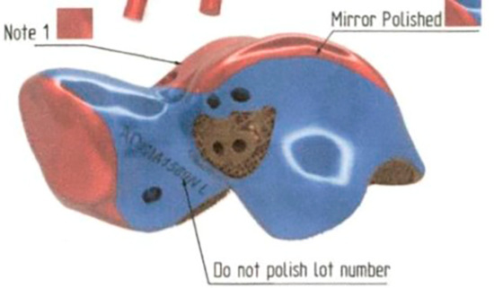
Figure 6.
Preoperative 3D rendering demonstrating mirror polished finish located at ankle and navicular cuneiform joint.
Further studies with increased follow-up time and more patients will be needed to further determine if a custom implant is viable option for SONNT. Overall, for our case, a constrained total talonavicular replacement with subtalar fusion appears to have demonstrated satisfactory results and may remain an option for treatment on SONNT.
Financial Disclosure: None reported.
Conflict of Interest: None reported.
References
- Mohiuddin T, Jennison T, Damany D: Müller-Weiss disease - review of current knowledge. Foot Ankle Surg 20: 79, 2014.
- Gelberman RH, Mortensen WW: The arterial anatomy of the talus. Foot Ankle 4: 64, 1983.
- Miller AN, Prasarn ML, Dyke JP, et al: Quantitative assessment of the vascularity of the talus with gadolinium-enhanced magnetic resonance imaging. J Bone Joint Surg Am 93: 1116, 2011.
- Clougherty CO, Razzante M, Leatham PR, et al: “3D-printed solutions for avascular necrosis of the talonavicular joint,” in Clinical Applications of 3D Printing in Foot and Ankle Surgery, 1st Ed, edited by PD Highlander, p 129, Elsevier, Philadelphia, 2024.
- Baron M, Paltiel H, Lander P: Aseptic necrosis of the talus and calcaneal insufficiency fractures in a patient with pancreatitis, subcutaneous fat necrosis, and arthritis. Arthritis Rheum 27: 1309, 1984.
- Trauth J, Bläsius K: Talus necrosis and its treatment. Aktuelle Traumatol 18: 152, 1988.
- Mont MA, Schon LC, Hungerford MW, et al: Avascular necrosis of the talus treated by core decompression. J Bone Joint Surg Br 78: 827, 1996.
- Vaishya R, Kumar V, Agarwal AK, et al: Spontaneous osteonecrosis of navicular and talus (SONNT). J Clin Orthop Trauma 7: 83, 2016.
- Solomon L: Mechanisms of idiopathic osteonecrosis. Orthop Clin North Am 16: 655, 1985.
- Fernández de Retana P, Maceira E, Fernández-Valencia JA, et al: Arthrodesis of the talonavicular-cuneiform joints in Müller-Weiss disease. Foot Ankle Clin 9: 65, 2004.
- Maceira E, Rochera R: Müller-Weiss disease: clinical and biomechanical features. Foot Ankle Clin 9: 105, 2004.
- Harnroongroj T, Vanadurongwan V: The talar body prosthesis. J Bone Joint Surg Am 79: 1313, 1997.
- Taniguchi A, Takakura Y, Sugimoto K, et al: The use of a ceramic talar body prosthesis in patients with aseptic necrosis of the talus. J Bone Joint Surg Br 94: 1529, 2012.
- Dekker TJ, Steele JR, Federer AE, et al: Use of patient-specific 3D-printed titanium implants for complex foot and ankle limb salvage, deformity correction, and arthrodesis procedures. Foot Ankle Int 39: 916, 2018.
- West TA, Rush SM: Total talus replacement: case series and literature review. J Foot Ankle Surg 60: 187, 2021.
- Adams SB, Danilkowicz RM: Talonavicular joint-sparing 3D printed navicular replacement for osteonecrosis of the navicular. Foot Ankle Int 42: 1197, 2021.
- Giannini S, Cadossi M, Mazzotti A, et al: Custom-made total talonavicular replacement in a professional rock climber. J Foot Ankle Surg 55: 1271, 2016.
- Leardini A, Benedetti MG, Berti L, et al: Rear-foot, mid-foot and fore-foot motion during the stance phase of gait. Gait Posture 25: 453, 2007.
- Angthong C, Younger ASE, Chuckpaiwong B, et al: A novel update on the management of Müller-Weiss disease: presentation of a treatment algorithm. Cartilage 15: 65, 2024.
- Klassen LJ, Shi E, Weinraub GM, et al: Comparative nonunion rates in triple arthrodesis. J Foot Ankle Surg 57: 1154, 2018.
- Gross CE, Haughom B, Chahal J, et al: Treatments for avascular necrosis of the talus: a systematic review. Foot Ankle Spec 7: 387, 2014.
- Jeng CL, Campbell JT, Tang EY, et al: Tibiotalocalcaneal arthrodesis with bulk femoral head allograft for salvage of large defects in the ankle. Foot Ankle Int 34: 1256, 2013.
- Plecko M, Sievert C, Andermatt D, et al: Osseointegration and biocompatibility of different metal implants-a comparative experimental investigation in sheep. BMC Musculoskelet Disord 13: 1, 2012.
- Hara D, Nakashima Y, Sato T, et al: Bone bonding strength of diamond-structured porous titanium-alloy implants manufactured using the electron beam-melting technique. Mater Sci Eng C Mater Biol Appl 59: 1047, 2016.
- Ogawa M, Tohma Y, Ohgushi H, et al: Early fixation of cobalt-chromium based alloy surgical implants to bone using a tissue-engineering approach. Int J Mol Sci 13: 5528, 2012.
- Bertolotti L, Sharma A, Parekh SG: Electron beam manufacturing 3D printed titanium with titanium nitride coating shows favorable overall performance when compared to selective laser sintering cobalt chrome in patient specific talus implants. Nat J Clin Orthop 6: 22, 2022.
© 2025 American Podiatric Medical Association