


Abstract
Os subtibiale is a rare accessory ossicle of the ankle, not considered to be clinically significant. However, its presence in trauma patients may cause a misdiagnosis such as malleolar fracture and over-treatment. Because the ossicle is attached to the deltoid ligament, ankle trauma that causes a medial compartment injury may detach the os subtibiale from the medial malleolus. In these situations, magnetic resonance imaging may help to diagnose the deltoid injury, demonstrate the features of the ossicle, and guide the treatment process.
Os subtibiale, first described by Bircher in 1918, is a rare accessory ossicle located inferiorly to the medial malleolus [1–3]. Some reports argue that os subtibiale originates from the failure of fusion of the secondary ossification center to medial malleolus [1,2,4]. Others claim that os subtibiale is a distinct entity and should be distinguished from the D-shaped persistent secondary ossification center [5–7]. There are several reports in the literature that discuss this ossicle [6–9]. Although os subtibiale is generally not of clinical significance, misdiagnosis of the ossicle as a malleolar fracture, symptomatic os subtibiale with chronic pain, and posterior tibial impingement due to os subtibiale have been reported [2,8–10]. However, to our knowledge, there is no report of displacement of the os subtibiale in the literature.
Ankle injuries caused by the mechanism of pronation-external rotation (PER) can lead to stretching of the structures in the medial ankle, rupture of the deltoid ligament, or transverse avulsion fractures of the medial malleolus [11]. In the presence of os subtibiale, the accessory ossicle attached to the deltoid ligament may be avulsed from the medial malleolus by the mechanism of pronation and eversion.
In this case report, we present a patient with PER ankle injury who was diagnosed and treated with suprasyndesmotic fibular fracture, syndesmosis injury, and displacement of os subtibiale between the talus and the medial malleolus.
Case Report
A 33-year-old male patient was admitted to our hospital after a distortion injury in his left ankle during a soccer match. Physical examination revealed deformity and swelling of the left ankle with intact neurovascular examination.
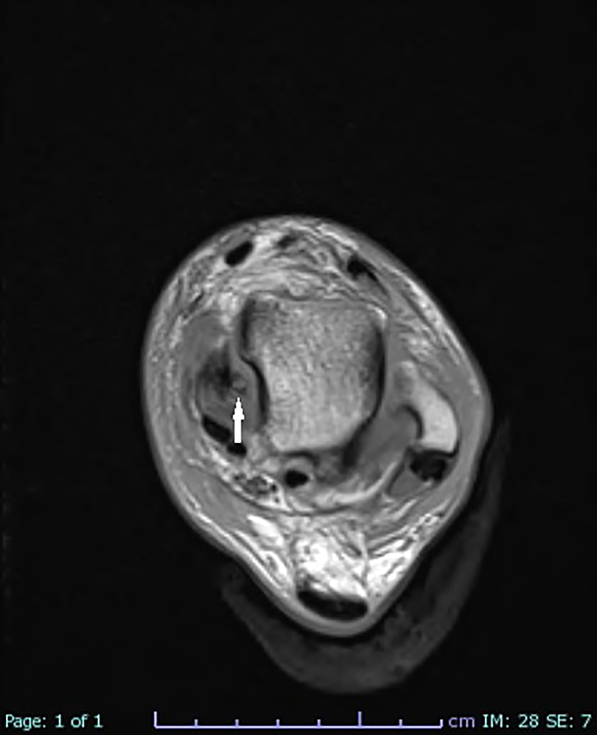
Radiographic images demonstrated a suprasyndesmotic fibular fracture and an ovoid bony structure between the talus and the medial malleolus. A 4.6-mm widening of the medial clear space was also noted (Fig. 1). Due to the nature of the trauma, magnetic resonance imaging (MRI) was performed to examine the deltoid ligament integrity. The MRI revealed a suprasyndesmotic fibular fracture, syndesmotic injury, partially ruptured deltoid ligament, and a well-corticated bony structure between the talus and the medial malleolus (Figs. 2 and 3). Cortical integrity of the medial malleolus was normal, and no fracture line was detected. The T1-weighted image demonstrated decreased medullary signal intensity consistent with reactive bone marrow edema (Fig. 3).
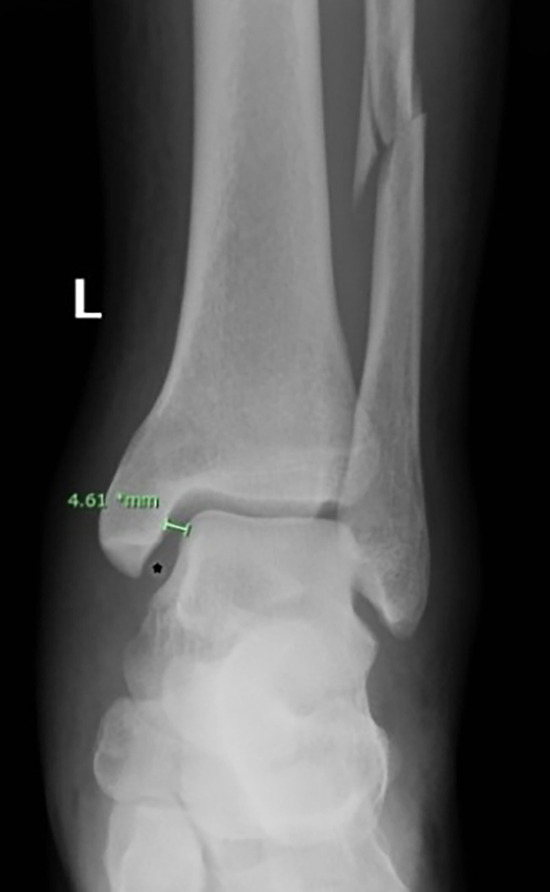
Figure 1.
Anteroposterior left ankle radiograph shows a suprasyndesmotic fibular fracture, widened medial clear space, and an ovoid bony structure (asterix) between the talus and the medial malleolus.
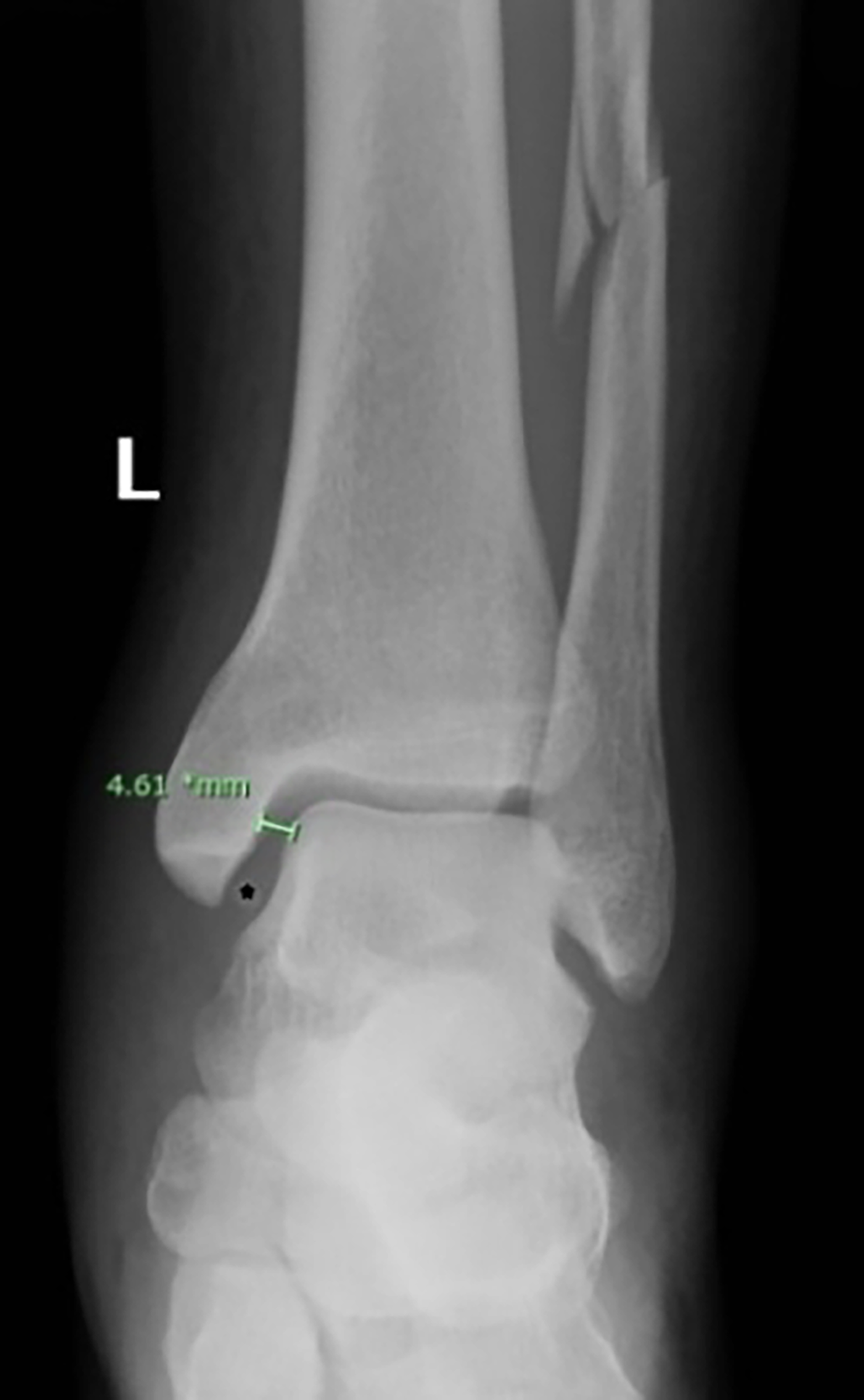
Figure 2.
Coronal T2-weighted fat-saturated magnetic resonance image (MRI) shows the partially ruptured deltoid ligament (white arrow) and a bony structure between the talus and the medial malleolus (open arrow).
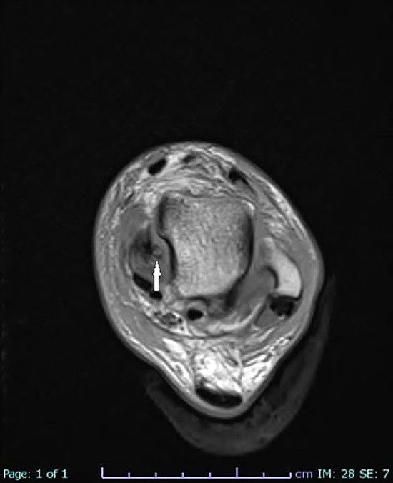
Figure 3.
Axial T1-weighted MRI shows a well-corticated bony structure between the talus and the medial malleolus (arrow).
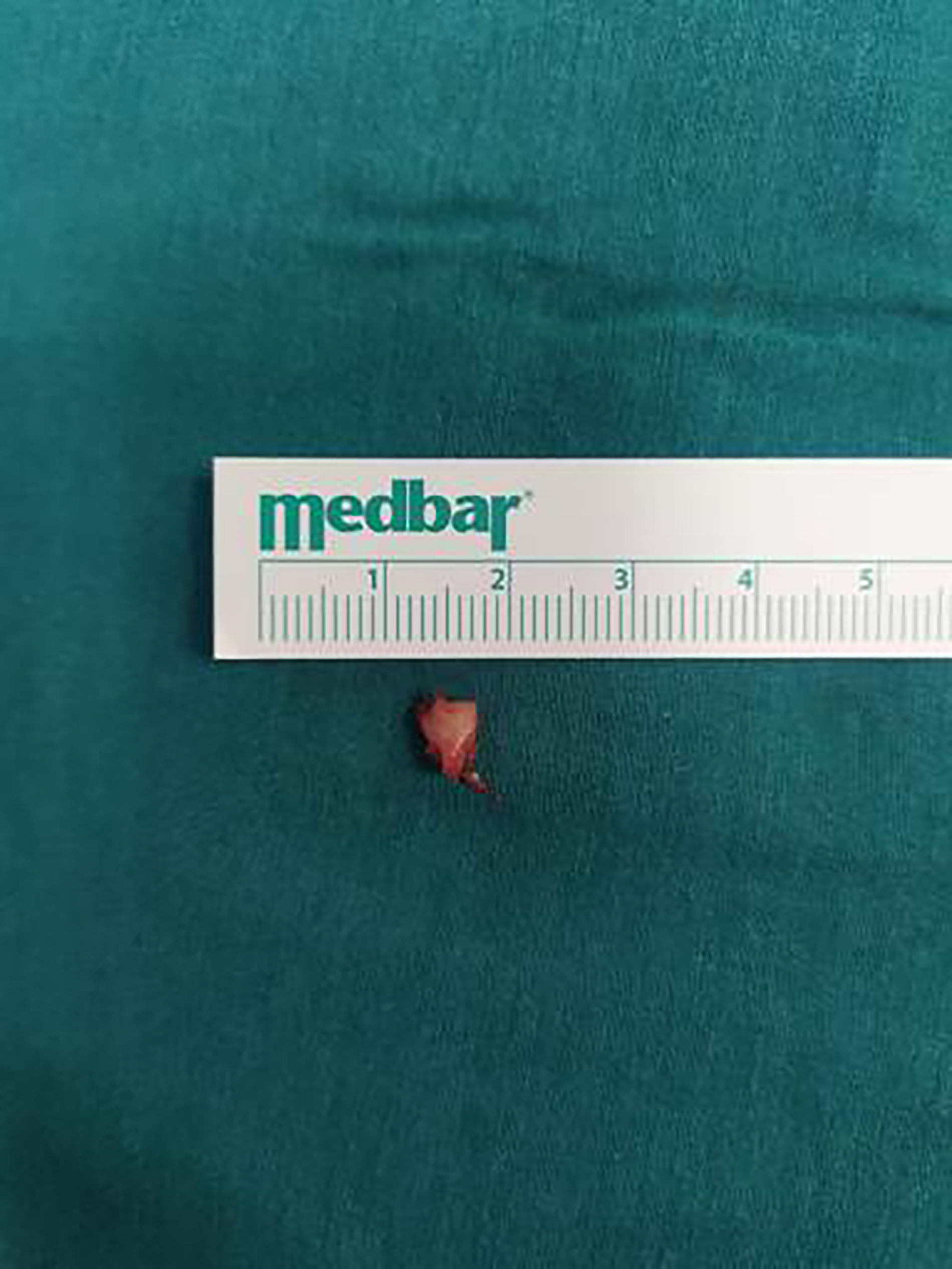
In light of the radiologic findings, surgical treatment was planned for the patient. After informed written consent was obtained from the patient, the operation was performed the day after the trauma. The suprasyndesmotic fibular fracture was reduced and fixed with a one-third tubular plate. Then, the medial clear space was approached by open exposure anterior to the medial malleolus. The deltoid ligament was noted to be ruptured and entrapped at the medial joint space and was released. A 5-mm os subtibiale attached to the ruptured deltoid ligament was seen and excised (Fig. 4). Syndesmosis examination was then performed under fluoroscopy imaging and the unstable syndesmosis was fixed with a tricortical screw placed over the plate. Finally, the deltoid ligament was repaired with sutures passed through bone tunnels on the medial malleolus.
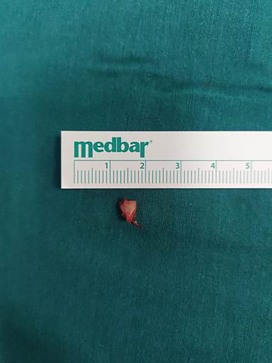
Figure 4.
Photograph of the 5-mm os subtibiale that was excised from the patient.
The patient was not allowed to bear weight on his left foot for 6 weeks, and the screw was removed 6 weeks after the operation. Painless full weightbearing and full range of motion were achieved in the third posteoperative month. The patient had no problems in the 6-month, 1-year, and 2-year follow-ups.
Discussion
This case report is the first in the literature to discuss the displacement of an os subtibiale secondary to PER ankle injury. Deltoid ligament injury in our patient caused detachment and displacement of the os subtibiale from the medial malleolus. To restore the stability of the joint, the os subtibiale was excised and the deltoid ligament was repaired with surgery.
A secondary ossification center at the tip of the medial malleolus can be seen at 7 to 10 years of age. This consolidates with the malleolus by the age of 11 to 12 years, and failure of fusion results in os subtibiale in adulthood [12,13]. The estimated prevalence of os subtibiale is reported between 0% and 4%, varying in different studies [1,13–16].
In trauma patients, os subtibiale can be confused with medial malleolus fractures. There are features to differentiate these ossicles from avulsion fractures. The edges of the ossicle are smooth with a thin cortex. But the edges of the fragment are usually irregular, and there is a cortical interruption at the fragment and the main body of the malleolus [15,17]. There are reports of os subtibiale presenting as symptomatic ossicles or ossicles that are misdiagnosed and treated as fractures. Han et al [18] reported os subtibiale as the source of pain in four of their 24 patients, claiming that the pain was due to the recurrent irritation of an ossicle on the surrounding soft tissue. Iliev et al [2] performed surgery on an 18-year-old triathlete with os subtibiale with complaints of localized pain and swelling on the medial side of the ankle. Turan et al [10] reported tibialis posterior tendon impingement caused by os subtibiale. Bandyopadhyay [19] reports os subtibiale misdiagnosed as a fragment with unsuccessful union.
Traditionally, the lateral compartment was felt to be the key for stabilization of the ankle joint. But in the past few decades, studies showed that medial column integrity (medial malleolus and deltoid ligament) is more important for ankle stability [20–22]. Pronation-external rotation injuries of the ankle joint may cause deltoid ligament rupture and widening of the medial clear space [11]. In these situations, an MRI may be helpful to recognize ligamentous injury. In the presence of os subtibiale, as so in our case, deltoid ligament rupture may cause detachment of the os subtibiale from the medial malleolus and displacement to the medial clear space.
In conclusion, both the clinician and the radiologist must take into consideration the mechanism of ligamentous injury in patients with a history of ankle trauma. In addition, the locations and features of the accessory ossicles around the ankle must be well-known. An MRI can be useful to determine the ligamentous injury and demonstrate the characteristics of the osseous fragments seen in the radiograph.
Financial Disclosure
None reported.
Conflict of Interest
None reported.
References
- Kalbouneh H, Alajoulin O, Shawaqfeh J, et al: Accessory ossicles in the region of the foot and ankle: an epidemiologic survey in a jordanian population. Medicina 57: 1178, 2021.
- Iliev AA, Georgiev GP, Landzhov BV, et al: Symptomatic os subtibiale associated with chronic pain around the medial malleolus in a young athlete. Folia Med (Plovdiv) 58: 60, 2016.
- LaMont L, Ladenhauf HN, Edobor-Osula F, et al: Secondary ossification centers in the development of the medial malleolus. J Pediatr Orthop 35: 314, 2015.
- Bellapianta JM, Andrews JR, Ostrander RV: Bilateral os subtibiale and talocalcaneal coalitions in a college soccer player: a case report. J Foot Ankle Surg 50: 462, 2011.
- Turan A, Kilicaslan OF, Kose O: Os subtibiale and secondary ossification center of medial malleolus are two different entities. Am J Emerg Med 35: 929, 2017.
- Shinohara Y, Tanaka M, Yokoi K, et al: Arthroscopic resection of symptomatic ossicle of the medial malleolus: a case report. J Foot Ankle Surg 55: 1302, 2016.
- Mellado JM, Ramos A, Salvado E, et al: Accessory ossicles and sesamoid bones of the ankle and foot: imaging findings, clinical significance, and differential diagnosis. Eur Radiol 13: 164, 2003.
- Topal M, Kose A, Dincer R, et al: Os subtibiale: mimicking medial malleolar fracture. Am J Emerg Med 35: 940, 2017.
- O’Loughlin PF, Murawski CD, Egan C, et al: Ankle instability in sports. Phys Sportsmed 37: 93, 2009.
- Turan A, Kose O, Acar B, et al: Posterior tibial tendon impingement due to os subtibiale: a case report and up-to-date review. Skeletal Radiol 46: 705, 2017.
- Lampridis V, Gougoulias N, Sakellariou A: Stability in ankle fractures: diagnosis and treatment. EFORT Open Rev 3: 294, 2018.
- Aydin D: Extra ossification center at the tip of the medial malleolus suspected as fracture: a clinical clue. J Foot Ankle Surg 55: 317, 2016.
- Kose O: The accessory ossicles of the foot and ankle; a diagnostic pitfall in emergency department in context of foot and ankle trauma. J Acad Emerg Med 11: 106, 2012.
- Coral A: The radiology of skeletal elements in the subtibial region: incidence and significance. Skeletal Radiol 16: 298, 1987.
- Keles-Celik N, Kose O, Sekerci R, et al: Accessory ossicles of the foot and ankle: disorders and a review of the literature. Cureus 9: 1881, 2017.
- Vega J, Marimón J, Golanó P, et al: True submalleolar accessory ossicles causing impingement of the ankle. Knee Surg Sports Traumatol Arthrosc 18: 254, 2010.
- Madhuri V, Poonnoose PM, Lurstep W: Accessory os subtibiale: a case report of misdiagnose fracture. Foot Ankle Online J 2: 3, 2009.
- Han SH, Choi WJ, Kim S, et al: Ossicles associated with chronic pain around the malleoli of the ankle. J Bone Joint Surg 90: 1049, 2008.
- Bandyopadhyay A: Symptomatic os subtibiale following injury: a case report of failed conservative treatment, leading to differential diagnosis made intraoperatively. J Orthop Case Rep 11: 6, 2021.
- Michelsen JD, Ahn UM, Helgemo SL: Motion of the ankle in a simulated supination-external rotation fracture model. J Bone Joint Surg Am 78: 1024, 1996.
- Gougoulias N, Sakellariou A: When is a simple fracture of the lateral malleolus not so simple? How to assess stability, which ones to fix and the role of the deltoid ligament. Bone Joint J 99-B: 851, 2017.
- DeAngelis NA, Eskander MS, French BG: Does medial tenderness predict deep deltoid ligament incompetence in supination-external rotation type ankle fractures? J Orthop Trauma 21: 244, 2007.
© 2025 American Podiatric Medical Association