


Abstract
Background: The ideal suture technique and type in tendon repair remain unclear. This biomechanical study aimed to assess the biomechanical characteristics of three techniques—modified Kessler (mKE), modified Krackow (mKR), and modified tension Bunnell (mtBU)—in sheep Achilles tendon tear repair using three suture types—polypropylene, polyester, and ultra-high-molecular-weight polyethylene (UHMWPE)—which are also compared. Methods: Sixty-three Achilles tendons harvested from sheep were transversely hacked as a replacement for rupture in a standardized measure and repaired using mKE, mKR, and mtBU techniques with No. 2 polypropylene, polyester, and UHMWPE sutures. Biomechanical parameters, such as Young’s modulus, ultimate strength, and strength to the 5-mm gap, were recorded for statistical analysis. Results: The mtBU technique with UHMWPE use resulted in increased ultimate strength, strength to 5-mm gap, Young’s modulus, and quantity of specimens with low clinical failure modes compared with the other techniques with other suture materials. Furthermore, mtBU has the lowest thickness at the repair side of the tendons. This approach showed tendon failure during maximal traction testing, whereas the mKE and mKR techniques had polyethylene and polyester suture failures. Conclusions: The UHMWPE suture was significantly superior to the other sutures in each technique in terms of strength and durability. The mtBU technique using UHMWPE suture showed better biomechanical results, implying that this repair might be more appropriate to obtain early mobilization after tendon ruptures.
The Achilles tendon is the thickest and strongest tendon in the human body and is one of the most frequently ruptured [1]. Although surgical tendon repair is often indicated, the ideal suture technique remains controversial [2,3]. The modified Krackow (mKR), modified tension Bunnell (mtBU), and modified Kessler (mKE) techniques are well-known tissue-grabbing sutures used in open and percutaneous Achilles tendon repairs, and their usefulness has been proved in various biomechanical and clinical studies. The resistance to gap development is critical when using these sutures for tendon and ligament restoration. There will be an increase in granulation tissue, adhesions, and delayed collagen maturation if there is a gap in the healing.
The larger the gap, the more delayed the healing; the result is a weaker, more attenuated repair. Of these current techniques, the one that minimizes gap formation is the mtBU. Although the highest tensile strength is achieved with the mKR suture technique, the bulkiest tendon reconstruction ends will also occur with it. On the contrary, the mKe suture technique has the least bulkiness at the tendon reconstruction ends among the others [4–6].
To achieve favorable clinical results in tendon repairs, the qualities of suture materials are just as crucial as the repair techniques themselves; nevertheless, several types of sutures have been modified to meet varied requirements in clinical situations. An optimal tendon repair suture should have sufficient tensile strength throughout the critical healing phase, good knot security and handling properties, a low and tolerable foreign body reaction, and better tissue regeneration [7].
Suture in the No. 2/0 to No. 5 range is advised for Achilles tendon repair, depending on the repair technique [8,9], with braided nonabsorbable polyester and monofilament absorbable polyglactin being two of the most widely used suture materials in Achilles tendon repair [10].
Ultra-high-molecular-weight polyethylene (UHMWPE) suture is a nonabsorbable braided surgical suture prepared from fibers of UHMWPE [11]. Moreover, UHMWPE is a subset of thermoplastic polyethylene and has extremely long chains. They are strong, and their softness and chemical inertness minimize tissue inflammation and irritation in applications [12].
Reconstructions of Achilles, patellar, and quadriceps tendon ruptures often cause serious difficulties for surgeons because they also affect the flexion mechanisms of the lower extremity. Although surgical tendon repair is often indicated, the ideal suture technique and type remain unclear. Suture pullout and/or rupture, gap development, and knot failure were all clinical failure types in tendon repair surgeries including suture materials, and they were all related to the mechanical characteristics of the sutures compressively [13]. Moreover, the functional outcomes of tendon repair surgery are strongly influenced by the surgical suturation technique. Hence, it is essential to investigate the best combination of suture technique and type in a biomechanical aspect.
Using three distinct suture materials—polypropylene, polyester, and UHMWPE—this study aimed to analyze and compare the efficacy of the mtBU, mKe, and mKR methods in terms of their biomechanical characteristics in a sheep Achilles tendon repair model.
Methods
Study Design
Sixty-three male sheep Achilles tendons were harvested from sheep butchered for commercial purposes at a local slaughterhouse. At the time of slaughter, the sheep were between 1 and 1.5 years old and weighed between 60 and 72 kg. A muscle-tendon junction proximally and a portion of the bony calcaneus, including the enthesis area distally, were found in each specimen. They were immediately frozen to a temperature of 24°C after gathering until the experiments were completed. Each specimen was gradually frozen and kept hydrated at room temperature (∼18°C) for 12 hours before the surgical intervention. After biomechanical testing, all of the specimens were disposed of according to the Republic of Turkey’s Ministry of Environment and Urbanization biological waste disposal guidelines.
A comparative study with block randomization was conducted. The tendon specimens (N = 63) were first allocated numbers at random. Tendons were then randomly assigned to one of three surgical technique groups (mKR, mKE, and mtBU) with three subgroups under each group (polypropylene, polyester, and UHMWPE sutures). Each subgroup consisted of seven specimens. Three surgeons (F.D., K.G., B.K.) with similar training and clinical expertise used all three surgical procedures to execute timed tendons repairs. The first surgeon used the mKE technique with three different types of suture material, the second one used the mtBU technique with three different types of suture material, and the third one used the mKR technique with three different types of suture materials.
Specimen Demographics and Preparation
One senior research assistant of veterinary anatomy, DVM and a specialist experienced at slaughterhouse veterinary, DVM inspected the specimens taken from the sheep via macroscopic evaluation to ensure that there were no tendon pathologies just after they were slaughtered and before being freshly frozen. The length from the calcaneal enthesis to the musculotendinous junction of the tendon and the distal and proximal distances of the narrowest part of the tendon that was planned to divide sharply into two in the transverse plane replicating rupture with a No. 11 scalpel blade were measured with a digital caliper and marked in each specimen. The suture needle entry and exit sites, based on the surgical technique to which the sample was allocated, were marked. Then, surgical repair was performed as shown in Figure 1 A-C.
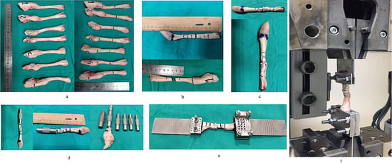
Figure 1.
Preparation and measurements of the specimens (A–D) and views of biomechanical testing apparatuses (E and F). A, Measurements of width, height, and length. B, Marking the needle entrance point at 1-cm intervals, and determination of incision localization at the midpoint of each specimen’s length, respectively. C, View of a sample specimen after suturation. D, Measurement of each specimen regarding the width, height, length, and bulkiness of the repair area after the suturation techniques were applied. E, View of a sample specimen applied to two custom-made clamps; one is fixed from the bony part with Kirschner wires and the other is fixed via bolt nuts. F, View of the sample specimen fixed to the biomechanical testing device.
Each specimen's repair bulkiness volume was determined by calculating the native tendon diameter and volume, then passing the native tendon through an anterior cruciate ligament graft sizing guide block and repairing it. The block's dimensions are preset in 0.5-mm increments; the best size was selected based on the repair bulkiness. Owing to the wide variation in tendon diameters and morphologies between samples, these values are represented as ratios. To compare tendon cross-sectional area (CSA) for three types of suture techniques, calculation was performed by dividing the repaired site’s CSA by the presutured adjacent proximal and distal parts of one of each specimen. This was performed to standardly quantify which tendon suturation was bulkiest (Figure 1D). The CSA, in square millimeters, of each tendon was calculated using the following formula:
× π, where the width (W) and height (H), both in millimeters, were obtained from measurements by an electronic gauge (CH-10-AT; Dasqua, Chengdu, Sichuan, People’s Republic of China) before and after suturing.
Surgical Techniques
All of the surgical operations were performed by three orthopedic foot and ankle surgeons (F.D., B.K., Y.K.). The mKE technique is a gripping suture technique in which a single loop of suture encircles the tendon. The suture is placed 1 cm proximal and distal to the rupture on both sides. To produce an end-to-end repair, the sutures are tightened and tied in a series of double and five knots. Three locking loops are put 1 cm from either side of the rupture in the mKR procedure. To achieve end-to-end repair, the loops are tightened and knotted with one double and five knots in a row. Interlacing sutures are used in the mtBU technique. A zigzag stitch is used to repair both sides of the torn tendons. Three loops are put 1 cm on either side of the rupture. To achieve end-to-end repair, the loops are tightened and knotted with one double and five knots in a row. To allow for examination of the biomechanical properties of two-strand core sutures and the bulk of the repair side of the tendon, no epitendinous suture augmentation was performed in any specimen (Figure 2).
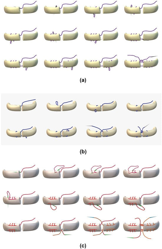
Figure 2.
The three suturation techniques used in the study. A, Modified tension Bunnell technique. B, Modified Kessler technique. C, Modified Krackow technique.
The modified versions of the original suture techniques used in this research (mKE, mKR, and mtBU), which had two strands crossing with knots located between the ends of the tendon repair site for mKE and mtBU and four strands crossing with knots located between the ends of the tendon repair site for mKR, are illustrated in Figure 2.
Mechanical Testing
All of the tests were performed in a controlled environment (mean ± SD temperature, 20°C ± 1°C; relative humidity, 50%). A universal testing apparatus (Z100; ZwickRoell GmbH, Ulm, Germany) with testXpert II, Version 3.2 software was used to conduct biomechanical testing. Two custom-made tendon clamps were used to mount specimens on the testing system, which were primed with a force of 10 N before the test and subsequently loaded to failure at a displacement rate of 20 mm/min (Figure 1 E and F). Ultimate strength and strength to a 5-mm gap were determined using stress-strain curves. The testing device software was used to calculate the Young's modulus (YM) values. After completion of the biomechanical tests, the fault location and mode in each sample were macroscopically identified and recorded.
During biomechanical testing, the maximum load delivered before a steep fall leading to failure is referred to as the ultimate strength [14]. The magnitude of displacement was ignored in these measurements. The quantity of load required to establish a 5-mm gap at the repair site is known as the strength to 5-mm gap. This is a critical criterion for attaining tendon regeneration close to its original length and determining the top limit for tolerable tendon elongation [15]. The measure of elastic deformation of materials under load is YM, which is defined by the slope of the stress-strain curve [16].
Statistics
All of the measurements of all of the specimens, including the tendon dimension measurement and measurement of tendon repaired bulk, were repeated twice at an interval of 1 hour by two orthopedic specialists (F.D., K.G.) with 5 years of professional experience, and intraobserver and interobserver reliabilities were evaluated. A two-way mixed effects model was used to evaluate the agreement and differences between intraobserver and interobserver measurements.
Statistical analysis to investigate differences between the groups was performed using a statistical software program (IBM SPSS Statistics for Windows, Version 21.0; IBM Corp, Armonk, New York). The Shapiro-Wilk test showed normally distributed data; homogeneity was verified by analysis with the Levene test. One-way analysis of variance, Tukey test, and t test were performed for multiple and independent group comparisons and pairwise comparisons, respectively. A P < .05 was considered statistically significant.
Results
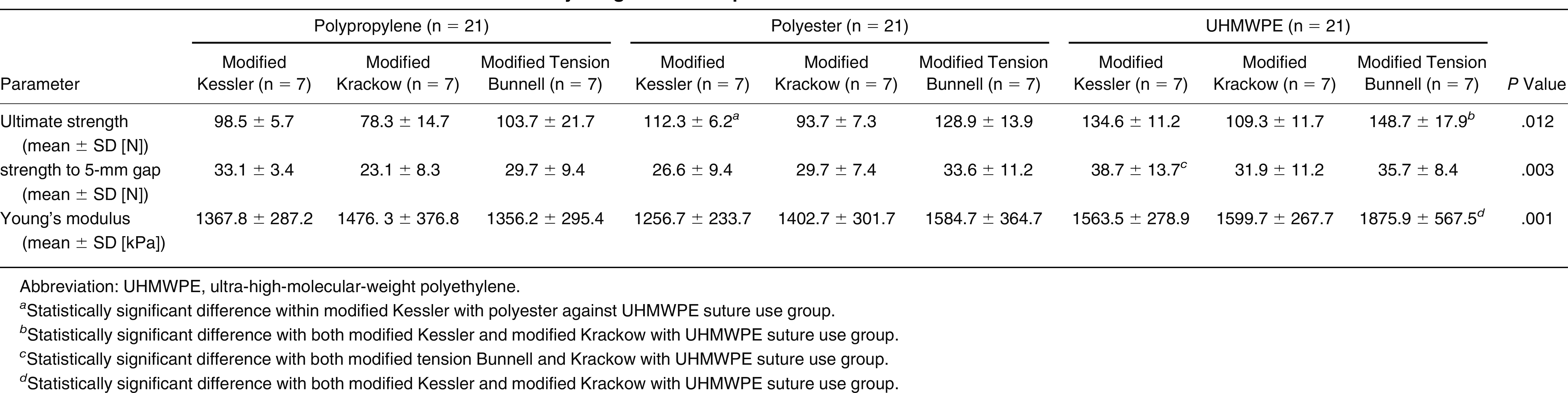
In terms of mean specimen dimensions (length, width, and thickness) and biomechanical parameters (ultimate strength, strength to 5-mm gap, and YM), the three groups with three subgroups were compared (Table 1). Table 2 shows the distribution of the failure mechanisms in the groups, and Table 3 compares the three repair strategies and three suture materials.

Table 1.
Dimensions and Biomechanical Parameters of the Specimens by Suturation Technique Group
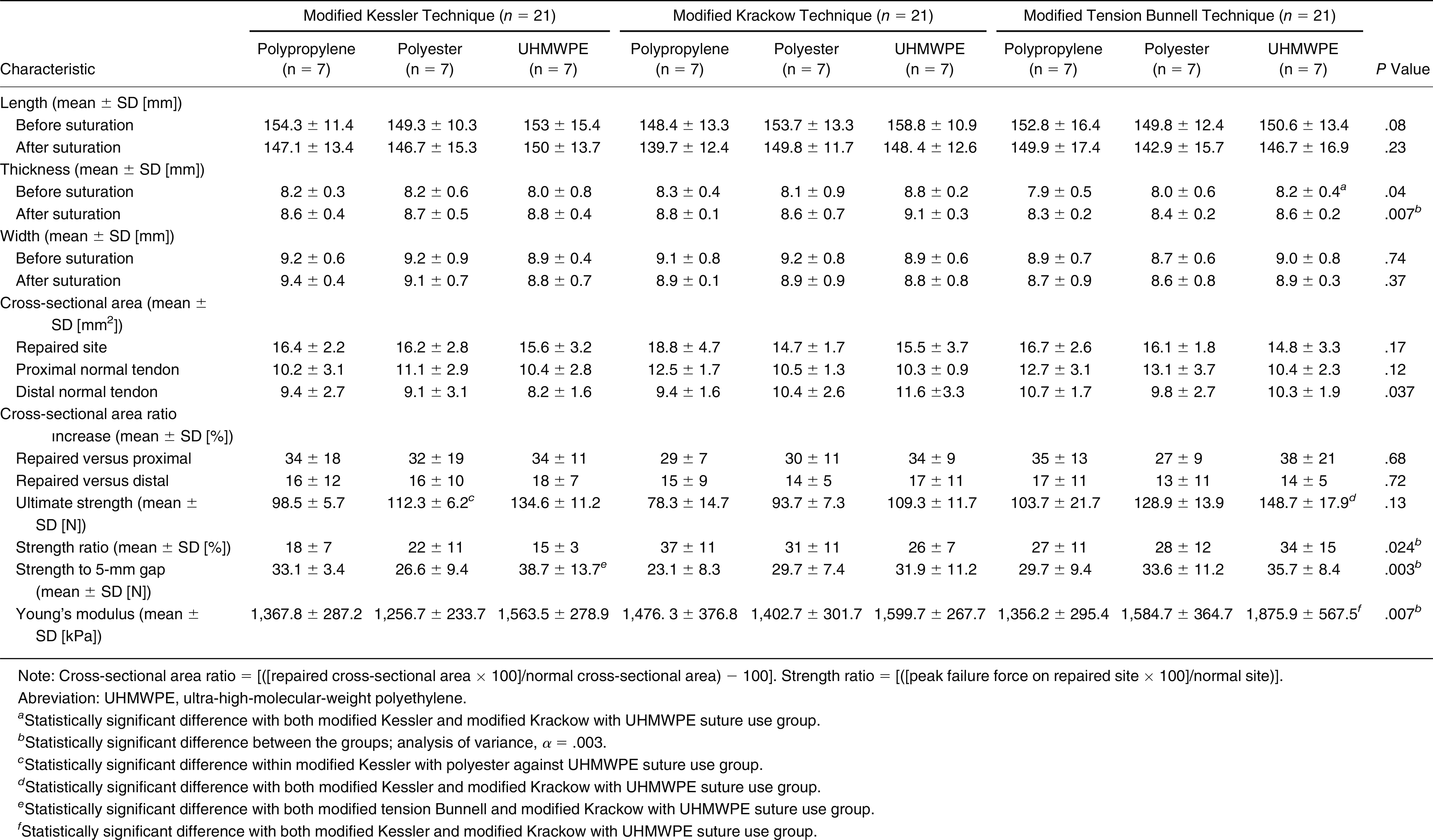
Table 2.
Failure Mode Distribution of Suture Materials in Each Suturation Technique Group

Table 3.
Biomechanical Parameters of the Suture Materials by Surgical Technique
A two-way mixed effects model was used to evaluate the agreement and differences between intraobserver and interobserver measurements. They were evaluated with the intraclass correlation coefficient (ICC). The intraobserver and interobserver reliabilities were determined to be excellent (ICC, 0.905–0.976) and good (ICC, 0.889–0.991), respectively, in all of the steps of specimen preparation before biomechanical testing.
Ultimate Strength
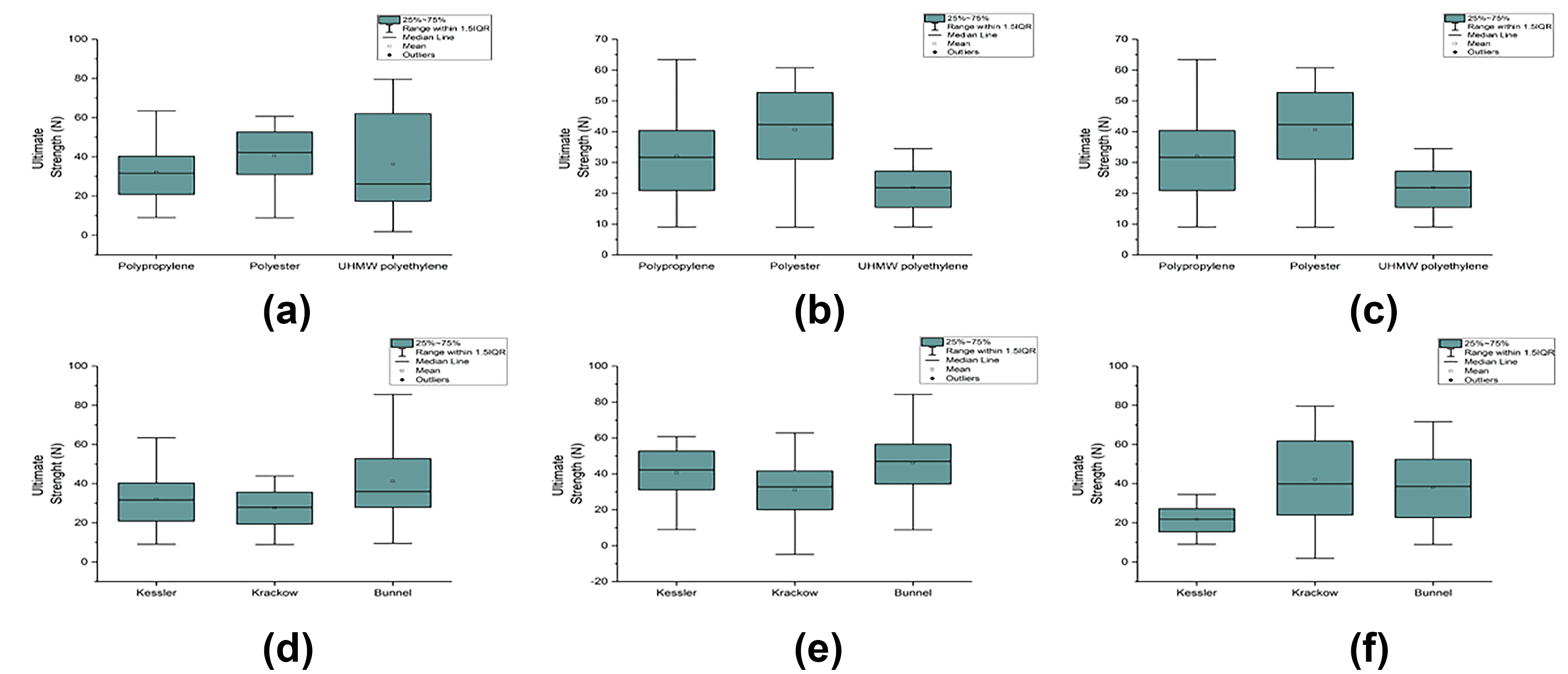
The mtBU-UHMWPE group had the highest mean ultimate strength value, although it was not significant (P = .03) (Table 1). For both the polypropylene (P = .15) and polyester (P = .95) sutures, the differences between the repair groups were negligible. In each suture technique, the UHMWPE suture was significantly superior to the other sutures (mKE: P = .024, mKR: P = .044, and mtBU: P = .047), and the polyester suture was superior to the polypropylene suture without significance in the mKR (P = .33) and mKE (P = .29) techniques; however, the polyester suture was significantly superior to the polypropylene suture in the mtBU technique (P = .017) (Figure 3)
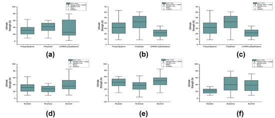
Figure 3.
Comparison of each suturation technique in terms of the ultimate strength of each suture material (A–C) and comparison of each suture material in terms of the ultimate strength of each suturation technique (D–F). A, Modified Kessler suturation technique versus each suture material. B, Modified Krackow suturation technique versus each suture material. C, Modified tension Bunnell suturation technique versus each suture material. D, Polypropylene suture material versus each suturation technique. E, Polyester suture material versus each suturation technique. F, Ultra-high-molecular-weight polyethylene (UHMWPE) suture material versus each suturation technique.
Strength to 5-mm Gap
The mKE-UHMWPE group had the highest mean strength to 5-mm gap value (P = .003) (Table 1). The suture technique groups were ordered in descending order of strength to 5-mm gap mean values in UHMWPE suture repairs, and the difference between the suture method groups was significant (P = .022). The difference between groups in polypropylene sutures was not significant (P = .32). The UHMWPE suture was considerably superior to other sutures in the suture technique groups in terms of strength to 5-mm gap mean values (P = .001).
Young's Modulus
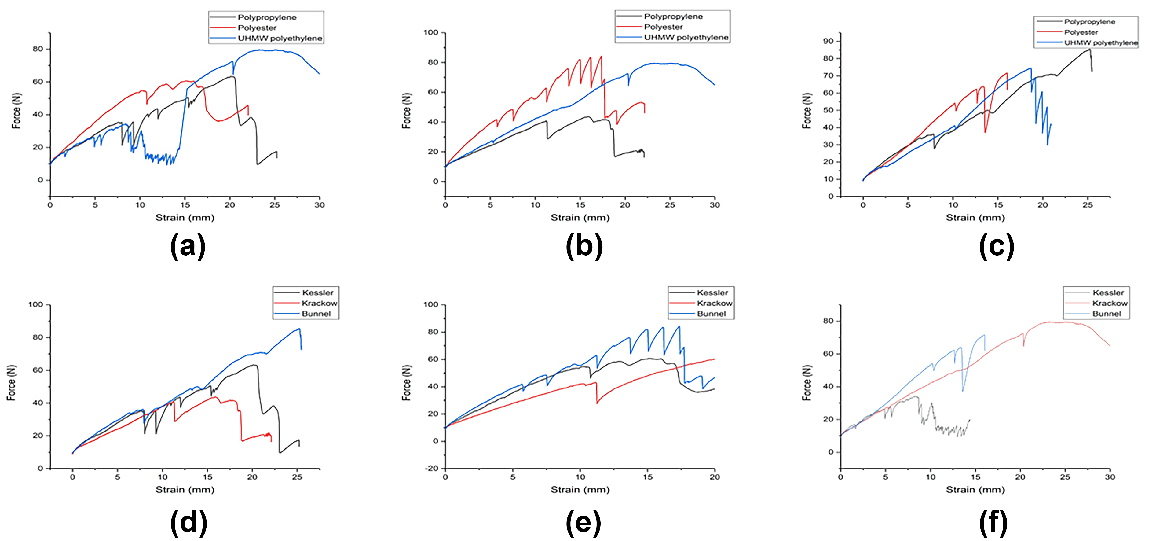
The mean YM value for the mtBU-UHMWPE group was the highest (P = .007). The groups were sorted in descending order of YM mean values as mKR, mKE, and mtBU procedures in polypropylene repairs, and a nonsignificant difference (P = .38) was found. The difference between groups in polyester repairs was also not significant (P = .33). In mKE repairs, the polyester suture outperformed the polypropylene suture by a significant margin (P = .023) (Figure 4).
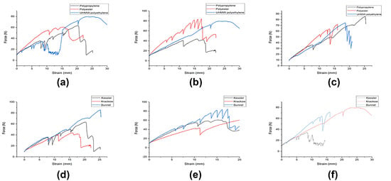
Figure 4.
Force-strain graphs of each suturation technique versus suture materials (A–C) and of suture materials versus each suturation technique (D–F). A, Modified Kessler technique versus each suture material. B, Modified Krackow technique versus each suture material. C, Modified tension Bunnell suturation technique versus each suture material. D, Polypropylene suture material versus each suturation technique. E, Polyester suture material versus each suturation technique. F, Ultra-high-molecular-weight polyethylene (UHMWPE) suture material versus each suturation technique.
Discussion
For almost all of the variables related to mechanical and dimensional properties, UHMWPE also has favorable results for the mtBU technique. The mtBU technique using UHMWPE seems to be a good alternative surgical solution for Achilles tendon repair biomechanically.
The ideal tendon repair should be biomechanically reliable to enable early range-of-motion exercises, be easily performed, minimize injury to vascular supply of the tendon, and not be bulky to provide smooth gliding of the tendon [17,18]. Many different suture techniques have been developed to minimize this bulkiness. Among these, the Kessler, Bunnell, and Krackow techniques are the most frequently used [19,20]. In this study, when three different suture materials—polypropylene, polyester, and UHMWPE—were used in each of these techniques, the lowest bulkiness was obtained with the mtBU surgical technique, UHMWPE suture material, and the mtBU technique with UHMWPE.
It is critical that a tendon repair suture not fail prematurely before reaching strength values close to the suture material’s simple straight-pull test yield values. Suture failure by pullout is an example of premature failure because the suture does not break, indicating that the force required to fail is less than the suture material’s yield strength [18]. Suture breakage adjacent to the knots is another example of premature failure because knots create a weak point in the suture [21]. Of all of the techniques and suture materials evaluated in this biomechanical study, UHMWPE suture material was observed with pullout from the proximal mode of failure in only two of 21 specimens in all of the applied techniques. This biomechanical finding has led to the assumption that it may reduce the rate of rerupture observed, especially after Achilles tendon open surgery, because in a prospective randomized study by Zhou et al [22], the rupture rate was calculated to be 1.4% (range, 0%–7.1%) after surgical treatment (0.7% after simple end-to-end repair) and 13.4% (range, 3.9%–50%) after conservative treatment. Zhang et al [23] found rupture rates of 2.8% in patients who underwent surgical treatment and 11.7% in patients who were followed conservatively in their study.
Another hugely influential biomechanical study, by McCoy and Haddad [24], found that there was no significant difference in strength among the Krackow, Bunnell, and Kessler suture techniques when performed with a double suture weave in a laboratory model of cadaveric Achilles tendon repairs. This body of research, which appears to be unfavorable, in essence lends support to the current study. In the present study, when the ultimate strength parameter was examined, a statistically significant difference was found among the mKR, mtBU, and mKE suture techniques. The reason for using modified techniques in the present study was based on the study by McCoy and Haddad [24].
The recent advancement of UHMWPE suture material, as well as its use in meniscal and rotator cuff repair techniques, is notable. Sutures and suture-based repair devices used in meniscal and rotator cuff repair in the past were more likely to break during tensioning and knot tying [25,26].
One of the main goals of treatment in Achilles tendon ruptures is to enable the patient to return to work as soon as possible. Another goal that applies to many patients is to return to the sports level they had before the injury. Providing virtually entire range of motion in the ankle joint can only be accomplished with early rehabilitation and weightbearing. This is the most serious complication that develops in patients during the early postoperative period, and it is also the factor that causes the most morbidity. The key to early rehabilitation is surgery performed with biomechanically reliable, robust suture material. Postoperative treatment after classic Achilles tendon surgical repair consists of immobilization in a cast for 6 weeks without weight. During this period, ankle joint range of motion is restricted, and this increases the time for the patient to return to work and return to the sports level they had before the injury. Costa et al [27] compared the immobilization protocol with a 6-week cast with a carbon-fiber ankle orthosis (1.5-cm heel support) and early weight group in a randomized prospective study of 48 patients. In the early weight group, they found shortened normal walking and stair climbing times. Tendon rupture recurred as a result of falling and not restricting activity in two patients in the early weight group. The authors emphasized the importance of patient selection, being careful in the selection of patients for whom the early weightbearing protocol will be applied and recommend weightbearing [27]. In light of the biomechanical findings obtained in the present study, it is important to conduct in vivo studies on the use of UHMWPE in Achilles tendon rupture surgery, although it is also dependent on the surgical technique.
The mtBU suture technique with UHMWPE suture use has possibly proved to be a reliable choice for tendon repairs, but as research is progressing, a new focus should be given to research on the histopathologically evaluated tendon repairs in an in vivo setting.
A limitation of this study is that the data are only an indication of the performance of an appliance and cannot be linked to clinical improvements. The amount of reparable strength and the long-term effects of materials used in these devices have still not been determined. Any of these repair instruments could be clinically suitable, and ultimately, the choice of the surgeon should be based on clinical results.
Conclusions
The UHMWPE suture was significantly superior to the other sutures in terms of durability and strength. Moreover, this study, which was conducted to evaluate and compare the biomechanical features of the mKE, mKR, and mtBU suture techniques, conceptualizes the mtBU suture technique with the use of UHMWPE to be the most effective technique in biomechanical terms.
Acknowledgment: The USKIM Center at Kahramanmaras Sutcu Imam University for its technical support in biomechanical tests; Mr Onur Öztürk for his tireless assistance with taking photographs and transportation; and Mr Bünyamin Vırıt for providing the sheep Achilles tendons butchered by his local slaughterhouse, Akerman Butcher, for commercial purposes.
Financial Disclosure: None reported.
Conflict of Interest: None reported.
References
- Wong J, Barrass V, Maffulli N: Quantitative review of operative and nonoperative management of Achilles tendon ruptures. Am J Sports Med 30: 565, 2002.
- Del Buono A, Volpin A, Maffulli N: Minimally invasive versus open surgery for acute Achilles’ tendon rupture: a systematic review. Br Med Bull 109: 45, 2014.
- Ortiz C, Wagner E, Mocoçain P, et al: Biomechanical comparison of four methods of repair of the Achilles tendon: a laboratory study with bovine tendons. J Bone Joint Surg Br 94: 663, 2012.
- Jordan MC, Hölscher-Doht S, Jakubietz MG, et al: Suture material for flexor tendon repair: 3-0 V-Loc versus 3-0 Stratafix in a biomechanical comparison ex vivo. J Orthop Surg Res 9: 72, 2014.
- Frosch S, Buchhorn G, Hawellek T, et al: Comparison of the double loop knot stitch and Kessler stitch for Achilles’ tendon repair: a biomechanical cadaver study. PLoS One 15: e0243306, 2020.
- Nguyen TP, Keyt LK, Herfat S, et al: Biomechanical study of a multifilament stainless steel cable crimp system versus a multistrand ultra-high molecular weight polyethylene polyester suture Krackow technique for Achilles’ tendon rupture repair. J Foot Ankle Surg 59: 86, 2020.
- Dennis C, Sethu S, Nayak S, et al: Suture materials: current and emerging trends. J Biomed Mater Res 104: 1544, 2016.
- Reda Y, Farouk A, Abdelmonem I, et al: Surgical versus non-surgical treatment for acute Achilles’ tendon rupture. a systematic review of literature and meta-analysis. Foot Ankle Surg 26: 280, 2020.
- Ozan F, Dogar F, Gurbuz K, et al: Chronic Achilles tendon rupture reconstruction using the Lindholm method and the Vulpius method. J Clin Med Res 9: 573, 2017.
- Seow D, Yasui Y, Calder JD, et al: Treatment of acute Achilles’ tendon ruptures: a systematic review and meta-analysis of complication rates with best- and worst-case analyses for rerupture rates. Am J Sports Med 49: 3728, 2021.
- Zhang Q, Li C, Luan J, et al: Polycaprolactone/ultra-high molecular weight polyethylene partially absorbable suture with improved mechanical performances for tendon and ligament repair. Text Res J 90: 2123, 2020.
- Teng Y, Da L, Zhang X, et al: Whether sutures reduce the graft laceration caused by interference screw in anterior cruciate ligament reconstruction a biomechanical study in vitro. PLoS One 2021.
- Ergün S, Akgün U, Barber FA, et al: The clinical and biomechanical performance of all-suture anchors: a systematic review. Sports Med Arthrosc Rehabil 2: e263, 2020.
- Tian J, Rui Y, Xu Y, et al: A biomechanical comparison of Achilles tendon suture repair techniques: locking block modified Krackow, Kessler, and percutaneous Achilles repair system with the early rehabilitation program in vitro bovine model. Arch Orthop Trauma Surg 140: 1775, 2020.
- Shepard ME, Lindsey DP, Chou LB: Biomechanical testing of epitenon suture strength in Achilles’ tendon repairs. Foot Ankle Int 28: 1074, 2007.
- Askeland DR, Fulay PP, Wright WJ: The Science and Engineering of Materials, 6th Ed, p 196, Cengage Learning, Stamford, CT, 2010.
- Wu YF, Tang JB: Recent developments in flexor tendon repair techniques and factors influencing strength of the tendon repair. J Hand Surg Eur Vol 39: 6, 2014.
- Müller SA, Todorov A, Heisterbach PE, et al: Tendon healing: an overview of physiology, biology, and pathology of tendon healing and systematic review of state of the art in tendon bioengineering. Knee Surg Sports Traumatol Arthrosc 23: 2097, 2015.
- Joyce CW, Sugrue C, Chan JC, et al: A barbed suture repair for flexor tendons: a novel technique with no exposed barbs. Plast Reconstr Surg 2: e237, 2014.
- Clemente A, Bergamin F, Surace C, et al: Barbed suture vs conventional tenorrhaphy: biomechanical analysis in an animal model. J Orthop Traumatol 16: 251, 2015.
- Shah A, Rowlands M, Au A: Barbed sutures and tendon repair: a review. Hand 10: 6, 2015.
- Zhou K, Song L, Zhang P, et al: Surgical versus non-surgical methods for acute Achilles tendon rupture: a meta-analysis of randomized controlled trials. J Foot Ankle Surg 57: 1191, 2018.
- Zhang YJ, Zhang C, Wang Q, et al: Augmented versus nonaugmented repair of acute Achilles tendon rupture: a systematic review and meta-analysis. Am J Sports Med 46: 1767, 2018.
- McCoy BW, Haddad SL: The strength of Achilles tendon repair: a comparison of three suture techniques in human cadaver tendons. Foot Ankle Int 31: 701, 2010.
- Barber FA, Howard MS, Ashraf W, et al: The biomechanical performance of the latest all-inside meniscal repair devices. Arthroscopy 36: 3001, 2020.
- Trofa DP, Ruder JA, Yeatts NC, et al: Cyclic and load-to-failure properties of all-suture anchors in human cadaveric shoulder greater tuberosities. Arthroscopy 36: 2805, 2020.
- Costa ML, MacMillan K, Halliday D, et al: Randomised controlled trials of immediate weight-bearing mobilization for rupture of the tendo Achillis. J Bone Joint Surg Br 88: 69, 2006.
© 2024 American Podiatric Medical Association