


Abstract
Background: The Sanders classification is a widely used method for classifying calcaneal fractures. Type IV fractures (>4 fragments) are known to vary in the number of fracture fragments. However, all relevant cases are classified as type IV irrespective of the number of fragments. We investigated the need for evaluation of postoperative prognoses based on radiologic factors and subtypes of Sanders classification type IV fractures.Methods: Fifty-six Sanders type IV calcaneal fractures were enrolled between 2010 and 2018. Patients were divided into two groups according to the number of fragments: four fragments (group 1) and more than four fragments (group 2). Radiologic evaluation was performed using a postoperative recovery percentage calculated from postoperative reduction of the Böhler angle, Gissane angle, and vertical height. Radiologic evaluation was divided into two groups according to postoperative recovery: good and bad recovery groups. American Orthopaedic Foot and Ankle Society (AOFAS) ankle-hindfoot score and visual analog scale (VAS) score were used for clinical outcome evaluation. Results: There was no significant difference in AOFAS ankle-hindfoot score (P = .909) or VAS score (P = .963) between groups 1 and 2. However, there was a significant (P = .001) difference in AOFAS ankle-hindfoot score or VAS score between good and bad recovery groups. Conclusions: Clinical results of Sanders type IV fractures were not related to the number of bone fragments but to the degree of injury to the Böhler angle, Gissane angle, and vertical height. Therefore, subtype classification of type IV calcaneal fractures is superfluous, and it is important to try to restore these parameters during surgery.
Calcaneal fracture is one of the most common foot fractures, accounting for a large proportion of lower-extremity fractures. Calcaneal fractures are the most common of tarsal bone fractures and currently account for approximately 2% of all fractures [1]. Multiple reports of calcaneal fractures reflected the poor functional outcome historically associated with these injuries. Böhler, who provided an in-depth analysis of the pathoanatomical features of calcaneal fractures that is still relevant today, changed his treatment regimen no less than 12 times across 30 years [2,3]. These calcaneal fractures can lead to complications such as arthritis of the subtalar joint, malunion, and so on, causing functional disability. Therefore, proper classification is important to define the treatment method and prognosis.
The currently known calcaneal fracture classification is the Essex-Lopresti classification first presented in 1952 [4]. It is divided into joint depression type and tongue type according to the mechanism of injury. Different fixing methods are needed for each type. After the development of computed tomography (CT), the Sanders calcaneal fracture classification, based on the articular surface of the CT coronal plane, was proposed. It is now commonly used [5]. However, the Sanders classification is based only on the number of bone fragments and the location of the fracture of the joint surface without reflecting the shape or severity of the fracture. Type IV fractures with more than four fragments in joints are not divided into more subgroups. If the number of fragments increases, anatomical reduction is often more difficult, and the prognosis after surgery is expected to vary [6]. However, reports on this are limited.
Therefore, the objective of this study was to investigate the need for the evaluation of postoperative prognoses based on radiologic factors and subtypes of Sanders classification type IV fractures and retrospectively analyze preoperative and postoperative radiographs as well as preoperative CT images of Sanders type IV calcaneal fractures of patients. Clinical outcomes were also analyzed according to the number of bone fragments. In addition, this study analyzed whether the Böhler angle, Gissane angle, and vertical height were correlated with clinical outcomes.
Materials and Methods
Study Design and Ethical Considerations
An independent reviewer performed a retrospective medical record review of relevant clinical and radiographic data. This study was performed after obtaining approval from the institutional review board of Jeonbuk National University Research Council (Jeonju, Korea).
Participants
Of 189 patients who underwent surgical treatment of calcaneal fractures from June 1, 2010, to May 31, 2018, 47 (39 males and 8 females) were classified as having Sanders calcaneal fracture type IV and were followed up for more than 2 year; 56 cases (right: 22; left: 16; both: 9) were retrospectively studied. All of the patients provided informed consent. All of the surgeries were performed by one surgeon (K.-B.L.). Surgeries were performed using either the sinus tarsi approach (n = 5) or the extensile lateral approach (n = 51). The mean age of patients was 43.1 years (range, 19–66 years). Mean follow-up was 29.5 months (range, 24–42 months).
Sample Size
Fifty-six cases (47 patients) were divided into two groups according to the number of fracture fragments by CT: group 1, four fragments (n = 30); and group 2, five or more fragments (n = 26). The mean patient age was 42.8 years (range, 22–66 years) for group 1 and 42.2 years (range, 19–51 years) for group 2. The male-to-female ratio was 5:1 in group 1 and 23:3 in group 2.
Radiologic Evaluation
Radiologic evaluation was performed by comparing lateral plain radiographs of the fractured calcaneus with those of the uninjured side. A postoperative recovery percentage for radiologic evaluation was calculated from the postoperative reduction of the Böhler angle, Gissane angle, and vertical height. It was divided into two group: good and bad recovery. We arranged the total patient values for angle and height and then divided the top 50% and bottom 50% into good and bad recovery groups, respectively. Results were compared with clinical outcomes at more than one year after treatment.
Clinical Outcome Measures
All of the patients were assessed for American Orthopaedic Foot and Ankle Society (AOFAS) ankle-hindfoot score and visual analog scale (VAS) score. Data collection and measurement were performed by two orthopedic surgeons (D.-G.S. and K.-B.L.). Clinical outcomes at more than 2-year follow-up after treatment were compared. It was completed at each patient's final follow-up.
Statistical Analysis
Data are expressed as mean ± SD. Significance of differences between groups was determined using independent-samples t tests. All of the statistical analyses were performed with IBM SPSS Statistics for Windows, Version 20.0 (IBM Corp, Armonk, New York). Statistical significance was defined at P < .05.
Results
Clinical Outcomes According to the Number of Fracture Fragments
For 56 cases, the mean AOFAS ankle-hindfoot score was 66.9 (range, 35–90), and the mean VAS score was 3.3 (range, 0–9). The mean ± SD AOFAS ankle-hindfoot and VAS scores were 67.2 ± 14.9 and 3.3 ± 1.9 for group 1 (four fragments [30 cases]) and 66.7 ± 15.7 and 3.3 ± 2.2 for group 2 (>4 fragments [26 cases]), respectively. There was no significant difference in AOFAS ankle-hindfoot score (P = .909) or VAS score (P = .963) between the two groups.
Clinical Outcomes According to Postoperative Radiologic Recovery
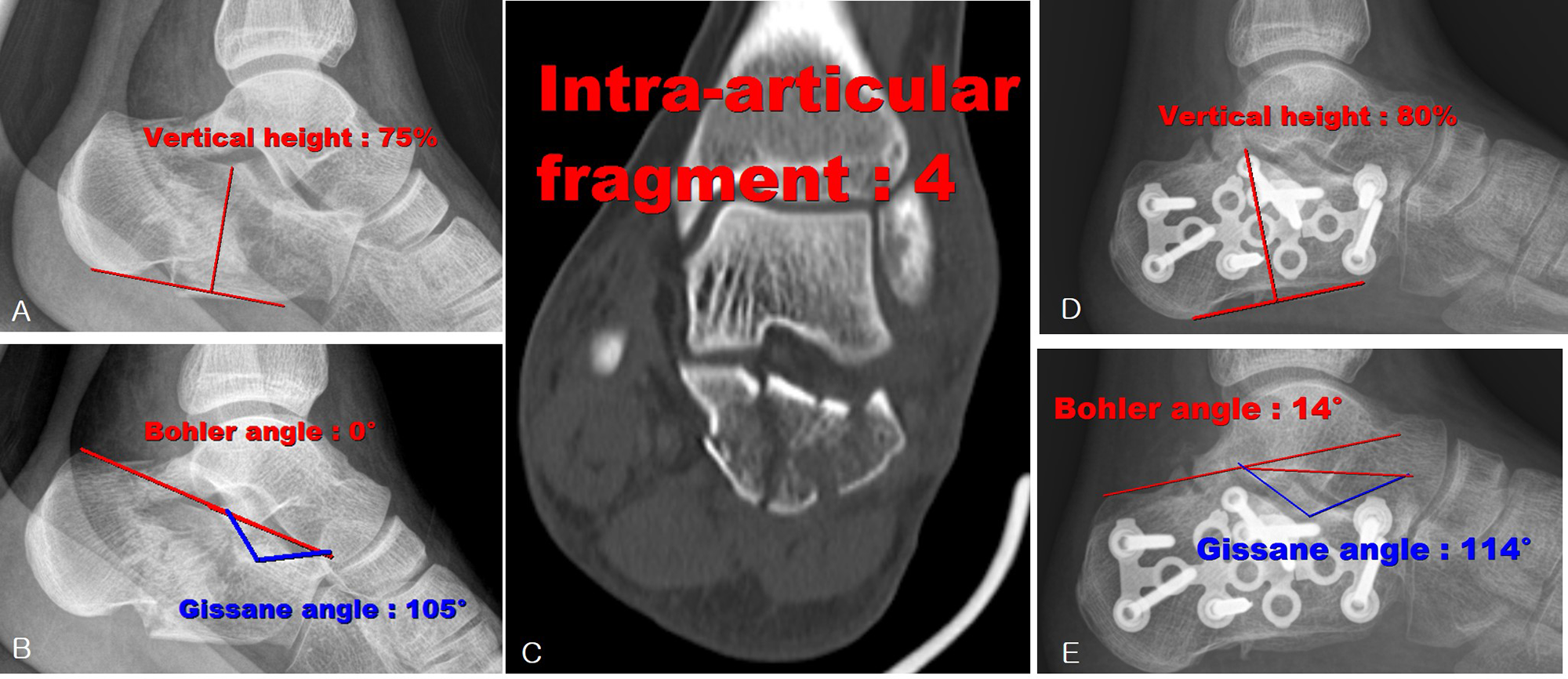
The Böhler angle, Gissane angle, and vertical height of the injured side were compared with those of the uninjured side, and the results were classified into good recovery (Fig. 1) and bad recovery (Fig. 2) groups. For the Böhler angle, mean ± SD AOFAS ankle-hindfoot and VAS scores were 56.1 ± 12.8 and 4.6 ± 2.1 for the good recovery group (n = 19) and 78.4 ± 10.2 and 2.1 ± 1.4 for the bad recovery group (n = 19), respectively. There were significant differences in these scores between the two groups (P = .001 for both).
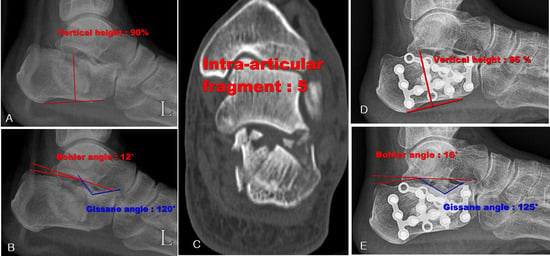
Figure 1.
A 71-year-old man underwent open reduction and internal fixation with good clinical outcomes (American Orthopaedic Foot and Ankle Society ankle-hindfoot score of 84). Preoperative plain lateral radiographs (A and B) and a coronal computed tomographic scan (C) show Sanders type IV with five fragments. D and E, Postoperative and last follow-up plain radiographs show good recovery of the Böhler angle, Gissane angle, and vertical height.
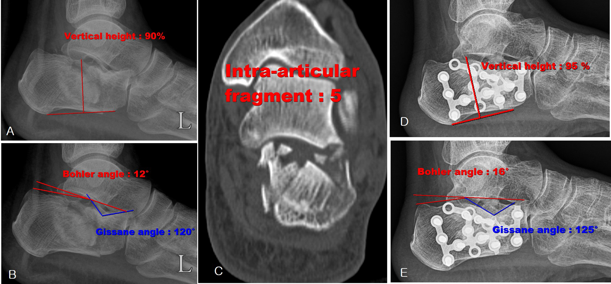
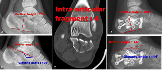
Figure 2.
A 28-year-old woman underwent open reduction and internal fixation with bad clinical outcomes (American Orthopaedic Foot and Ankle Society ankle-hindfoot score of 55). Preoperative plain lateral radiographs (A and B) and a coronal computed tomographic scan (C) show Sanders type IV with four fragments. D and E, Postoperative and last follow-up plain radiographs show bad recovery of the Böhler angle, Gissane angle, and vertical height.
For the Gissane angle, mean ± SD AOFAS ankle-hindfoot and VAS scores were 55.5 ± 12.0 and 4.8 ± 2.0 for the good recovery group (n = 19) and 79.0 ± 9.7 and 1.8 ± 1.1 for the bad recovery group (n = 19), respectively. There were significant differences in these scores between the two groups (P = .001 for both).
For the vertical height, mean ± SD AOFAS ankle-hindfoot and VAS scores were 56.6 ± 12.0 and 4.7 ± 1.9 for the good recovery group (19 patients) and 77.8 ± 12.2 and 1.9 ± 1.5 for the bad recovery group (19 patients), respectively. There were significant differences in these scores between the two groups (P = .001 for both).
Discussion
Although many studies have attempted to classify the exact pattern and type of fracture, there are still controversies due to the complex shape of the calcaneus and the variety of fracture patterns. The Sanders classification system is the most used system for classifying intra-articular calcaneal fractures by orthopedic surgeons. In many studies, the posterior facet fracture pattern of the calcaneus used in the Sanders classification is the most important prognostic factor [7,8].
The present study found significant differences in clinical outcomes between groups with four fragments versus more than four fragments. This is because Sanders type IV fractures with at least three fracture lines across the posterior subtalar facet usually result from high-energy injuries that show poor outcomes with nonoperative or operative management [9,10]. Despite carefully planned open reduction and near anatomical reconstruction, a group of patients may proceed to have painful subtalar arthritis that needs delayed subtalar fusion [10–12]. Various studies [10,13–15] have reported that the prevalence of poor results after open reduction and internal fixation (ORIF) increases with the severity of comminution [9]. A large Canadian prospective study [11] showed that 47% of the late fusion group had Sanders type IV fractures and that 20 of 81 available patients with Sanders type IV injury required fusion within 2 years of presentation. They also found that patients with Sanders type IV fractures were 5.5 times more likely to need subtalar fusion than those with fractures of lesser complexity (Sanders type II). Results of delayed subtalar fusion were good, and patient satisfaction was high regardless of initial nonoperative or operative treatment for the fracture. A high percentage of patients with Sanders type IV fractures do poorly with operative reduction [8,9,15]. For those with type IV fractures, outcomes could be different according to fracture patterns and the degree of fractures. However, the results of this study did not show significant differences in outcomes.
The ideal goal of ORIF for these fractures is to recreate calcaneal width, height, and length; subtalar joint congruency; and soft-tissue balancing. Although several previous studies have suggested that operative treatment of displaced intra-articular fracture of Sanders type IV is difficult [16–18], some authors have suggested that ORIF should be applied to type IV calcaneal fractures [17,19–21]. These studies in general report that surgery can reduce the time to return to work and the progression to subtalar fusion with less expense [22].
In the present study, no statistically significant difference was found between groups according to the number of fracture fragments (four fragments versus more than four fragments, P > .05). However, this study found that the clinical results differed according to the degree of injury to and recovery of the Böhler angle, Gissane angle, and vertical height. Some authors found an improvement in the Bohler angle in the surgical group treated with ORIF. Makki et al [23] concluded that restoration of the Bohler angle was associated with better functional outcomes in 45 patients treated with ORIF and that rapid osteosynthesis should be considered in displaced intra-articular calcaneal fracture to correct the hindfoot angle and the Bohler angle. In conservative treatment of displaced intra-articular calcaneal fractures, the fact that the Bohler angle can be reduced to 11° in the first year after trauma should be taken into consideration. For this reason, anatomical restoration of the Böhler angle, Gissane angle, and vertical height is more important than the number of fracture fragments to attain a better clinical outcome. Therefore, orthopedic surgeons should do their best to achieve anatomical restoration of the Böhler angle, Gissane angle, and vertical height through surgery.
This study has some limitations. First, different surgical procedures were performed. Second, the prognosis was assessed at different time points for each patient across 1 year after surgery. Third, this is a retrospective study and only one surgeon performed the surgeries, which will likely add inherent bias to the study.
Conclusions
The results of this study suggest that clinical results of Sanders type IV fractures were not related to the number of bone fragments but to the degree of injury to the Böhler angle, Gissane angle, and vertical height. Therefore, subtype classification of type IV calcaneal fractures is superfluous, and it is important to try to restore these parameters during surgery. It can also be seen that these parameters are important factors in predicting the prognosis. Further research enrolling more patients is needed to confirm the results of this study.
This research was supported by a grant from the Korea Health Technology R&D Project through the Korea Health Industry Development Institute (KHIDI), funded by the Ministry of Health & Welfare, Republic of Korea (grant number: HR22C1832).
None reported.
References
- Slätis P, Kiviluoto O, Santavirta S, et al: Fractures of the calcaneum.J Trauma 19: 939, 1979.
- Böhler L: The Treatment of Fractures, 4th Ed, John Wright & Sons, Bristol, United Kingdom, 1935.
- Borrelli J Jr, Lashgari C: Vascularity of the lateral calcaneal flap: a cadaveric injection study.J Orthop Trauma 13: 73, 1999.
- Essex-Lopresti P: The mechanism, reduction technique, and results in fractures of the os calcis.Br J Surg 39: 395, 1952.
- Sanders R, Fortin P, DiPasquale T, et al: Operative treatment of 120 displaced intraarticular fractures of the calcaneus.Clin Orthop 290: 87, 1993.
- Crosby LA, Fitzgibbons T: Computerized tomography scanning of acute intraarticular fractures of the calcaneus.J Bone Joint Surg Am 72: 852, 1990.
- Andermahr J, Jesch AB, Helling HJ, et al: CT morphometry for calcaneal fractures and comparison of the Zwipp and Sanders classifications [in German].Z Orthop Ihre Grenzgeb 140: 339, 2002
- Furey A, Stone C, Squire D, et al: Os calcis fractures: analysis of interobserver variability in using Sanders classification.J Foot Ankle Surg 42: 21, 2003.
- Buckley RE, Tough S, McCormack R, et al: Operative compared with nonoperative treatment of displaced intra-articular calcaneal fractures.J Bone Joint Surg Am 84: 1733, 2002.
- Lindsay R, Dewar F. Fractures of the oscalcis.Am J Surg 95: 555, 1958.
- Radnay CS, Clare MP, Sanders RW: Subtalar fusion after displaced intraarticular calcaneal fractures: does initial operative treatment matter?J Bone Joint Surg Am 91: 541, 2009.
- Schepers T, Kieboom BC, Bessems GH, et al: Subtalar versus triple arthrodesis after intra-articular calcaneal fractures.Strategies Trauma Limb Reconstr 5: 97, 2010.
- Tscherne H, Zwipp H: “Calcaneal Fractures,” in Major Fractures of the Pilon, Talus, and the Calcaneus: Current Concepts in Treatment, ed by HTscherne, JSchatzker, p 153, Springer-Verlag, Berlin, 1993.
- Sanders R, Hansen ST, McReynolds IS: “Trauma to the Calcaneus and Its Tendon,” in Disorders of the Foot and Ankle: Medical and Surgical Management, 2nd Ed, ed by Jahss MH, p 2326, WB Saunders, Philadelphia, 1991.
- Csizy M, Buckley R, Tough S, et al: Displaced intraarticular calcaneal fractures: variables predicting late subtalar fusion.J Orthop Trauma 17: 106, 2003.
- Ebraheim NA, Elgafy H, Sabry FF, et al: Sinus tarsi approach with trans-articular fixation for displaced intra-articular fractures of the calcaneus.Foot Ankle Int 21: 105, 2000.
- Hetsroni I, Nyska M, Ben-Sira D, et al: Analysis of foot and ankle kinematics after operative reduction of high-grade intra-articular fractures of the calcaneus.J Trauma 70: 1234, 2011.
- Stulik J, Stehlik J, Rysavy M, et al: Minimally invasive treatment of intraarticular fractures of the calcaneum.J Bone Joint Surg Br 88: 1634, 2006.
- Jian G-J, Chen F-R, Lin J-D: Surgical treatment of calcaneus comminuted fractures involving calcaneal-talar joint [in Chinese].Zhongguo Gu Shang 23: 808, 2010.
- Su Y, Chen W, Zhang Q, et al: Bony destructive injuries of the calcaneus: long-term results of a minimally invasive procedure followed by early functional exercise: a retrospective study.BMC Surg 14: 19, 2014.
- Zhang T, Su Y, Chen W, et al: Displaced intra-articular calcaneal fractures treated in a minimally invasive fashion: longitudinal approach versus sinus tarsi approach.J Bone Joint Surg Am 96: 302, 2014.
- Gurkan V, Dursun M, Orhun H, et al: Long-term results of conservative treatment of Sanders type 4 fractures of the calcaneum: a series of 64 cases.J Bone Joint Surg Br 93: 975, 2011.
- Makki D, Alnajjar HM, Walkay S, et al: Osteosynthesis of displaced intra-articular fractures of the calcaneum: a long-term review of 47 cases.J Bone Joint Surg Br 92: 693, 2010.
- Bakker B, Halm JA, Van Lieshout EM, et al: The fate of Böhler’s angle in conservatively-treated displaced intraarticular calcaneal fractures.Int Orthop 36: 2495, 2012.
© 2023 American Podiatric Medical Association