


Abstract
Background: Supination-adduction (SAD) type injuries are pylon variant injuries and lie between partial intra-articular pylon fractures and rotational ankle fractures. We aimed to evaluate functional outcomes of SAD type 2 bimalleolar fractures in comparison to supination–external rotation (SER) type 4 fractures. Methods: We retrospectively reviewed data of 42 cases with SER type 4 and 20 cases with SAD type 2 injuries. Patients with a history of rheumatic disease, open fractures, pathologic fractures, nonbimalleolar fractures, neuropathic disease, and talus osteochondral lesion, and those operated on after greater than 72 hours because of skin lesion or managed with a two-stage surgical protocol after external fixation, were not included in the study. We compared these two groups in terms of the mean age, follow-up time, visual analog scale pain and American Orthopedic Foot and Ankle Society scores, Kellgren-Lawrence arthrosis classification, union time, and complications. Results: The groups did not differ in terms of mean age (P = .115) and sex (P = .573). There was no significant difference in terms of union time between the groups (P = .686). American Orthopedic Foot and Ankle Society score was significantly higher in the SER group (91.2 ± 9.9) than in the SAD group (86.1 ± 13.2; P = .034). Visual analog scale pain scores were similar in the SAD (0.3 ± 0.92) and the SER (0.26 ± 0.7) groups (P = .897). Conclusions: Supination-adduction bimalleolar fractures may have worse functional outcomes in the intermediate term than do SER bimalleolar fractures, implying pylon variant fractures as a mechanism of injury. Supination-adduction bimalleolar fractures might be associated with a high rate of intra-articular cartilage impaction, resulting in varus deformity after surgery.
Ankle fractures are among the most common fractures of the lower extremity.[1] Bimalleolar fractures constitute approximately 25% of all ankle fractures.[2] These fractures are typically unstable and often require open reduction and internal fixation.[3]
Ankle fractures are usually categorized according to the Lauge-Hansen classification, which primarily considers the position of the foot at the time of injury and the deforming force on the ankle.[4] This classification also provides information about possible treatment method.[5] Supination-adduction (SAD) type injuries are pylon variant injuries, remaining between partial intra-articular pylon fractures and rotational ankle fractures.[6] These injuries are mostly associated with high-energy trauma and polytrauma, whereas supination-external rotation (SER) type injuries accompany low-energy trauma and monotrauma.[7]
Supination-adduction type injuries are rare injuries and but have high rates of cartilage impaction.[6] Cartilage impaction is associated with ankle arthrosis and poor joint survival.[6,7] The target population of ankle-related studies in the literature consists mostly of patients with low-energy trauma injuries involving sports-related and rotational injuries.[7] As the different types of ankle fractures were not typically evaluated separately in the literature, studies on these fractures may reflect the composite outcomes of SER and pronation-external rotation (PER) type injuries caused by monotrauma.[7]
Compared to SER/PER type fractures, there are limited data on the functional and radiologic results of SAD type fractures. Our hypothesis is these fractures may have worse functional and radiologic outcomes than SER type fractures because of joint impaction and cartilage damage in bimalleolar fractures. Therefore, we aimed to compare functional and radiologic outcomes of the SAD type 2 versus SER type 4 injuries among the patients undergoing surgery because of diagnosis of the isolated bimalleolar fracture.
Methods
The files of 62 patients who were operated on because of ankle fracture between 2013 and 2018 were examined retrospectively. The study protocol was approved by the institutional review board ethics committee (November 8, 2020; no. 331). Written informed consent was obtained from each patient. The study was conducted in accordance with the principles of the Declaration of Helsinki. We examined preoperative computed tomographic (CT) scans of the cases and selected only the images of the cases with bimalleolar fractures (lateral and medial). We divided the cases into two groups based on the Lauge-Hansen classification, which was performed on CT images independently by two surgeons. If an inconsistency was noticed between the two observers, the images were examined jointly by these two investigators to reach a common decision.[8] The cases where no consensus was achieved were not included in the study. Eventually, 42 cases of SER type 4 and 20 cases of SAD type 2 were identified. Among 62 patients, men constituted 60.0% and 52.4% of the cases in the SAD and SER groups, respectively. The mean age of the patients was 39.0 ± 12.6 years in the SAD group and 44.5 ± 12.8 years in the SER group. Patients with a history of rheumatic disease, open fractures, pathologic fractures, nonbimalleolar fractures, fracture of posterior malleolus (ie, patients with a posterior malleolus fracture were not included because it affects functional results[9–11]), neuropathic disease, talus osteochondral lesion, and those operated on after greater than 72 hours because of skin lesion or managed with a two-stage surgical protocol after external fixation were not included in the study.
Fracture fixation was achieved in all cases by using a lateral and medial double skin incision. Posterolateral anatomical distal fibula plate (TST Medical Devices Company, Istanbul, Turkey) was used for stabilization of the lateral malleolus. Medial malleolar fracture was fixed differently in the study groups: through two malleolar screws or tension band method in the SER group, and through the anatomical medial malleolar plate (TST-Türk Spinal Travma) in the SAD group. If syndesmosis failure was detected after fracture stabilization in the cotton test or the external rotation test, syndesmosis stabilization was achieved by a syndesmosis screw introduced through the appropriate hole on the plate. Then, syndesmosis was reevaluated by means of the cotton or external rotation test and the operation was terminated. A short leg splint was applied for 7 to 14 days; then, the splint was removed and joint movements were allowed. Nonweightbearing walking was allowed up to 6 weeks postoperatively, followed by weightbearing walking as much as the patient could tolerate. The loading was increased over time to reach full weightbearing. The use of the short leg splint was extended up to 4 to 6 weeks in the presence of reduced cartilage impaction.
In patients with cartilage impaction in the SAD group, the medial malleolus was reduced and fixed after the impacted fragment was reduced. No bone grafts were used in any patient. In addition, whereas medial malleolar fracture was reduced, materials within the joint were removed by intra-articular washing. The presence of plafond impaction on preoperative CT scan was determined by the method described by Haller et al.[6]
Postoperative plain radiographic and fluoroscopic images were assessed by using the following criteria: tibiofibular clear space (normal, <6 mm), anteroposterior tibiofibular overlap (normal, >6 mm), mortis tibiofibular overlap (normal, >1 mm), and medial clear space (normal, <5 mm). Cases with values outside the normal range belonging to any of these criteria were accepted as tibiofibular malreduction.[12] The same criteria had been used for the evaluation of the intraoperative syndesmosis joint.
On anteroposterior radiographs, the lateral distal tibial angle was accepted as normal between 86° and 92°. Cases with a lateral distal tibial angle greater than 92° were regarded as varus deformity, and those with less than 86° were regarded as valgus deformity.[13]
The cases were operated on by two different surgeons. All surgeons had more than 5 years of trauma surgery experience. The number of surgical ankle fracture interventions was above 15 annually within the past 3 years.
Preoperative and postoperative radiographic measurements were performed by means of the INFINITT PACS (Picture Archiving and Communication Systems) version 3.0.11.4 (BN13) imaging software (Seoul, Republic of Korea). Postoperative measurements, obtained from the radiographic images taken at the latest outpatient visit, were assessed by a senior radiologist experienced in the musculoskeletal system. Clinical assessments were performed by the senior orthopedic surgeon.
Demographic and clinical data of patients in each of the study groups were collected, including age; sex; fracture side; follow-up duration; mechanism of injury; time to union; presence of syndesmosis joint injury; lateral and medial malleolus fracture type according to the Denis-Weber classification and the Herscovici classification, respectively; VAS pain and AOFAS ankle function scores; Kellgren-Lawrence arthrosis classification; presence of postoperative malalignment (varus-valgus); and postoperative complications. The study groups were compared in terms of these mentioned characteristics.
Statistical analysis was performed using IBM SPSS Version 24.0 (IBM Corp, Armonk, New York). Descriptive variables were expressed as the mean ± standard deviation (SD) or numbers and percentages, where appropriate. The normality of the continuous data was tested with the Shapiro-Wilk test. The minimal clinically important difference (MCID) for ankle fracture patients is not defined for the AOFAS score. In earlier studies, the MCID has been suggested to be 50% of the SD. The SD was set to 12 points based on data from a previous trial with a similar population,[18] giving a MCID of the AOFAS score of 6 points.[14] These parametric variables between the two groups were compared by the Student t test and the Mann-Whitney U test for normally and nonnormally distributed data, respectively. Categorical variables were compared through the χ2 test. A value of P < .05 was considered statistically significant.
Results
Time to union was 7.7 ± 1.8 weeks in the SAD group and 8.1 ± 2.3 weeks in the SER group. The groups were similar in terms of the mean age, sex, time to union, duration of follow-up, and VAS scores. The mean AOFAS score was significantly lower in the SAD group (86.1 ± 13.2) compared with the SER group (91.2 ± 9.9; P = .034) (Table 1).

Table 1.
Comparison of the Groups in Terms of the Mean Age, Duration of Union and Follow-Up, and Clinical Scores
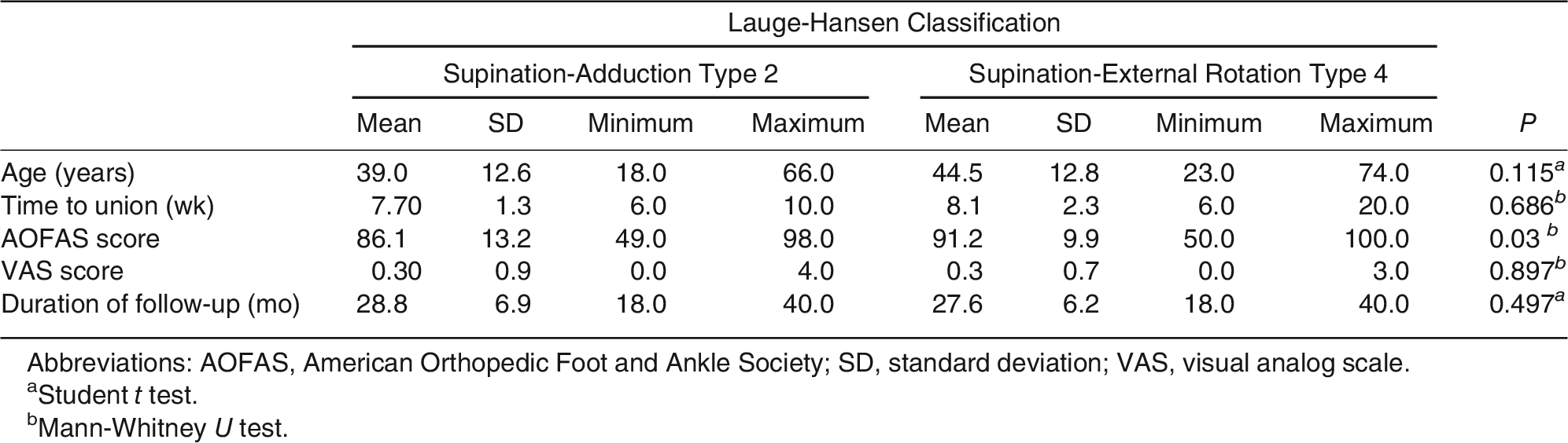
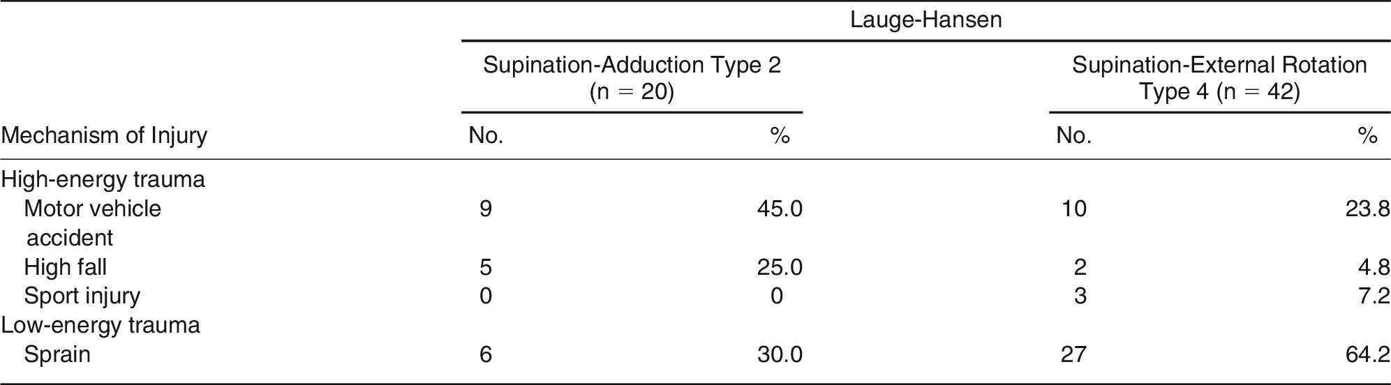
Table 2 shows the distribution of cases included in the study according to the Denis-Weber, Herscovici, and Kellgren-Lawrence classifications. Table 3 shows the mechanisms for injuries of the cases included in the study.
Table 2.
Comparison of the Groups in Terms of Their Other Classification Systems
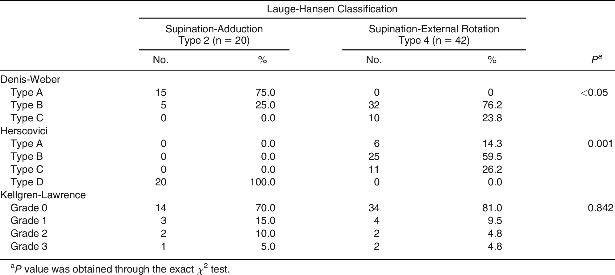
Table 3.
Mechanism of Injury in the Study Groups
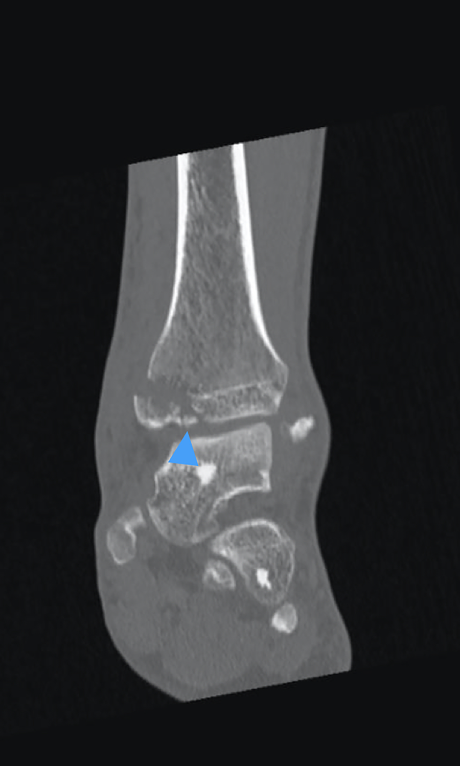
In the SAD group, three cases (15%) received syndesmosis screws; three cortices were penetrated in all cases. In contrast, a syndesmosis screw was applied in 37 cases (88.1%) in the SER group, where four cortices were penetrated with the screw only in one patient. Preoperative CT images in the SAD group showed cartilage impaction in 11 patients (55.0%). On postoperative radiographs, three cases of varus deformities were detected in the SAD group (Fig. 1). Whereas two of these cases had cartilage impaction in the postoperative period, the other with good postoperative reduction developed collapse at the cartilage fragment during follow-up. There was syndesmosis instability in seven patients in the SER group postoperatively. Preoperative CT images revealed intra-articular free fragments in 10 and 11 cases of the SAD group and the SER group, respectively (Fig. 2). Whereas fixation was performed by means of medial malleolar plate in all cases of the SAD group, this was achieved through malleolar screw (71.4%) and tension band (28.6%) in the SER group.
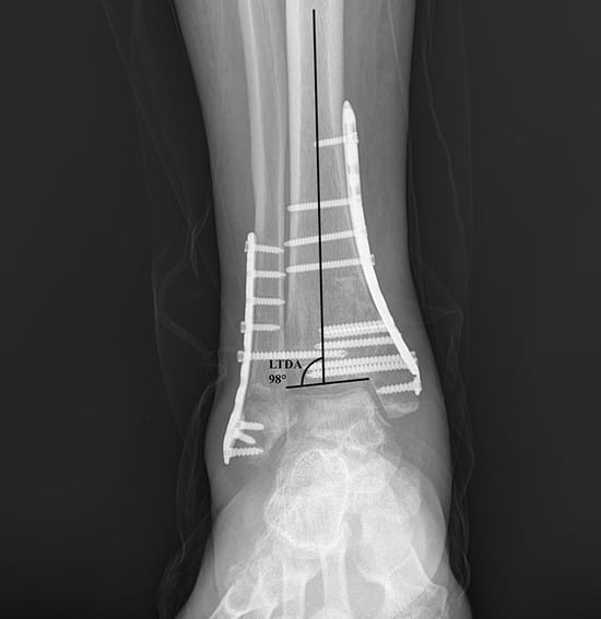
Figure 1.
A 54-year-old male patient treated for supination-adduction type 2 fracture has varus deformity due to the lateral distal tibial angle measured at 6 months postoperatively being higher than 92°.
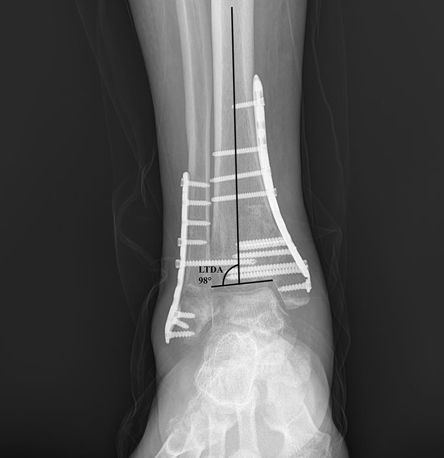
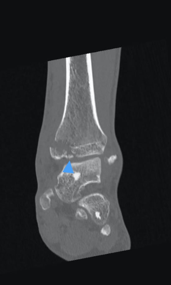
Figure 2.
Preoperative computed tomographic image of a 49-year-old male patient with a left bimalleolar fracture (supination adduction type 2). Blue arrow indicates free intra-articular part.
Superficial infection responding to oral antibiotic therapy was seen in two patients (10%) in the SAD group and three patients (7.1%) in the SER group. Physical therapy was required on the diagnosis of reflex sympathetic dystrophy in two patients (10%) in the SAD group and two patients (4.8%) in the SER group. Delayed union was detected in four patients in the SAD group and in two patients in the SER group.
Discussion
It is important to know the exact mechanism of ankle fractures, as it helps surgeons in evaluating the fracture pattern and soft-tissue pattern. The Lauge-Hansen classification system, which determines the order of injury in bone and soft tissues, assists surgeons both as the initial assessment tool and as the guidance for the treatment by determining the mechanism of injury.[5] Among the subtypes of the Lauge-Hansen classification, SER was the most common fracture mechanism and SAD is the least common injury type.[4,5,8] In addition, bimalleolar fractures are typically unstable and often require open reduction and internal fixation.[15] Marginal impaction is common in SAD type fractures, and the severity of the injury is somewhere between the rotational ankle fractures and partial articular pylon fractures.[6] Moreover, the presence of medial malleolar fracture—especially vertical type—and intra-articular cartilage impaction may be associated with poor outcomes.[16,17] Studies on the ankle fractures in the literature mostly were reported to consist of findings in patients with low-energy trauma including rotational injuries (SER and PER), whereas some advocated that SAD type fractures needed more explanation, as the patterns of such fractures were different from typical ankle fractures described in the literature.[7,18] This might result from undervaluation of the SAD type fractures because of their rare occurrence.
SAD fractures comprise vertical shift fracture of the medial malleolus and lower level avulsion fracture of the lateral malleolus, and are often associated with impaction injury of the medial tibial plafond or free osteochondral fragment or medial tibial plafond, which should be handled appropriately during surgical fixation.[18] These fractures may accompany plafond impaction in up to 61% of cases.[6,19] Consistently, we observed the rate of plafond impaction on preoperative CT scan to be 55% in our study.
Articular impaction has been associated with cartilage injury and poorer joint survival.[6] In our study, there was no difference between the two groups in terms of radiologic arthrosis and VAS scores. Considering that pain is associated with arthrosis and the latter would require many years to develop after the trauma, the lack of radiologic differences between the two groups may be because the follow-up time in our study was quite shorter than the mean time to clinical or radiologic evidence of arthrosis.[20]
It was reported that unfavorable outcomes following medial malleolar fractures were less important than those following lateral or posterior malleolar fractures. In fact, poor functional outcomes caused by synthesis incompatibility were emphasized. However, long-term follow-up of medial malleolar fractures (especially vertical fractures) might be associated with posttraumatic poor functional outcomes.[16,17,21,22] Intra-articular chondral lesions often accompany acute ankle fractures, where poor ankle function may be secondary to such intra-articular injury.[23] In addition, cartilage lesions in the medial malleolus were also reported to be associated with poor functional results.[24] Briet et al[7] reported that SAD was caused by high-energy trauma, whereas SER was associated with low-energy trauma, with no mention of functional outcomes in their study. In our study, the fracture occurred because of a high-energy mechanism in 70% of the cases in the SAD group and 35.7% in the SER group. In AOFAS scoring, the SAD group had significantly poor functional results compared with the SER group. This might be attributed to the vertical nature of the fracture or articular impaction, or the injury caused by the underlying high-energy trauma on the articular cartilage of the weightbearing plafond.
Changes in the ankle joint alignment cause formation of contact stress, and are associated with the development of osteoarthritis.[25] In SAD type fractures where medial malleolar fractures are complicated by intra-articular impaction and fragmentation, a gap may remain on the surface of the joint after the outer cortex—as a guide—is exposed, debrided, and anatomically fixed. This may be associated with poorer results by leading to varus alignment.[2,17] In SAD type fractures, marginal impaction is common, but it was not reported to be associated with increased rates of ankle arthrosis or ankle joint failure if the impaction was appropriately treated by surgical intervention.[6] The recommended treatment involves lifting the collapsed joint segment and filling the defect with bone graft to prevent joint instability and varus tendency of the talus.[2] In our study, varus deformity was present in three of 11 patients (27.3%) with articular impaction in the SAD group. One of these three cases had a collapse during the postoperative follow-up. No grafting was performed in any patients. It may be suggested that routine reduction of the impacted joint segment followed by bone autografting might be an essential tool to avoid varus deformity and poor functional outcomes in such cases.
In bimalleolar fractures, the union time of fracture was reported to range from 8 to 12 weeks.[24] Postoperative infection rates vary from 4% to 10%.[6,26] The union time and infection rates were similar in our study groups and consistent with those reported in the literature.
In SAD injuries, lateral malleolar fractures usually correspond to Denis-Weber type A injuries and rarely accompany syndesmosis injury. In SER, however, concurrent syndesmosis injury often accompanies lateral malleolar fracture, which generally corresponds to Denis-Weber type B fractures.[27] According to this classification, 76.2% of the patients in the SER group had type B fracture, whereas 75% of the patients were categorized as type B in the SAD group. We detected syndesmosis injury in 15% of the SAD group and 88.1% of those in the SER group, supporting the findings reported in the literature.
The findings of our study should be interpreted with its limitations, including its retrospective design, the small size of the study sample, and shorter follow-up duration than is needed to observe arthrosis development. We believe that this could be clarified with larger case series with a longer duration of follow-up. In addition, further studies with a prospective design that considers negative prognostic factors including body mass index, smoking, arthroscopic documentation of cartilage injury, and others that adversely affect the ankle functions and promote arthrosis development would yield better findings.
Conclusions
SAD type bimalleolar fractures, with a pilon variant fracture as mechanism of injury, may have poorer functional outcomes than SER type fractures in the intermediate term. SAD type fractures may cause postoperative varus deformity, likely because of intra-articular cartilage impaction.
Financial Disclosure: None reported. | |
Conflict of Interest: None reported. |
References
- Tomé-Bermejo F, Arévalo AS, Micó NR: Open reduction and internal fixation of displaced ankle fractures in patients older than 65 years of age. Analysis of results at five-year follow-up. Rev Esp Cir Ortop Traumatol 60: 99, 2016.
- Rammelt S, Boszczyk A: Computed tomography in the diagnosis and treatment of ankle fractures: a critical analysis review. JBJS Rev 6: e7, 2018.
- Still GP, Atwood TC: Operative outcome of 41 ankle fractures: a retrospective analysis. J Foot Ankle Surg 48: 330, 2009.
- Alluri RK, Hill JR, Donohoe S: Radiographic detection of marginal impaction in supination-adduction ankle fractures. Foot Ankle Int 38: 1005, 2017.
- Russo A, Reginelli A, Zappia M: Ankle fracture: radiographic approach according to the Lauge-Hansen classification. Musculoskelet Surg 97: 155, 2013.
- Haller JM, Ross H, Jacobson K: Supination adduction ankle fractures: ankle fracture or pilon variant? Injury 51: 759, 2020.
- Briet JP, Houwert RM, Smeeing DPJ: Differences in classification between mono- and polytrauma and low- and high-energy trauma patients with an ankle fracture: a retrospective cohort study. J Foot Ankle Surg 56: 793, 2017.
- Warner SJ, Garner MR, Hinds RM: Correlation between the Lauge-Hansen classification and ligament injuries in ankle fractures. J Orthop Trauma 29: 574, 2015.
- Tejwani NC, Pahk B, Egol KA Effect of posterior malleolus fracture on outcome after unstable ankle fracture. J Trauma 69: 666, 2010.
- Langenhuijsen JF, Heetveld MJ, Ultee JM: Results of ankle fractures with involvement of the posterior tibial margin. J Trauma 53: 55, 2002.
- Jaskulka RA, Ittner G, Schedl R: Fractures of the posterior tibial margin: their role in the prognosis of malleolar fractures. J Trauma 29: 1565, 1989.
- Gardner MJ, Demetrakopoulos D, Briggs SM: Malreduction of the tibiofibular syndesmosis in ankle fractures. Foot Ankle Int 27: 788, 2006.
- Cakmak M, Civan M: “Ankle Deformities,” in Basic Techniques for Extremity Reconstruction, edited by M Cakmak, C Sen, L Eralp, p 413, Springer, Cham, Switzerland, 2018.
- Ræder BW, Figved W, Madsen JE: Better outcome for suture button compared with single syndesmotic screw for syndesmosis injury: five-year results of a randomized controlled trial. Bone Joint J 102: 212, 2020.
- Still GP, Atwood TC: Operative outcome of 41 ankle fractures: a retrospective analysis. J Foot Ankle Surg 48: 330, 2009.
- Bhandari M, Sprague S, Hanson B: Health-related quality of life following operative treatment of unstable ankle fractures: a prospective observational study. J Orthop Trauma 18: 338, 2004.
- Hu J, Zhang C, Zhu K: Adverse radiographic outcomes following operative treatment of medial malleolar fractures. Foot Ankle Int 39: 1301, 2018.
- Khan UZ, Muhammad W, Qureshi AR: Ankle fractures; an epidemiological study on pattern of ankle fractures in a tertiary hospital; a one-year prospective observation. Professional Med J 25: 1160, 2018.
- McConnell T, Tornetta P III: Marginal plafond impaction in association with supination-adduction ankle fractures: a report of eight cases. J Orthop Trauma 15: 447, 2001.
- Horisberger M, Valderrabano V, Hintermann B: Posttraumatic ankle osteoarthritis after ankle-related fractures. J Orthop Trauma 23: 60, 2009.
- Cherney SM, Haynes JA, Spraggs-Hughes A: In vivo syndesmotic over-compression after fixation of ankle fractures with a syndesmotic injury. J Orthop Trauma 29: 414, 2015.
- Verhage SM, Schipper IB, Hoogendoorn JM: Long-term functional and radiographic outcomes in 243 operated ankle fractures. J Foot Ankle Res 8: 45, 2015.
- Leontaritis N, Hinojosa L, Panchbhavi VK: Arthroscopically detected intra-articular lesions associated with acute ankle fractures. J Bone Joint Surg Am 91: 333, 2009.
- Stufkens SA, Knupp M, Horisberger M: Cartilage lesions and the development of osteoarthritis after internal fixation of ankle fractures: a prospective study. J Bone Joint Surg Am 92: 279, 2010.
- Ebinger T, Goetz J, Dolan L: 3D model analysis of existing CT syndesmosis measurements. Iowa Orthop J 33: 40, 2013.
- Shah ZA, Arif U: Surgical management of bimalleolar fractures of ankle. Pak J Med Health Sci 7: 471, 2013.
- Chun D-I, Kim J, Kim YS: Relationship between fracture morphology of lateral malleolus and syndesmotic stability after supination-external rotation type ankle fractures. Injury 50: 1382, 2019.
© 2023 American Podiatric Medical Association