


Abstract
For diabetic foot ulcers with “pockets” (wound edge undermining), a surgical incision to the undermining space and debridement are required primarily. Osteomyelitis should be treated if present. Furthermore, it is necessary for the foot to be able to withstand load after controlling the infection and necrosis. We report a case of a patient treated with a skin-covering pocket as a local flap and negative pressure wound therapy with instillation and dwell time and negative pressure wound therapy to relieve osteomyelitis and promote wound contraction. There was no involvement of other body parts, and only the pocket consisting of skin at the sole of the foot was used, and load could be withstood.
Callus or ulcers beneath the callus may form on the foot due to diabetes [1]. Chronic pressure on the protruding part of the bone forms an ulcer, causing infection and necrosis [2]. Ulcers are sometimes accompanied by wound edge undermining, which is commonly termed a “pocket” among Japanese wound care professionals [3]. In cases of pressure ulcers accompanied by a pocket, incision of the skin to the pocket space or removal of the skin covering the pocket is recommended to facilitate easier removal of deeply located necrotic and infected tissues [3,4]. Similarly, we believe that pocket incision or resection is required to control the infection and necrotic tissue when diabetic foot ulcers are accompanied by a pocket.
If concomitant osteomyelitis is present, it needs to be treated. The wound requires closure using a procedure that would enable load to be withstood after controlling the infection and necrosis. We report a case of diabetic foot ulcer with calcaneal osteomyelitis that was treated using a skin-covering pocket as a local flap and then negative pressure wound therapy (NPWT) with instillation and dwell time (NPWTi-d)/NPWT.
Case Report
A 72-year-old man presented with an intractable ulcer. Approximately 9 years earlier he had undergone Chopart joint amputation due to left diabetic foot gangrene and has been living with a foot orthosis since then. Ulceration in the heel had started a few months earlier, and he consulted our department. The ulcer had enlarged 2 weeks before admission. The patient was admitted to the hospital because he developed a fever and lost consciousness, and he was subsequently admitted to our department.
At the time of admission, he had a fever of 100.4°F, and although mild tachycardia was observed, no further findings were found via other vital signs. In clinical and ultrasound examinations, blood flow in the lower limbs was maintained. Physical examination revealed ulceration with a 7-cm pocket in the heel; the internal space was filled with necrotic tissue and the calcaneus was exposed (Fig. 1). Blood test results revealed a white blood cell count of 141,410/μL (neutrophils, 96.3%), and a C-reactive protein level of 6.84 mg/dL.
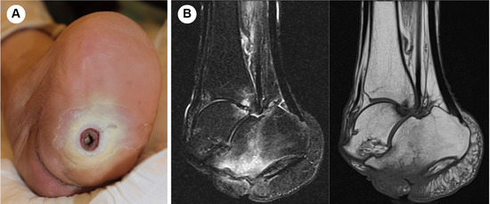
Figure 1.
A, Heel ulcer with a 7-cm pocket after Chopart amputation; the internal space was filled with necrotic tissue and the calcaneus was exposed. B, Magnetic resonance images of the calcaneus showing low signal intensity on a T1-weighted image (left) and high signal intensity on a short tau inversion recovery image (right). Findings suggest calcaneal osteomyelitis.
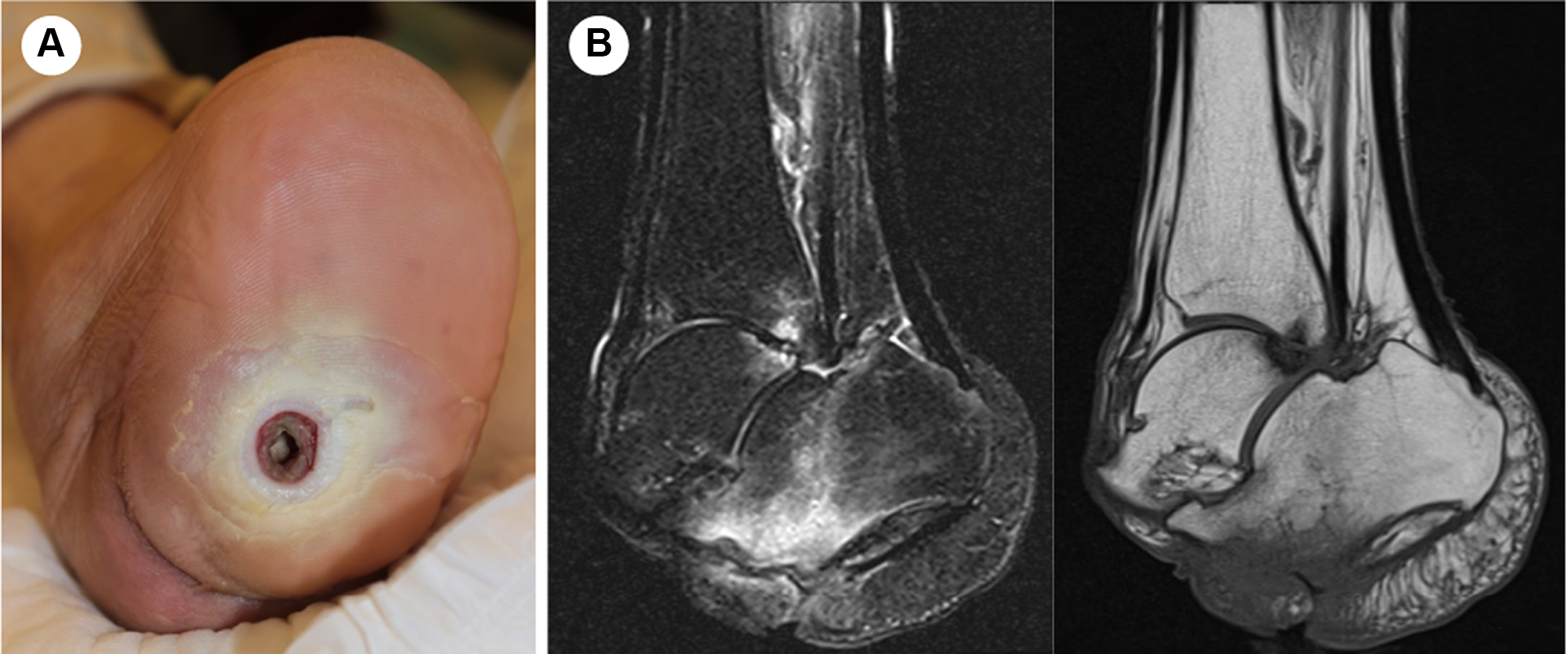
First, we performed a pocket incision due to infection control. An incision was made to allow the skin covering the pocket to be harvested as a flap. T1- and T2-weighted magnetic resonance imaging revealed findings suggestive of calcaneal osteomyelitis (Fig. 1). Eight days after the pocket incision, debridement was performed inside the pocket and the calcaneus was shaved until proper blood flow was achieved. Bone cortex of the calcaneus had become brittle. At the same time, the flap of the pocket was positioned to cover the exposed bone. The donor site of the flap was an open wound, and we simultaneously performed “the shoelace technique” [5] to fasten the skin at the edge of the wound of the donor site. For the technique, we fixed staples that were opposed on the edge of the wound of the donor side to provide gradual wound contraction with an elastic vessel loop subsequently (Fig. 2).
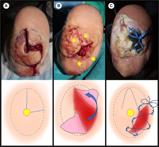
Figure 2.
A, After making a pocket incision. The blue line delineates the pocket incision. The yellow circle represents the opening of the pocket. B, Harvested pocket flap; the flap was transferred and sutured. A skin stapler was used on the wound edge, and the shoelace technique was used for wound contraction. C, Wound contraction with the shoelace technique using elastic vessel loops under negative pressure wound therapy with instillation and dwell time; the form was positioned on the calcaneus.
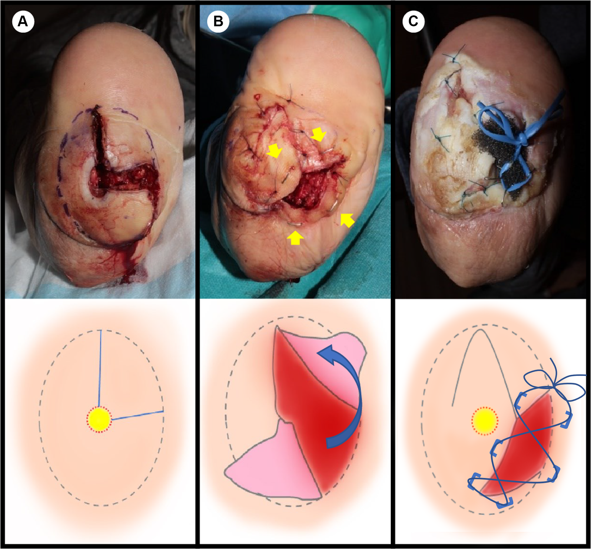
We performed the aforementioned operation under a lower-extremity peripheral nerve block. On postoperative day 1, we performed NPWTi-d using normal saline (24 mL) with 30 sec of dwell time, and NPWT (−100 mm Hg) for 3.5 hours. The form that was used in NPWTi-d was located on the calcaneus beneath the pocket flap. Simultaneously, we performed the shoelace technique with an elastic vessel loop and staples. The vessel loop was passed through the staples crisscross and tied so as to pull the edge of the wound closer (Fig. 2C). On postoperative day 2, the instillation solution was reduced to 12 mL. We achieved contraction of the open wound at the donor site and good formation of granulation tissue. From postoperative day 9, we performed NPWT at –100 mm Hg without instillation.
During each exchange of the NPWTi-d/NPWT, we fastened the vessel loop to provide wound closure. During NPWT, the wound edge was gradually pulled by tying elastic vessel loops so that the wound contracted horizontally according to the shoelace technique. The wound edge was sutured after the width of the wound was reduced. The wound was slit-shaped, with only a slight raw surface, and treatment with povidone-iodine sugar ointment was performed 20 days after the operation. The wound was closed on postoperative day 34. The results of a bacterial culture of a wound swab showed the presence of Streptococcus agalactiae, Streptococcus anginosus, Actinomyces odonolyticus, and Candida sp. Blood cultures were negative as the patient had a fever and lost consciousness. Tazobactam and piperacillin were administered intravenously for 12 days, and subsequently amoxicillin/clavulanate (2:1) was orally administered for 23 days. The NPWTi-d/NPWT with the shoelace technique and suturing of the wound were performed at the bedside. After adjusting the orthosis, the patient was able to walk by himself with the orthosis. The local flap, consisting of the skin from the sole of the foot, was able to withstand the load. In addition, there were neither scars on the protruding bones nor the formation of a callus. He was able to wear the orthosis without the shape of the foot becoming bulky.
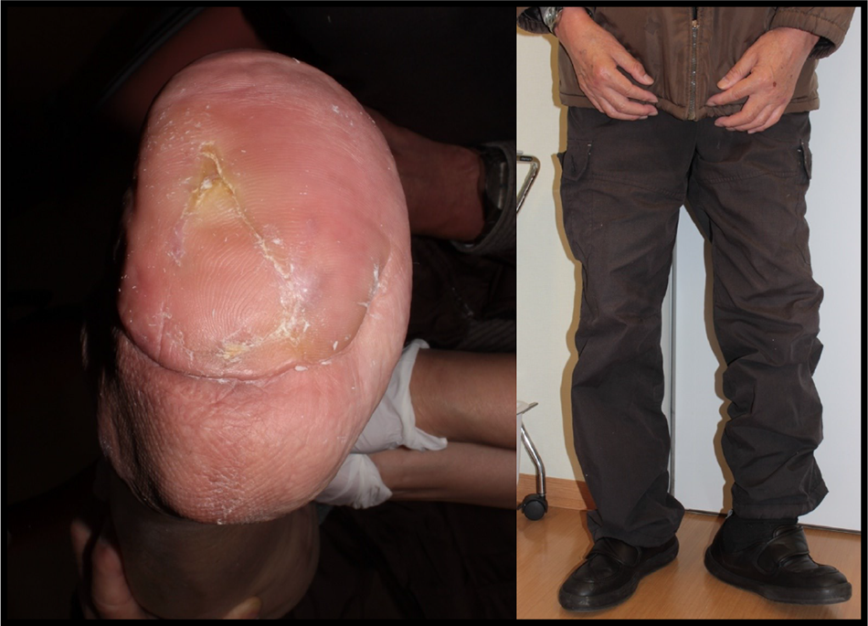
We followed-up with the patient 7 months after the operation. After that, he was followed in the foot care outpatient clinic in our hospital. There was no recurrence of the ulcer (Fig. 3).
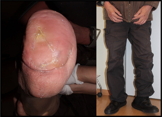
Figure 3.
Six months after surgery, the patient was able to walk with an orthosis. No recurrence of the ulcer was observed.
Discussion
Callus or ulcers beneath the callus may form on the foot due to diabetes [1]. When diabetic foot ulcers with wound edge undermining (pockets [3]) occur on the weightbearing plantar area, a pocket incision and internal debridement are required. If concomitant osteomyelitis is present, it needs to be treated.
Subsequently, wound closure and reconstruction of the foot is considered once the infection is under control and the necrotic tissue has been removed. Various reconstructive procedures have been reported for the weightbearing area of the plantar foot, and weightbearing reconstruction is necessary to prevent ulcer recurrence [6]. Based on the condition of the patient and the defect area and range, reconstruction using local, pedicled, or free flaps has been reported [7]. Therefore, when a pocket and an ulcer with infection are formed on the plantar load portion, it is necessary to consider wound closure to release the pocket infection and prevent recurrence.
A simple excision of the pocket will result in a larger defect. In addition, it is necessary to bring tissue from outside the ulcer, and invasion is added to other parts. Also, even if a pocket incision is simply added to heal the scar, it will form a scar on the protruding part of the bone, which may lead to the formation of an ulcer again. We reconstructed the plantar load portion using a local flap from the subcutaneous skin tissue of the pocket, which is usually simply incised and excised.
We considered covering the calcaneus with the appropriate flap to prevent ulcer recurrence, and we used a local flap from the skin and subcutaneous tissue of the pocket. Tsukamoto et al. [8] reported a less invasive and short-term wound closure procedure in refractory pressure ulcers by using undermined skin as a local flap and simultaneously preserving a skin-covered pocket.
In the present case, the pocket of the opening was small, but there was ulceration with a large pocket in the heel, and the internal space was filled with necrotic tissue (Fig. 1). The skin and subcutaneous tissue covering the pocket were relatively thick, and we concluded that the tissue covering the pocket was appropriate for use as a local flap. We performed the pocket incision so that we could subsequently use the tissue covering the pocket as a local flap. After debridement of the bone, the exposed area of the calcaneus was covered with the tissue of the pocket. On the other hand, a local flap may also have risks and complications. Attention should be given to the possibility of flap necrosis and wound dehiscence due to poor blood flow of the flap and too much tension of wound margins.
We did not perform the delay procedure, but if the blood flow of the flap was of concern, we considered gradually raising the flap of the pocket with the delay procedure.
Because the flap can be raised by incising the pocket, the inside debridement can be easily performed in parallel with the wound closure. At the same time, the osteomyelitis site could be treated with NPWTi-d/NPWT.
The NPWTi-d was applied from the first postoperative day after debridement to treat the calcaneal osteomyelitis. The NPWT promotes wound healing by inducing a negative pressure on the wound, and it can aspirate excess effusion, promote granulation, and contract wound edges. The NPWTi-d is NPWT with intermittent instillation and dwellings of irrigation solution [9]. It periodically irrigates the wounds. Previous studies have reported the efficacy of NPWT with instillation for osteomyelitis [10,11].
In the present case, we irrigated the inside of the pocket through the flap-harvested area using NPWTi-d, and in addition, the open wound was contracted using the shoelace technique [5]. The shoelace technique is a procedure in which the wound edges are pulled with vascular tape and gradually tightened like a shoelace, which can promote horizontal wound contraction and closure [5]. The technique is a dermatotraction procedure through constant tension of the wound margins [12]. Constant tension to the skin allows for the elongation of skin beyond its intrinsic extensibility by realigning the collagen fibers and fragmenting the elastic fibers [13,14]. Horizontal wound contraction using the shoelace technique with a vessel loop and vertical wound contraction with NPWT could be achieved.
Using a pocket as a local flap and NPWTi-d/NPWT combined with the shoelace technique, it was possible to promote wound closure and simultaneously control infection and necrotic tissue.
Because only the pocket of the sole was used, there was no invasion to other parts of the body, and reconstruction capable of withstanding load was possible. Placing a flap on the protruding part of the bone can prevent recurrence of the ulcer. We believe that treatment with a skin-covering pocket as a local flap and simultaneous treatment using NPWTi-d/NPWT in diabetic foot ulcers with calcaneal osteomyelitis is a less invasive procedure that enables load to be withstood.
Conclusions
Diabetic foot ulcers with a pocket in the weightbearing plantar area may occur. We conclude that treatment using a skin-covering pocket as a local flap with simultaneous NPWTi-d/NPWT in diabetic foot ulcers with calcaneal osteomyelitis is an effective procedure that is less invasive and enables load bearing.
Financial Disclosures: None reported. | |
Conflict of Interest: None reported. |
References
- Murray HJ, Young MJ, Hollis S, et al.: The association between callus formation, high pressures and neuropathy in diabetic foot ulceration. Diabet Med 13: 979, 1996.
- Hasenstein TA, Greene T, Van JC, et al.: Soft tissue reconstruction with diabetic foot tissue loss. Clin Podiatr Med Surg 36: 425, 2019.
- Nagase T, Iizaka S, Kato H, et al.: Undermining incision and healing of deep pressure ulcers: a prospective cohort study of pressure ulcers by the Japanese National Hospital Organization. Wound Repair Regen 21: 512, 2013.
- Schiffman J, Golinko MS, Yan A, et al.: Operative debridement of pressure ulcers. World J Surg 33: 1396, 2009.
- Berman SS, Schilling JD, McIntyre KE, et al.: Shoelace technique for delayed primary closure of fasciotomies. Am J Surg 167: 435, 1994.
- Oh SJ, Moon M, Cha J, et al.: Weight-bearing plantar reconstruction using versatile medial plantar sensate flap. J Plast Reconstr Aesthet Surg 64: 248, 2011.
- Crowe CS, Cho DY, Kneib CJ, et al.: Strategies for reconstruction of the plantar surface of the foot: a systematic review of the literature. Plast Reconstr Surg 143: 1223, 2019.
- Hirokazu T, Yoshihiko K, Yoshiko Nagao: A simple way to close refractory pressure ulcers: the simultaneous use of the undermined skin as a local flap and negative pressure wound therapy. J Jpn P.R.S. 37: 117, 2017.
- Faust E, Opoku-Agyeman JL, Behnam AB: Use of negative-pressure wound therapy with instillation and dwell time: an overview. Plast Reconstr Surg 147: 16S, 2021.
- Leffler M, Horch RE, Dragu A, et al.: Instillation therapy and chronic osteomyelitis: preliminary results with the VAC instill therapy. Infection 37: 24, 2009.
- Timmers MS, Graafland N, Bernards AT, et al.: Negative pressure wound treatment with polyvinyl alcohol foam and polyhexanide antiseptic solution instillation in posttraumatic osteomyelitis. Wound Repair Regen 17: 278, 2009.
- Edgardo RC, Adam F, Tom C, et al.: Orthoplastic Techniques in Lower Extremity Reconstruction. Clin Surg 2: 1632, 2017.
- Wilhelmi BJ, Blackwell SJ, Mancoll JS, et al.: Creep vs. stretch: a review of the viscoelastic properties of skin. Ann Plast Surg 41: 215, 1998.
- Eid A, Elsoufy M: Shoelace wound closure for the management of fracture-related fasciotomy wounds. ISRN Orthop 2012: 528386, 2012.
© 2023 American Podiatric Medical Association