


Abstract
A talar body fracture is typically associated with talar body dislocation, resulting in ankle and subtalar joint subluxation and/or dislocation. A talar body fracture with talar head dislocation is a very rare injury. In this report, we describe the case of a 23-year-old man who suffered a coronal shearing fracture of the talar body with talar head dislocation related to a navicular compression fracture. The patient sought orthopedic consultation 6 weeks after injury. The injury was managed with open reduction, talar head relocation, internal fixation of the talar body fracture with Kirschner wires and cancellous screws and talonavicular fusion with an iliac bone autograft. Follow-up 4 years after operative treatment showed that the patient had satisfactory treatment results, including good bone healing with minimal changes in the anterior talotibial compartment and excellent function of the hindfoot and ankle.
The incidence of talar fractures is quite low but has increased recently. Talar fractures previously constituted 0.85% to 1% of all fractures, but a recent study showed that these fractures may constitute almost 2% of all fractures [1]. Talar fractures are classified based on the anatomical region—the head, neck, or body—in which they occur. Talar body fractures account for 53% to 60% of all talar fractures [1]. Talar body fractures are generally caused by axial forces on a dorsiflexed foot because of high-energy trauma, such as falls from a height or motorcycle accidents. According to the Sneppen classification [2], talar body fractures include five groups: compression, shearing, posterior tubercle, lateral tubercle, and crush fractures. Shearing fractures of the talar body are classified in different planes according to Boyd and Knight [3]. Following the Delee’s classification [4], coronal and sagittal shearing fractures of the talar body include four groups: nondisplaced fracture, fracture with trochlear articular surface displacement, fracture with trochlear articular surface displacement and subtalar dislocation, and displaced fracture with subtalar and ankle dislocation. However, there are a variety of shearing fractures of the talar body that do not fit into any of the above classifications. Fracture dislocation of the talar head from the navicular articulation with an undisplaced talar body inside the ankle mortise is extremely rare. Only a single case of talar body fracture with talar head dislocation and undisplaced talar body inside the ankle mortise was found in the English literature. To our knowledge, coronal shearing fracture of the talar body with talar head dislocation, in combination with intact ankle and subtalar joint, and a navicular compression fracture, has not been described yet.
We describe an unusual case of a patient with a coronal shearing fracture of the talar body with talar head dislocation related to a navicular compression fracture. The patient was successfully treated with open reduction and internal fixation with Kirschner wires and cancellous screws.
Case Report
A 23-year-old Vietnamese man presented with a history of falling from a height of 4 m. He indicated that he primarily landed on the ground on his left foot but was unclear about the posture of his foot on landing. After the accident, he was admitted to a local hospital with a diagnosis of an unstable L2 vertebral fracture and left ankle trauma. His left ankle was immobilized using a short leg cast, and lumbar spinal stabilization was carried out. He was discharged from the local hospital after 1 week without any further treatment of the left ankle. Six weeks later, he came to our hospital for a left ankle consultation. Physical examination revealed an obvious deformity of the left midfoot with stable skin and soft tissue. No active motion of the left ankle was found. His neurovascular status was intact distal to the left ankle. He could not put weight on his left foot because of the pain he experienced when doing so. Preoperative radiographic images showed a left talar fracture combined with plantar talar head dislocation and a navicular fracture (Fig. 1). A preoperative computed tomographic scan revealed a coronal shearing fracture of the left talar body with plantar talar head dislocation laterally, intact ankle and subtalar joints, and compression fracture of the navicular bone (Fig. 2).
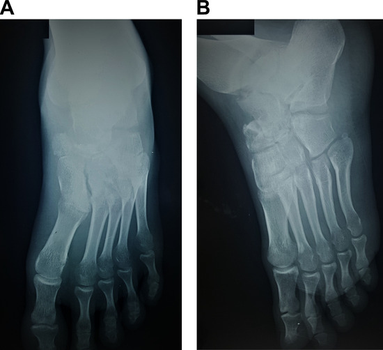
Figure 1.
Preoperative radiographic images showing a left talar fracture combined with plantar talar head dislocation and a navicular fracture: dorsoplantar (A) and oblique (B) views of the left foot.
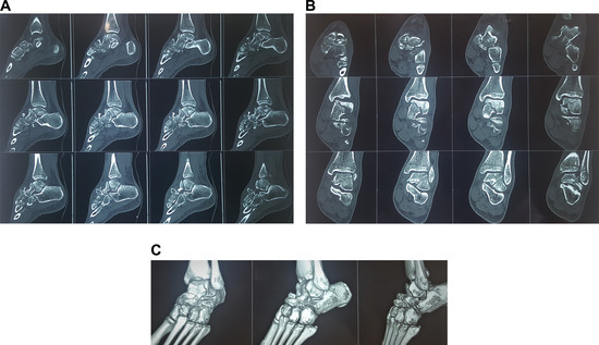
Figure 2.
The sagittal (A), coronal (B), and three-dimensional rendering (C) of left ankle computed tomographic scans showing a coronal shearing talar body fracture with talar head dislocation and a navicular compression fracture.
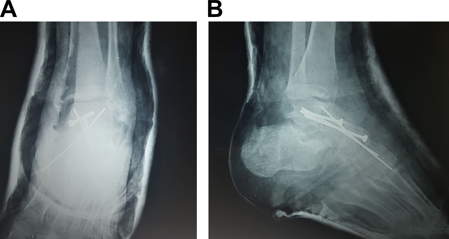
The planned operative treatment was performed. A dorsal approach was used. An 8-cm longitudinal incision between the tibialis anterior tendon and the extensor hallucis longus tendon was made, starting from distal tibia to medial cuneiform bone. The talar head was reduced. The talar body fracture was fixed with Kirschner wires and 4.0-mm cancellous screws. The talonavicular joint was relocated and fused with an iliac crest bone autograft by Kirschner wires and 4.0-mm cancellous screws (Fig. 3). A short leg cast was placed on the patient, and he was not to bear weight on the foot for 10 weeks. After 10 weeks had passed, he started to put his weight on the left foot. He was able to put his full weight on his foot after 20 weeks.
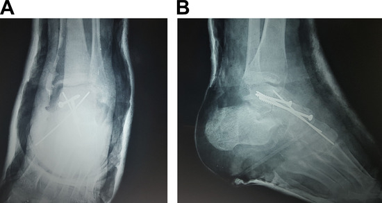
Figure 3.
Postoperative radiographic images showing an acceptable bone reduction and fixation: anteroposterior (A) and lateral (B) views of the left ankle.
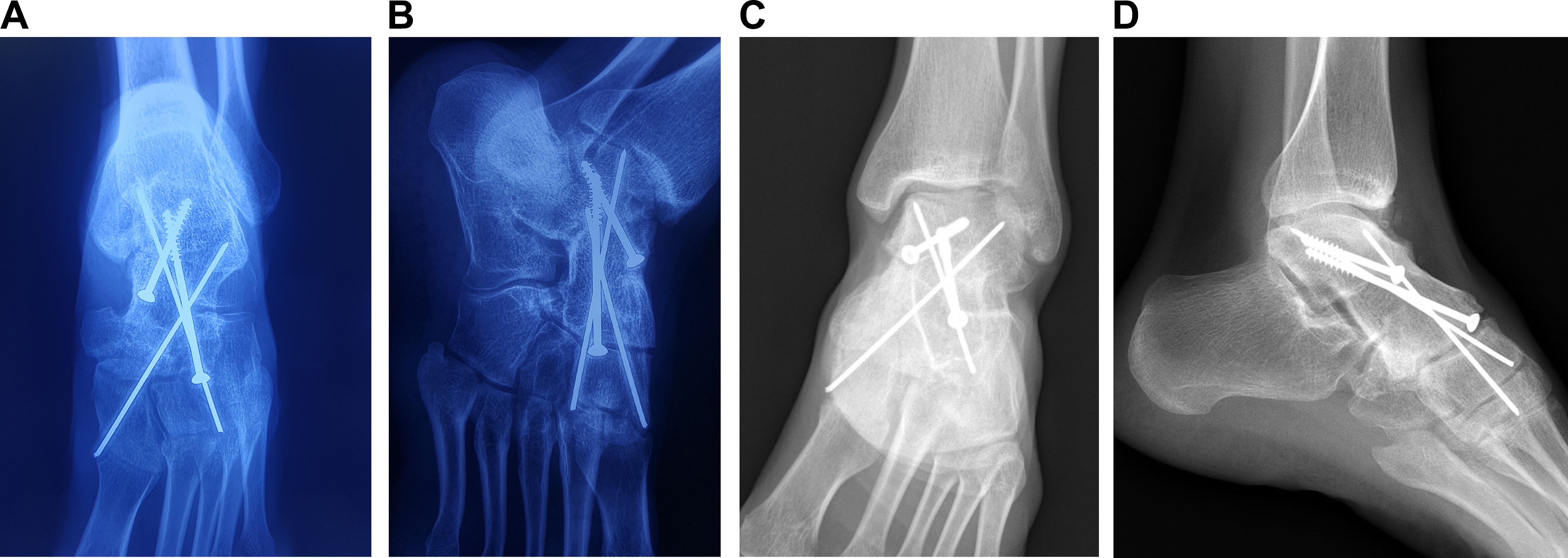
At the time of the present evaluation, 4 years had passed since the patient had the operation to repair the fracture. The fracture united well, with some small osteophytes within the anterior talotibial compartment and normal subtalar joint (Fig. 4). He had 40° of active plantarflexion, 20° of active dorsal flexion, and normal active inversion and eversion. He could walk and run without pain. The patient’s American Orthopedic Foot and Ankle Society hindfoot score was 99. The patient signed a consent form documenting that he agreed for his case to be reported to the public.
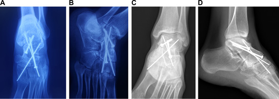
Figure 4.
Postoperative radiography at 4 years showed good fracture union with some small osteophytes within the anterior talotibial compartment and normal subtalar joint: dorsoplantar (A) and oblique (B) views of the left foot; anteroposterior (C) and lateral (D) views of the left ankle.
Discussion
Fracture of the talar body is an intraarticular fracture, whereas fracture of the talar neck is an extraarticular fracture. This distinction seems well defined, but these fracture locations are not easy to determine in some circumstances. Sometimes, talar neck fractures are extremely difficult to differentiate from coronal shearing fractures of the talar body. A fracture of the talar body is identified by a fracture line behind the lateral process of the talus that entered the articular surface of posterior facet [1]. In our case, it was difficult to determine which type of talar fracture occurred based on radiographic images alone. Computed tomographic images revealed that the fracture line from the articular surface of the distal tibia extended to the posterior facet of the talus.
Coronal shearing of the talar body fracture mechanism is similar to talar neck fracture and typically results from an axial force applied to a dorsiflexion foot in certain circumstances, such as falling from a height or motorcycle accident [4]. The specific mechanism of injury in our case was unclear because the patient did not know the position of his foot when he landed on the ground. In our opinion, none of the mechanisms of talar fracture described previously explain the variety of injuries in our case.
A shearing fracture of the talar body is usually related to talar body dislocation, resulting in talotibial joint and subtalar joint dislocation and/or subluxation. Sneppen et al [2] reported 17 patients with shearing fracture of the talar body among 51 patients with talar body fractures, including four patients with nondisplaced fracture, three patients with displaced fracture without any adjacent joint dislocation and/or subluxation, and 10 patients with displaced fracture with talotibial and subtalar joint dislocation and/or subluxation without any patients with talar head dislocation. Although Sneppen et al [2] described a systematic classification of talar body fractures, Boyd and Knight [3] and Delee (cited in Sanders [4]) classified shearing fractures of the talus, and a fracture of the talus was recently classified by Weber and Martin (cited in Sanders [4]) also, some unclassified varieties of talar body fractures have been reported in the scientific literature, such as talar body fracture with closed pantalar dislocation [5,6], and combined talar body fracture with malleolus fracture [7–10].
Fracture dislocation of the talar head from the navicular articulation with an undisplaced talar body inside the ankle mortise is extremely rare. A report of a series of 20 talar neck fractures by Pantazopoulos et al [11] included one case of an open talar neck fracture in which the head of the talus was superiorly dislocated from the talonavicular joint, with the body of the talus retaining its normal position in the ankle and subtalar joint. Khazim and Salo [12] wrote a case report of talar neck fracture with talar head dislocation and intact ankle and subtalar joints combined with an ipsilateral cuboid fracture. The talar head was dislocated out of the talonavicular joint and rotated approximately 180° around the tibialis posterior tendon. Both the talus and cuboid were reduced with the open approach and internal fixation with crews. Veerappa and Gopalkrishna [13] reported a rare case of talar neck fracture with talar head dislocation and intact ankle and subtalar joints. The talar head fragment rotated counterclockwise, and the fractured surface of the talar head was opposite the anterior facet of the subtalar joint. The patient was treated with open reduction and internal fixation with two cancellous screws and talonavicular ligament repair. Kapoor and Patra [14] encountered a case of talar neck fracture with talonavicular dislocation related to fracture of the posteromedial talar tubercle in combination with an intact ankle and subtalar joint subluxation. The talar head was dorsolaterally dislocated out of the talonavicular joint. The large posteromedial fragment was posteriorly displaced. The injuries were treated with open reduction and internal fixation with screws. At 28 months after operation, the fracture of talar neck was united but the fracture of the posteromedial talar tubercle remained unhealed. Hafez et al [15] described the first case of a closed talar body fracture with ankle and subtalar subluxation combined with talonavicular dislocation. The talar head was laterally rotated 90°. This patient was successfully treated with open reduction and internal fixation with screw and Kirschner wires. In our present case, the talar head was inferolaterally dislocated, resulting in a compression fracture of the navicular bone. The pattern of injury we discussed was a unique configuration of talar fracture dislocation.
We believe that the appropriate treatment of choice for our patient was stable anatomical reduction and fixation. The optimal treatment required restoration of the integrity of the talar articular surface and the congruity of the talonavicular joint. We decided to perform open reduction using a dorsal approach because the patient was evaluated late after the injury. Through this approach, we dealt with both the talar body fracture and talonavicular fracture dislocation. Because of the complexity of the talonavicular fracture dislocation, we performed primary talonavicular fusion with an iliac crest bone autograft. With our plan of treatment, a combined Kirschner wire and cancellous screw fixation was simple and effective. Although avascular necrosis and posttraumatic arthritis are the most common complications of talar body fractures, neither avascular necrosis nor remarkable posttraumatic arthritis was found after 4 years. The fracture united well, and the patient was satisfied with the operative outcome.
Conclusions
A coronal shearing fracture of the talar body with plantar talar head dislocation and intact ankle and subtalar joints associated with a navicular compression fracture is a very rare injury pattern. This injury was successfully treated with open reduction and internal fixation with Kirschner wires and screws and talonavicular joint fusion with an iliac crest bone graft performed by trained orthopedic surgeons.
Financial Disclosure
None.
Conflicts of Interest
None.
References
- LeeC, BrodkeD, PerduePW, : Talus fractures: evaluation and treatment. J Am Acad Orthop Surg28: e878, 2020.
- SneppenO, ChristensenSB, KrogsøeO, : Fracture of the body of the talus. Acta Orthop Scand48: 317, 1977.
- BoydH, KnightR: Fractures of the astragalus. South Med J35: 160, 1942.
- SandersR, LindvallE: “Fractures and Fracture—Dislocations of the Talus,” in Surgery of the Foot and Ankle, 8th ed, MJCoughlin, RAMann, CASaltzman (eds), 2075, Mosby, St. Louis, 2007.
- StirtonJB, EbraheimNA, RamineniSK: Medial peritalar fracture dislocation of the talar body. Trauma Case Rep1: 32, 2015.
- PatraSK, SwaroopS, PatroBP, : Talar body fracture with closed pan-talar dislocation. Cureus13: 15349, 2021.
- LahrachK, El KadiKI, MarzoukiA, :. Talar body fracture combined with medial malleolar fracture: a case report and literature review. Pan Afr Med J32: 57, 2019.
- OesmanI, NugrohoA: A rare combination of closed fracture of right talar body Sneppen 2 with associated medial malleolus: a case report. Int J Surg Case Rep65: 20, 2019.
- ChenK-J, KoC-Y, HoT-Y, : A combination of bimalleolar fracture and fracture on talar body and neck: a rare case report. Medicine (Baltimore)99: e20862, 2020.
- MogerNM, PragadeeshwaranJ, BanerjeeS, : Combined talus fracture with medial malleolar fracture: a case report with review of literature. J Orthop Case Rep10: 11, 2020.
- PantazopoulosT, GalanosP, VayanosE, . Fractures of the neck of the talus. Acta Orthop Scand45: 296, 1974.
- KhazimR, SaloPT: Talar neck fracture with talar head dislocation and intact ankle and subtalar joints: a case report. Foot Ankle Int16: 44, 1995.
- VeerappaLA, GopalkrishnaC: Talar neck fracture with talar head dislocation and intact ankle and subtalar joints—a rare case report. Foot20: 39, 2010.
- KapoorSK, PatraSR: Talar neck fracture associated with talonavicular dislocation and fracture of posteromedial process of talus. J Foot Ankle Surg50: 758, 2011.
- HafezMA, BawarishMA, GuvvalaR: Closed talar body fracture with talonavicular dislocation; a case report. Foot Ankle Int21: 599, 2000.
© 2023 American Podiatric Medical Association