


Abstract
Nevoid melanoma (NeM) is a rare variant of malignant melanoma characterized by slight cellular atypia, polymorphism, and incomplete maturation. It most frequently occurs on the trunk and arms but rarely on the foot. Here, we report a subungual NeM of the right hallux. A 65-year-old man presented with severe pain of 6 months’ duration to his right great toe following self-treatment for an ingrown nail. He was evaluated and treated with debridement of the toenail at an urgent medical center 3 months prior. However, this had not relieved his pain. The patient also noticed discoloration of his distal great toe over the past 3 months. Removal of part of the ingrown nail revealed a pigmented mass extending distally from the matrix. Surgical excision of the mass was performed because of the concern for malignancy. The diagnosis of NeM was based on histologic analysis along with enhanced diagnostic modalities. The patient was further treated with surgical amputation of the great toe and anti–programmed cell death-1 therapy. The patient had no relapse at 1-year follow-up. Nevoid melanoma is a rare variant of malignant melanoma on the toes, which needs to be differentiated from a nevus with atypia, with a variety of modalities including cellular and molecular profiling. The optimal treatment is amputation.
Nevoid melanoma (NeM) defines a rare variant of malignant melanoma. It accounts for less than 2% of all cutaneous melanomas.[1] It has been reported in all age groups, from children[2,3] to adults, and in both genders,[4,5] but it is predominantly found in male patients in their 50s and 60s.[6] Clinically, it has a variety of presentations, including nodule (nevus-like), plaque, papule, or mixed types.[7] Nevoid melanoma resembles an ordinary compound or intradermal melanocytic nevus, and there are no significant differences in sex, size, or anatomical distribution between nevus and NeM.[8] However, it is a malignant lesion with high rates of local recurrence (75%), metastasis (37.5%–45%), and mortality (24.2%–37.5%) within 3 to 5 years.[1,4] It is therefore essential to discriminate nevus from NeM.
Distinction between nevus and NeM relies on detailed histologic analysis with adjunct diagnostic modalities, including immunohistochemistry and molecular profiling. Histologically, NeM is characterized by slight atypia and polymorphism, incomplete maturation, increased mitoses, infiltration of adnexa, infiltrative growth, pigmented tumor cells, sharply demarcated tumor nests in the deeper dermis, and lack of a prominent intraepidermal component.[1,9] It has been histologically classified into several growth patterns, including “classic” NeM, superficial spreading melanomas with invasive NeM, lentigo maligna with invasive NeM, and deep penetrating nevus-like NeM.[7,10] Immunohistochemically, approximately 70% of NeMs exhibit complete loss of p16 expression, which is highly correlated with loss of the CDKN2A gene (chromosome 9p21) demonstrated by cytogenetic analysis.[8] Fluorescence in situ hybridization (FISH) analysis reveals NeMs but not mitotically active nevi having copy number abnormalities in either chromosome 6, 8, or 11.[10,11] Furthermore, next-generation sequencing analysis demonstrates that most NeMs carry hotspot mutations in NRAS and noncoding mutations.[12] These results suggest that FISH-targeting specific chromosomal loci on chromosomes 6, 8, 9, and 11 and molecular profiling play important roles in the differential diagnosis of NeM from nevi.[11]
Nevoid melanoma is most frequently localized on the trunk and arms,[6] but rarely on the foot. There are no reported NeMs on the toes in the English literature. We present a case of NeM, emanating from the subungual matrix of the right great toe in a 65-year-old man. We highlight its clinical features, diagnosis with different modalities, and treatment.
Case Report
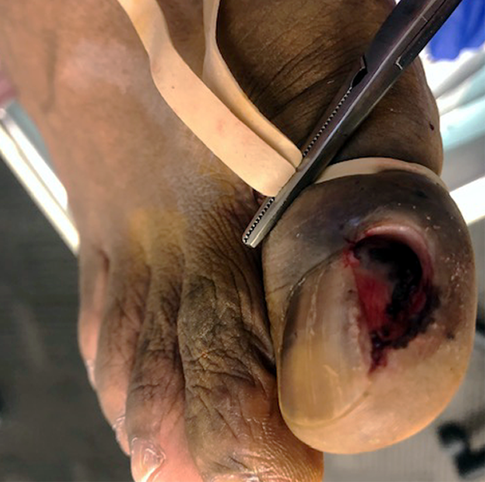
A 65-year-old man complained severe pain to his right great toe for 6 months after self-treatment of his ingrown nail. The patient was initially treated with debridement of his right great toenail, amoxicillin-potassium clavulanate for an infection, and hydrocodone-acetaminophen for his pain in an urgent care center 3 months prior. However, this did not relieve his pain. The patient continuously cut his nail away from the area of pain, but this only aggravated his pain. He also noticed the growth and discoloration of the distal skin of the right great toe over the previous 3 months. Physical examination revealed a suspected ingrown nail on the medial border, with evidence of previous debridement/wedge resection, and a pigmented granulation tissue noted distal to the remaining ingrown nail. Because of the concern for malignancy, the patient agreed to local excision of the mass and surrounding tissues for diagnosis. Surgical removal of the remaining portion of the ingrown nail revealed a 1.0 × 1.0-cm pigmented mass extending distally from the matrix. The mass and surrounding tissues were excised surgically (Fig. 1) and submitted to pathologic analysis.
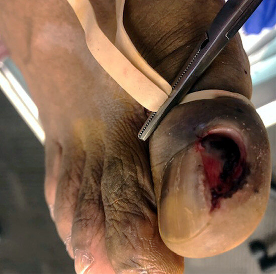
Figure 1.
Clinical presentation of the nevoid melanoma on the right hallux. Removing the top part of the pigmented mass revealed the remaining portion of the pigmented tumor tissue, distally extending from the matrix on the medial border of the toe.
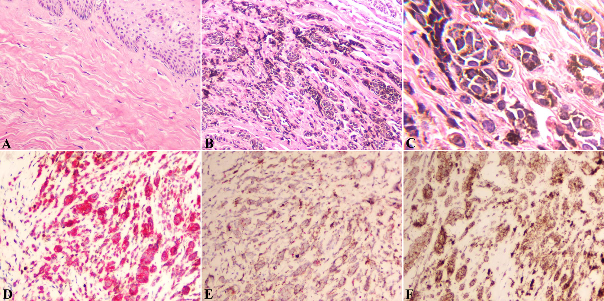
Histologic and immunostaining evaluations of the mass revealed nests and individual pigmented melanocytes within the dermis (Fig. 2), which suggested a diagnosis of an intradermal nevus with atypia. Further interphase FISH analysis performed on paraffin-embedded tissue slides, with MelanoCITE probe sets (NeoGenomics, Aliso Viejo, California) including RREB1 (6p25), c-MYC (8q24), CDKN2A (p16/CEN 9), and CCND1 (11q13),[4] used for the diagnostic discrimination of melanoma from nevus,[11,13] showed 38% of melanocytes having extra copies of RREB1 (6p25). Moreover, polymerase chain reaction–based high-sensitivity Sanger sequencing detected the BRAF mutation at nucleotide 1799 (T>A), which leads to a substitution of valine (V) for glutamic acid (E) at position 600 (p.V600E). These cellular and molecular analyses supported a final diagnosis of NeM.
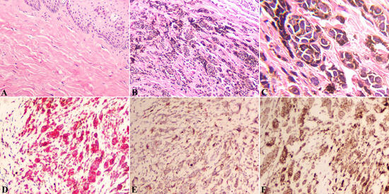
Figure 2.
Histopathology of the nevoid melanoma on the right hallux. A (HE, ×100), No junctional components are seen in the epidermis. B (H&E, ×100) and C (H&E, ×400), Single and nested melanocytes with finely distributed melanin pigments within the dermis showing slight cellular atypia with increased size and irregular shape but without obvious mitotic figures. D, Double immunostaining revealing the strong cytoplasmic expression of Malan-A (Alka Phospha red, [Ventana Medical Systems, Oro Valley, Arizona] ×100) and sporadic Ki-67 nuclear expression (diaminobenzidine, ×100) in the melanocytes. E, Patchy expression of HMB-45 in the melanocytes (Alka Phospha red, ×100). F, Cytoplasmic expression of BRAF in the melanocytes (diaminobenzidine, ×100).
The patient was further treated with right great toe amputation with lymphadenectomy at the Levine Cancer Institute (Charlotte, North Carolina). Pathologic examination revealed micrometastasis in the right sentinel lymph node (1/1) but not in other inguinal lymph nodes. The NeM was classified as pT3aN1aM0. The patient was subsequently treated with anti–programmed cell death-1 (PD1) antibody. The patient had no relapse or recurrence at a follow-up of 1 year.
Discussion
The differential diagnosis between intradermal nevus with atypia and NeM is crucial for this patient’s treatment. Histologic evaluation with immunohistochemistry is not enough to make an accurate diagnosis.[14] Other modalities, including FISH- and polymerase chain reaction–based sequencing, are needed.
Genetic study demonstrated 50% to 70% of melanomas harboring BRAF mutations, with 97% in codon 600.[15–17] BRAF-mutated melanomas frequently occur in younger patients and present with a superficial spreading or nodular morphology, aggregated in locations without chronic sun exposure, and associated with highly aggressive behavior with metastasis to the brain and shorter survival.[18–20] Functionally, BRAF mutations constitutively activate a BRAF-encoded cytoplasmic serine–threonine kinase, resulting in activating the downstream mitogen-activated protein kinase (MAPK) pathway involved in melanomagenesis.[21,22] Furthermore, the overall survival and progression-free survival rates in patients with BRAF V600E-mutant advanced melanoma were markedly improved with combined targeted therapies with BRAF V600E-mutated kinase inhibitors such as vemurafenib and dabrafenib, and downstream mitogen-activated protein kinase inhibitors including trametinib and cobimetinib.[23–27] Molecular testing of BRAF mutations thus becomes essential for not only diagnosis but also therapeutics in melanoma.
The standard treatment of NeM on the toes is surgical excision, which includes either wide local excision or toe amputation. The treatment option for the individual patient should be based on the tumor size, location, relationship with surrounding anatomical structures, and clinical staging.[28] The current case was treated with whole-toe amputation because of the subungual location and potential remaining tumor cells in the matrix after initial surgical excision. The patient was further treated with anti-PD1 antibody. A sustained recurrence-free survival benefit with anti-PD1 therapy was demonstrated in phase III trials in resected stage IIIB-C or IV melanoma patients.[29] This patient had no relapse or recurrence at a follow-up of 1 year.
Conclusions
Nevoid melanoma is a rare variant of melanoma on the toes, which clinically and histologically resembles a nevus with atypia. Multiple diagnostic modalities, including histologic evaluation with immunohistochemistry and molecular profiling, are needed to distinguish NeM from nevus with atypia. The best treatment is surgical excision with immunotherapy.
Financial Disclosures
None reported.
Conflicts of Interest
None reported.
References
- Schmoeckel C, Castro CE, Braun-Falco O: Nevoid malignant melanoma. Arch Dermatol Res 277: 362, 1985.
- Knöpfel N, Martín-Santiago A, Del Pozo LJ, : Amelanotic naevoid melanoma in a 16-month-old albino infant. Clin Exp Dermatol 42: 84, 2017.
- Cassarino DS, Fullen DR, Sondak VK, : Metastatic nevoid melanoma in a 4 1/2-year-old child. J Cutan Pathol 30: 647, 2003.
- Zembowicz A, McCusker M, Chiarelli C, : Morphological analysis of nevoid melanoma: a study of 20 cases with a review of the literature. Am J Dermatopathol 23: 167, 2001.
- Pampena R, Lai M, Lombardi M, : Clinical and dermoscopic features associated with difficult-to-recognize variants of cutaneous melanoma: a systematic review. JAMA Dermatol 156: 430, 2020.
- Idriss MH, Rizwan L, Sferuzza A, : Nevoid melanoma: a study of 43 cases with emphasis on growth pattern. J Am Acad Dermatol 73: 836, 2015.
- Longo C, Piana S, Marghoob A, : Morphological features of naevoid melanoma: results of a multicentre study of the International Dermoscopy Society. Br J Dermatol 172: 961, 2015.
- Mesbah Ardakani N, Singh S, Thomas C, : Mitotically active nevus and nevoid melanoma: a clinicopathological and molecular study. Am J Dermatopathol 43: 182, 2021.
- Magro CM, Crowson AN, Mihm MC: Unusual variants of malignant melanoma. Mod Pathol 19(suppl 2): S41, 2006.
- Yélamos O, Busam KJ, Lee C, : Morphologic clues and utility of fluorescence in situ hybridization for the diagnosis of nevoid melanoma. J Cutan Pathol 42: 796, 2015.
- Gerami P, Wass A, Mafee M, : Fluorescence in situ hybridization for distinguishing nevoid melanomas from mitotically active nevi. Am J Surg Pathol 33: 1783, 2009.
- Jackett LA, Colebatch AJ, Rawson RV, : Molecular profiling of noncoding mutations distinguishes nevoid melanomas from mitotically active nevi in pregnancy. Am J Surg Pathol 44: 357, 2020.
- Gerami P, Li G, Pouryazdanparast P, : A highly specific and discriminatory FISH assay for distinguishing between benign and malignant melanocytic neoplasms. Am J Surg Pathol 36: 808, 2012.
- Lezcano C, Jungbluth AA, Busam KJ: Comparison of immunohistochemistry for PRAME with cytogenetic test results in the evaluation of challenging melanocytic tumors. Am J Surg Pathol 44: 893, 2020.
- Ihle MA, Fassunke J, König K, : Comparison of high resolution melting analysis, pyrosequencing, next generation sequencing and immunohistochemistry to conventional Sanger sequencing for the detection of p.V600e and non-p.V600e BRAF mutations. BMC Cancer 14: 13, 2014.
- Richtig G, Hoeller C, Kashofer K, : Beyond the BRAFV600e hotspot: biology and clinical implications of rare BRAF gene mutations in melanoma patients. Br J Dermatol 177: 936, 2017.
- Cheng L, Lopez-Beltran A, Massari F, : Molecular testing for BRAF mutations to inform melanoma treatment decisions: a move toward precision medicine. Mod Pathol 31: 24, 2018.
- Long GV, Menzies AM, Nagrial AM, : Prognostic and clinicopathologic associations of oncogenic BRAF in metastatic melanoma. J Clin Oncol 29: 1239, 2011.
- Ribas A, Flaherty KT: BRAF targeted therapy changes the treatment paradigm in melanoma. Nat Rev Clin Oncol 8: 426, 2011.
- Hugdahl E, Kalvenes MB, Puntervoll HE, : BRAF-V600E expression in primary nodular melanoma is associated with aggressive tumour features and reduced survival. Br J Cancer 114: 801, 2016.
- Pratilas CA, Solit DB: Targeting the mitogen-activated protein kinase pathway: physiological feedback and drug response. Clin Cancer Res 16: 3329, 2010.
- Cohen C, Zavala-Pompa A, Sequeira JH, : Mitogen-actived protein kinase activation is an early event in melanoma progression. Clin Cancer Res 8: 3728, 2002.
- Long GV, Weber JS, Infante JR, : Overall survival and durable responses in patients with BRAF V600-mutant metastatic melanoma receiving dabrafenib combined with trametinib. J Clin Oncol 34: 871, 2016.
- Long GV, Stroyakovskiy D, Gogas H, : Dabrafenib and trametinib versus dabrafenib and placebo for Val600 BRAF-mutant melanoma: a multicentre, double-blind, phase 3 randomised controlled trial. Lancet 386: 444, 2015.
- Robert C, Karaszewska B, Schachter J, : Improved overall survival in melanoma with combined dabrafenib and trametinib. N Engl J Med 372: 30, 2015.
- Ascierto PA, McArthur GA, Dréno B, : Cobimetinib combined with vemurafenib in advanced BRAF(V600)-mutant melanoma (coBRIM): updated efficacy results from a randomised, double-blind, phase 3 trial. Lancet Oncol 17: 1248, 2016.
- McArthur GA, Chapman PB, Robert C, : Safety and efficacy of vemurafenib in BRAF(V600E) and BRAF(V600K) mutation-positive melanoma (BRIM-3): extended follow-up of a phase 3, randomised, open-label study. Lancet Oncol 15: 323, 2014.
- Hao X, Yim J, Chang S, : Acral lentiginous melanoma of foot and ankle: a clinicopathological study of 7 cases. Anticancer Res 39: 6175, 2019.
- Ascierto PA, Del Vecchio M, Mandalá M, : Adjuvant nivolumab versus ipilimumab in resected stage IIIB-C and stage IV melanoma (CheckMate 238): 4-year results from a multicentre, double-blind, randomised, controlled, phase 3 trial. Lancet Oncol 21: 1465, 2020.
© 2023 American Podiatric Medical Association