


Abstract
Eccrine porocarcinoma is a rare malignant tumor of the eccrine sweat gland. This malignancy occurs most commonly in the lower extremities. It tends to occur in patients aged 60 to 80 years, affecting men and women equally. We present the case of a 62-year-old man with a lesion on the left foot. The diagnosis of the initial biopsy was squamous cell carcinoma. Six months later, the lesion reoccurred, and a second biopsy confirmed it to be eccrine porocarcinoma.
Eccrine porocarcinoma is a rare malignant tumor of the eccrine sweat gland, accounting for 0.005% of skin tumors [1]. The first reported case dates to 1963 and was coined as epidermotropic eccrine carcinoma. There has been limited documentation since then due to lack of diagnosis. In 1969, the term eccrine porocarcinoma was introduced by Mishma and Morimoka [2]. The tumor can arise either de novo of the intraepithelial portion of the eccrine sweat gland, termed acrosyringeum, or as a malignant transformation of a benign poroma [3,4]. Although the malignancy can present at any age, it occurs most commonly between 60 and 80 years, with a mean age of 73 years [5,6]. It is not gender specific, and both sexes are usually equally affected [7,8].
Forty percent of reported cases occur in the lower extremity, which is where the malignancy is most common [4]. Other reported sites include the head, scalp, upper extremities, trunk, and abdomen [9]. Twenty percent of eccrine porocarcinoma will metastasize to regional lymph nodes, and 25% will reoccur [10]. The progression of eccrine porocarcinoma from a benign lesion to a malignancy is unpredictable.
The clinical appearance of eccrine porocarcinoma is variable. It can occur slowly, with a mean average of 8.5 years, or progress rapidly [11]. Appearance of the lesion is also an important fact to consider because the malignancy can present as nodules, papules, or plaquelike, with size varying from 1 to 130 cm [12]. Pigmentation of the lesions can be erythematous, violaceous, brownish, or skin-colored tones [13]. Due to its varied presentation, eccrine porocarcinoma can resemble many benign and malignant neoplasms, and clinical diagnosis cannot be based on appearance alone.
In this case study, we describe a 62-year-old man with a recurring lesion to the left fifth toe that was initially diagnosed as squamous cell carcinoma (SCC); however, the lesion reoccurred, which prompted further evaluation, which led to a diagnosis of eccrine porocarcinoma.
Case Report
A 62-year-old man with a history of type 2 diabetes mellitus, hyperlipidemia, hypertension, and vitamin D deficiency presented to the surgical oncology department of a local hospital with a lesion on the left fifth toe for the past 8 months. The patient stated that the lesion started as a blister, which he lanced with a sewing needle. The patient went to his primary care physician because he saw no improvement to the blister and had worsening pain. The patient was placed on antibiotic drug treatment for his skin infection, which showed notable improvement; however, the lesion reoccurred 6 months later. The patient was referred to a podiatric physician for further evaluation and treatment.
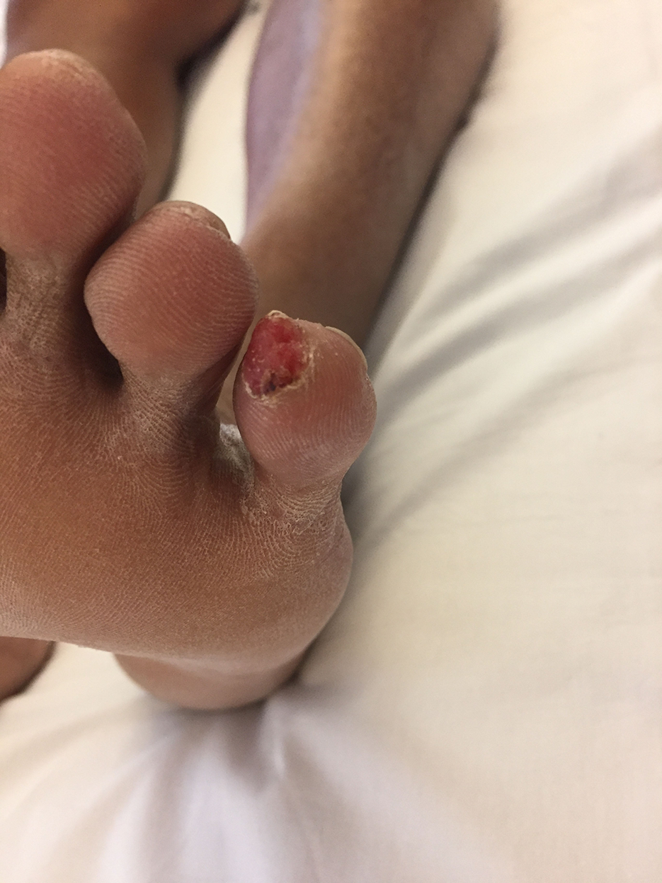
The podiatric physician referred the patient for follow-up care with the hospital for concerns of SCC indicated by punch biopsy findings. Magnetic resonance imaging indicated no bony involvement. The surgical oncology and dermatology departments were consulted regarding the biopsy results and noted a 1-cm, erythematous, fungating plaque on the fifth digit of the left foot (Fig. 1).
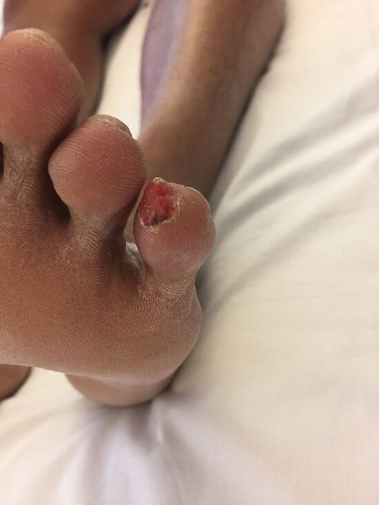
Figure 1.
A 1-cm, erythematous, fungating plaque on the fifth digit of the left foot.
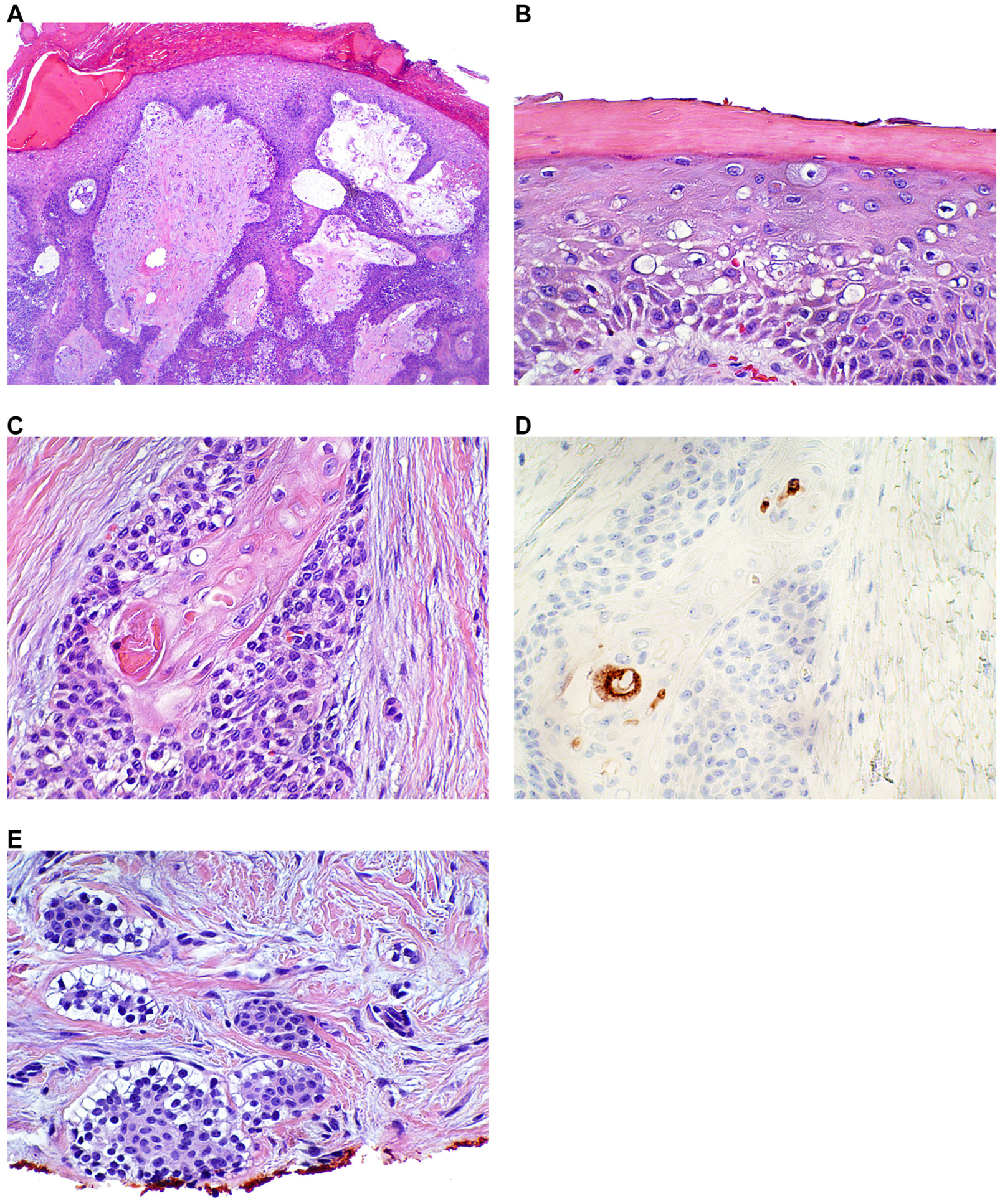
The pathology department reviewed the pathology slides and described variably sized aggregates of epithelium with clear changes and squamatization in an infiltrating pattern in a fibrotic stroma. Histopathologic analysis demonstrated an infiltrating tumor with multifocal attachment to the epidermis overlying in situ carcinoma with pagetoid spread and squamatized tumor islands with eccrine ductal differentiation confirmed by staining with carcinoembryonic antigen (CEA) (Fig. 2). The lesional cells were positive for p63, epithelial membrane antigen (EMA), and, focally, CEA, leading to a revised diagnosis consistent with eccrine porocarcinoma. Dermatopathologists recommended and performed a shave biopsy, which confirmed the presence of eccrine porocarcinoma.
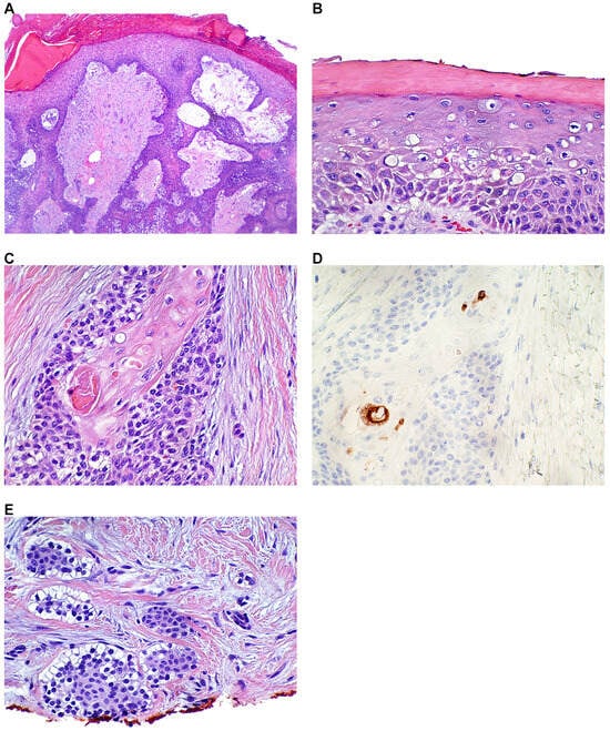
Figure 2.
Histopathologic analysis. A, Porocarcinoma demonstrating an infiltrating tumor with multifocal attachment to the epidermis. B, Overlying in situ carcinoma with pagetoid spread. C, Squamatized tumor islands with eccrine ductal differentiation (arrow). D, Confirmed by staining with carcinoembryonic antigen. E, Note clear cell change of the infiltrating islands at the deep specimen margin and the prominent fibromyxoid stroma.
A recommendation to amputate the affected digit to achieve adequate margins was discussed during a multidisciplinary cutaneous oncology conference. A sentinel lymph node biopsy was not advised due to unclear benefit. The patient underwent a left fifth-digit amputation, with the surgical specimen measuring 4.1 × 1.9 × 1.8 cm. The final pathology report confirmed eccrine porocarcinoma.
The patient had multiple follow-up visits with the multidisciplinary team and had healed well. Dermatology and surgical oncology followed the patient for 1 to 2 years after the surgery to monitor for possible recurrence. Further pathology reports were discussed, and all of the residual carcinoma was excised with negative margins. Overall, the patient tolerated the surgical amputation well, with no noted complications.
Discussion
Eccrine porocarcinoma is an aggressive malignant tumor that arises either de novo from the intraepithelial portion of eccrine sweat glands or from an eccrine porocarcinoma. There are few cases of eccrine porocarcinoma of the foot reported, and the progression and reappearance of this malignant lesion were extremely rapid, with a course of 6 months, which prompted further diagnostic testing for confirmation. Due to the rarity and common physical appearance, it was difficult to confirm a positive initial conformational diagnosis.
The clinical differential diagnosis included eccrine porocarcinoma, verruca vulgaris, seborrheic keratosis, pyogenic granuloma, SCC, basal cell carcinoma, malignant melanoma, cutaneous lymphoma, Bowen disease, and other primary skin appendage tumors [3]. Invasive porocarcinoma can be differentiated from SCC by the absence of ductal differentiation of SCC and can show continuity with the surface epithelium. It may be associated with a broad, pushing, deep margin or a more obvious infiltrative lower border and may show lymphovascular invasion [14].
Although morphological and histologic analyses are the mainstay for diagnosing porocarcinoma, an immunohistochemical panel that includes CEA, EMA, CAM5.2, CK7, CK15, CK19, BerEP4, and CD117 may help in distinguishing between the two carcinomas [3,15]. The lesion in the present patient showed p63, EMA, and, focally, CEA. Even with a confirmation diagnosis of CEA, treatment was not as effective.
Chemotherapy with methotrexate, cisplatin, anthracycline, bleomycin, or isotretinoin, and interferon alpha have been used with partial or little response. Radiotherapy and chemotherapy were also ineffective for controlling tumor recurrence or metastasis [9]. Due to the nature of how rapidly this tumor can grow and with such limited amount of information of this rare cancer, treatment has little success. With a detailed history and physical examination and early sentinel lymph node biopsy, this cancer can be identified and diagnosed early, which can lead to better treatment outcomes. As for this patient with histopathologic and immunohistochemical evidence, the lesion was surgically excised with wide margins to prevent reoccurrence.
Conclusions
With aggressive malignant tumors such as eccrine porocarcinoma, one must obtain a thorough history and physical examination to be able to distinguish from SCC and to determine which type of treatment is appropriate; in the present patient, the tumor was surgically excised. Radical excision of the tumor is effective in dealing with the rare forms of eccrine porocarcinoma.
Financial Disclosure: None reported.
Conflict of Interest: None reported.
References
- Pinkus H, Mehregan AH: Epidermotropic eccrine carcinoma. Arch Dermatol 88: 597, 1963.
- Mishma Y, Morioka S: Oncogenic differentiation of the intra-epidermal eccrine sweat duct: eccrine poroma, poro-epithelioma, and porocarcinoma. Dermatologica 138: 238, 1969.
- Margaritescu I, Shalin S, Chirita A: “Porocarcinoma,” in Rare Malignant Skin Tumors, edited by F Rongioletti, I Margaritescu, BR Smoller, p 107, Springer, New York, 2015.
- Robson A, Greene J, Ansari N, et al: Eccrine porocarcinoma (malignant eccrine poroma): a clinicopathologic study of 69 cases. Am J Surg Pathol 25: 710, 2001.
- Sawaya J, Khachemoune A: Poroma: A review of eccrine, apocrine, and malignant forms. Int J Dermatol 53: 1053, 2014.
- Berke A, Grant-Kels JM: Eccrine sweat gland disorders: Part I–Neoplasms. Int J Dermatol 33: 79, 1994.
- Ruffieux C, Ramelet AA: Porocarcinoma eccrine. Dermatologica 170: 202, 1985.
- Walsh MS: A case of eccrine poroma. J R Soc Med 83: 529, 1990.
- Chang O, Elnawawi A, Rimpel B, et al: Eccrine porocarcinoma of the lower extremity: A case report and review of literature. World J Surg Oncol 9: 94, 2011.
- Okada N, Ota J, Sato K et al: Metastasizing eccrine sweat gland carcinoma. Report of a case. Arch Dermatol 120: 768, 1984.
- Pylyser K, De Wolf-Peeters C, Marine K: The histology of eccrine poromas. A study of fourteen cases. Dermatologica 167: 243, 1983.
- Behbahani S, Malerba S, Karanfilian KM, et al: Demographics and outcomes of eccrine porocarcinoma: Results from the National Cancer Database. Br J Dermatol 183: 161, 2020.
- Shiohara J, Koga H, Uhara H. et al: Eccrine porocarcinoma: Clinical and pathological studies of 12 cases. J Dermatol 34: 516, 2007.
- Vaz Salgado MA, García CG, Lopez Martin JA. et al: Porocarcinoma: clinical evolution. Dermatol Surg 36: 264, 2010.
- Goto K, Takai T, Fukumoto T. et al: CD117 (KIT) is a useful immunohistochemical marker for differentiating porocarcinoma from squamous cell carcinoma. J Cutan Pathol 43: 219, 2016.
© 2023 American Podiatric Medical Association