


Abstract
Necrotizing fasciitis is a devastating inflammatory infection requiring emergent medical treatment and surgical intervention. Even with timely management, the mortality rate of necrotizing fasciitis approaches 25%. The causative bacteria invade fascial planes and express toxins that advance rapidly. Here, we document a rare case of necrotizing fasciitis from Serratia marcescens infection. Serratia marcescens is capable of inducing a necrotizing inflammatory cascade mediated by extracellular cytotoxin and lipase. In this case report, a 90-year-old man presented to our emergency department from a long-term care facility with a relatively benign-appearing ulcer with surrounding cellulitis on the right ankle. Blood cultures and wound cultures confirmed the organism to be S marcescens. A multidisciplinary team was consulted for management. The patient received antibiotic therapy and medical support, but because of his comorbid conditions and social situation, the designated medical decision maker opted for comfort care rather than aggressive surgical debridement. The patient progressed through the clinical stages of necrotizing fasciitis. Within 36 hours, the patient died as result of sepsis-induced organ failure.
Necrotizing fasciitis (NF) is a severe inflammatory infection requiring emergent medical treatment and often surgical intervention, with the potential for devastating consequences.[1,2] Even with timely management, the mortality rate of necrotizing fasciitis is cited to be 24% or higher.[1,2,3] The causative bacteria express toxins that advance rapidly along fascial planes.[1,2] Presentation of NF is rare, with an incidence of approximately 0.40 cases per 100,000 individuals.[2] The true pathogenesis of NF is under debate.[1,2,3] Patients are often immunosuppressed with comorbid conditions including diabetes mellitus (DM), peripheral vascular disease, and chronic lymphedema.[1,2] An initial traumatic episode may be identified in some patients’ history as the antecedent event.[1,2] The development of NF can be surreptitious, with clinically unimpressive wounds.[1,2] Two-thirds of such cases are seen in the lower extremities.[1,2]
The primary causative organism cited is group A streptococcus.[1,2] Although streptococcal and polymicrobial infections are more common, many other organisms have been identified.[4,5] NF may also be caused by Clostridium, Staphylococcus aureus, Vibrio vulnificus, and Aeromonas. Interestingly, the rates of gram-negative organisms causing NF may be increasing because of the growing elderly population with multiple medical comorbidities.[4,5] Serratia species are gram-negative facultative anaerobes, with Serratia marcescens being the most frequently implicated species in infections, and the causative agent of the present case. They are members of the order Enterobacterales and characterized by their red pigment (prodigiosin) on specific stains.[4,5,6] Serratia marcescens infections are often a result of contacting shared surfaces in a community setting. Serratia marcescens can induce a necrotizing inflammatory cascade mediated by extracellular cytotoxin and lipase.
A review of recent literature was conducted on S marcescens NF cases from 1980 to 2022. MEDLINE was searched by means of PubMed.gov for all English articles containing the key words “necrotizing fasciitis,” “necrotizing infection,” “Serratia,” and/or “Serratia marcescens.” Twenty-six articles with 27 total patients were identified between 1987 and 2022.[6,7,8,9,10] All publications were case reports, with the exception of one case series containing three patients who suffered S marcescens infections, two with NF.[5] In total, 18 cases involved the lower extremity, with only six specifying the distal leg or foot (22.2%). Of the 27 cases, five (18.5%) involved patients without any comorbidities. Eight (29.6%) involved patients with DM, and four (14.8%) with documented peripheral vascular disease. The mean age was 44.7 years. Twelve (44.4%) of the patients were male.
With regard to S marcescens in particular, the calculated pooled mortality rate of 27 patients across 42 years of publications was 48.1%. The overall mortality was 73.1% with medical treatment alone (namely, antibiotic therapy) and 40.7% when including a surgical debridement or amputation. The mortality rate of S marcescens NF has been observed to be considerably higher than other causative organisms.[8,9,10] A multidisciplinary approach with heightened vigilance is salient to differentiate any wound infection from NF as early as possible. Any delay in medical and surgical intervention is associated with increasing mortality.[3]
In this article, we report a case of necrotizing fasciitis of the leg caused by S marcescens, along with a review of the literature over the past 42 years. Pooled mortality was assessed, demonstrating the considerable virulence and risks associated with S marcescens necrotizing fasciitis.
Case Report
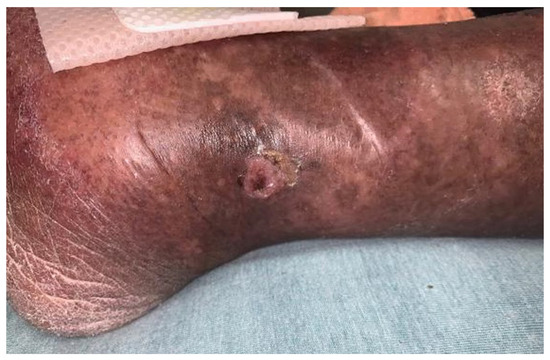
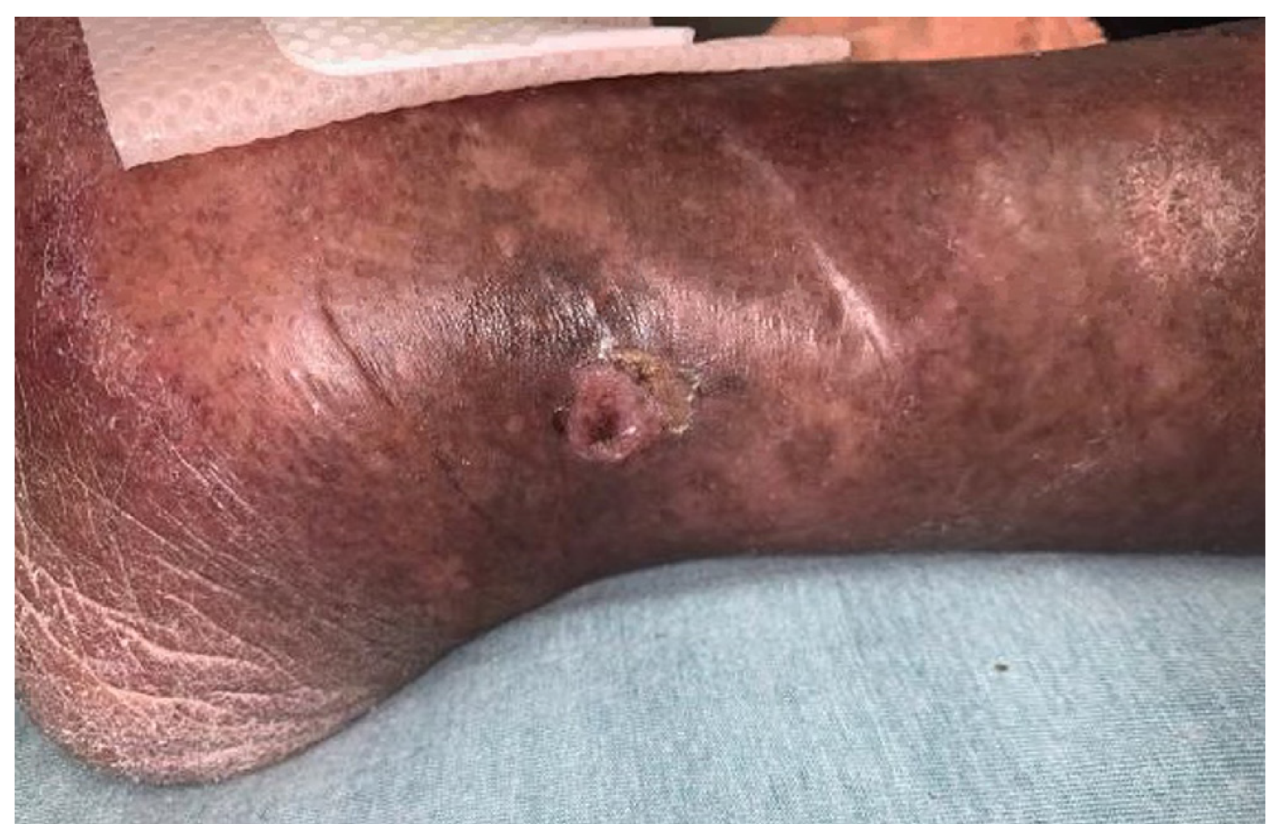
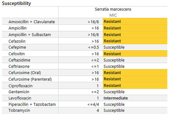
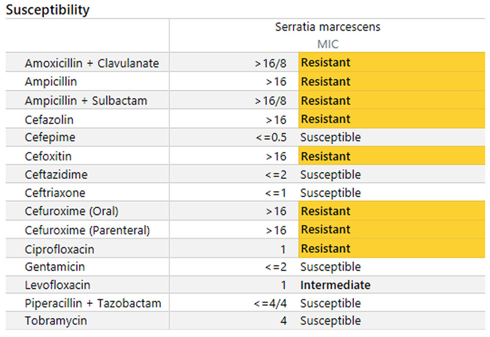
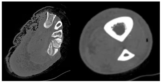
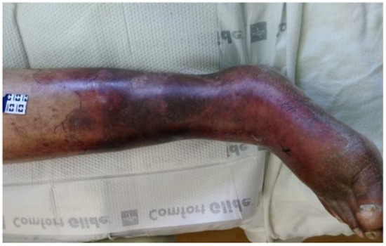
A 90-year-old man presented to the emergency department from a long-term care facility with increasing pain, erythema, and swelling surrounding a chronic right ankle wound. The patient had a medical history of dementia, hypertension, type 2 diabetes mellitus, chronic kidney disease, benign prostatic hyperplasia, chronic treated hepatitis C, and peripheral vascular disease. Physical examination showed moderate induration circumferentially about a full-thickness ulcer at the right medial malleolus as shown in Figure 1. Podiatry and vascular surgery were consulted for evaluation. Laboratory markers were significant for a venous lactate level of 10.1 mmol/L. The patient was started on broad-spectrum antibiotics in the form of vancomycin and piperacillin-tazobactam. Final antibiotic susceptibilities are visualized in Figure 2. Computed tomography (CT) imaging showed diffuse swelling consistent with cellulitis but with no drainable fluid collections or subcutaneous gas. Repeated CT imaging 4 hours later demonstrated subcutaneous emphysema tracking in the medial foot and lateral leg fascial planes (Figure 3). The patient also had an abdomen-pelvis CT scan to rule out any intra-abdominal process contributing to his lactic acidosis. Wound cultures were positive for gram-negative rods, with final culture results growing S marcescens. Blood cultures were also positive for S marcescens, with antibiotic sensitivities illustrated in Figure 3. At this time, a diagnosis of NF was made, given the progression of infection proximally about the fascial planes, as shown in Figure 4. After multidisciplinary discussion, the patient was deemed a poor surgical candidate and would have likely required an above-the-knee amputation. A goals of care discussion took place with the family and the decision was made to forego full care.
Figure 1.
Initial clinical appearance of patient’s right lower extremity.
Figure 2.
Antibiotic susceptibilities of the infecting pathogen.
Figure 3.
Computed tomographic images in the axial plane of ensuring soft-tissue emphysema (bone window setting).
Figure 4.
Clinical appearance of patient’s right lower extremity 8 hours from initial presentation.
The patient received intravenous fluid resuscitation but remained hypotensive, with lactic acidosis increasing to 16 mmol/L (Table 1), and the patient was transferred to the intensive care unit for pressor support. Palliative care was consulted given the poor prognosis without surgical intervention. The patient progressed through the clinical stages of necrotizing fasciitis over the next 36 hours, as described in Table 2. On the second hospital day, the patient died with comfort measures.
Table 1.
Patient’s Laboratory Values on Presentation and 8 Hours into Admission
Table 2.
Clinical Stages of Necrotizing Fasciitis According to Wang et ala
Discussion
To the authors’ knowledge, this is the second case of NF following S marcescens infection at our institution in the past 5 years.[4] The case itself began had relatively mild clinical features. The obscure manifestation of NF can make it difficult to readily identify and treat.[6] The benign presentation of the present patient and their rapid clinical deterioration demonstrates the insidious nature of NF.
The first diagnostic clue was likely a markedly elevated lactate at admission. Another item was the patient’s C-reactive protein, which was elevated at admission to 33.8 mg/L, another classic marker for inflammation and infection.[4,5] However, awareness of the serum lactate in what appears to be mild-tomoderate cellulitis may aid in early diagnosis of NF.
A study in 2013 of 53 patients noted that a serum lactate greater than 2.0 mmol/L had a sensitivity of 1 and a specificity of 0.76 (P # .0001).[11] In another analysis of 707 NF cases, high lactate level was an independent risk factor for in-hospital morality (OR, 1.17; P = .001). Furthermore, the patient’s age and comorbid diagnoses of hypertension, diabetes mellitus, and coronary artery disease are associated with poor prognosis in NF.[3] It is also possible this was a subacute presentation, as the duration of infection is unclear before arrival from the long-term care facility. Mortality of subacute NF has been found to be higher than in acute infections.[5,6,7] However, the workup may favor an acute presentation, as the initial CT imaging was negative, whereas a repeated scan after 4 hours illustrated an infectious process within the subcutaneous fascial planes.
Conclusions
One major limitation in the present case is the lack of formal tissue pathology. Previous authors have used permanent and fresh-frozen pathology to aid diagnosis of NF.[14] Some characteristic microscopic findings include microorganisms and polymorphonuclear infiltration of the dermis and fascia, fibrinous thrombi and angiitis of vessels, and necrosis of superficial fascia.[11,12] Nevertheless, NF remains a clinical diagnosis.[6,7,8,9,12] Although the initial presentation of our patient appeared grossly benign, classic findings manifested within hours. These clinical markers included tissue with a lack of bleeding and resistance, without contracting muscles, and distinct nonpitting fascial edema.[8,9,10] Ecchymotic bullae formed also within 8 hours (Table 2), with considerable dermal mottling. Sharp debridement may have demonstrated characteristic gray necrotic tissue and/or dishwater purulence.[10] The patient’s baseline medical and mental state played a key role in his course. Extensive surgical debridement was offered. The absence of a lower extremity debridement or amputation is certainly a unique aspect of this case. The psychological and social dynamics are important to consider in all care, and particularly with a morbid diagnosis. Multi-disciplinary discussion, with palliative care consulted, lead to a decision to transition to comfort care.
Funding
None reported.
Conflicts of Interest
None reported.
References
- EDLICH, RF; CROSS, CL; DAHLSTROM, JJ. ET AL: Modern concepts of the diagnosis and treatment of necrotizing fasciitis. J Emerg Med 2010, 39, 261. [Google Scholar] [CrossRef] [Scilit] [PubMed]
- LANCEROTTO, L; TOCCO, I; SALMASO, R. ET AL: Necrotizing fasciitis: classification, diagnosis, and management. J Trauma Acute Care 2012, 72, 560. [Google Scholar] [CrossRef] [Scilit]
- JABBOUR, G; EL-MENYAR, A; PERALTA, R. ET AL: Pattern and predictors of mortality in necrotizing fasciitis patients in a single tertiary hospital. World J Emerg Surg 2016, 11, 1. [Google Scholar] [CrossRef] [Scilit] [PubMed]
- MAJUMDAR, R. CRUM-CIANFLONE NF: Necrotizing fasciitis due to Serratia marcescens: case report and review of the literature. Infection 2016, 44, 371. [Google Scholar] [CrossRef] [Scilit] [PubMed]
- MARIN, L; ROWAN, R; MANTILLA, A. ET AL: Lower-extremity infections caused by Serratia marcescens: a report of three cases and a literature review. JAPMA 2017, 107, 231. [Google Scholar] [CrossRef] [Scilit]
- WANG, YS; WONG, CH; TAY, YK. Staging of necrotizing fasciitis based on the evolving cutaneous features. Int J Dermatol 2007, 46, 1036. [Google Scholar] [CrossRef] [Scilit]
- VERALDI, S; NAZZARO, G. Skin ulcers caused by Serratia marcescens: three cases and a review of the literature. Eur J Dermatol 2016, 26, 373. [Google Scholar] [CrossRef] [Scilit] [PubMed]
- ROBERTS, M; CRASTO, D; ROY, D. A case of subacute necrotizing fasciitis due to Serratia marcescens. J Clin Aesthet Dermatol 2021, 14, 55. [Google Scholar] [PubMed]
- CHIDAMBARAM, M; CHARAN, MS; SARAGA, K. ET AL: Necrotizing fasciitis due to Serratia marcescens infection following snake bite. Indian J Pediatr 2022, 89, 414. [Google Scholar] [CrossRef] [Scilit]
- HEIGH, EG; MALETTA-BAILEY, A; HAIGHT, J. ET AL: Necrotizing fasciitis of the lower extremity caused by Serratia marcescens: a case report. JAPMA 2016, 106, 144. [Google Scholar] [CrossRef] [Scilit] [PubMed]
- HAGIYA, H; OJIMA, M; YOSHIDA, T. ET AL: Necrotizing soft tissue infection caused by Serratia marcescens: a case report and literature review. J Infect Chemother 2016, 22, 335. [Google Scholar] [CrossRef] [Scilit] [PubMed]
- MURPHY, G; MARKESON, D; CHOA, R. ET AL: Raised serum lactate: a marker of necrotizing fasciitis? J Plast Reconstr Aesthet Surg 2013, 66, 1712. [Google Scholar] [CrossRef] [Scilit] [PubMed]
- CHANG, CP; FANN, WC; WU, SR; ET, AL. Lactate on emergency department arrival as a predictor of in-hospital mortality in necrotizing fasciitis: a retrospective study. J Orthop Surg Res 2019, 14, 73. [Google Scholar] [CrossRef] [Scilit] [PubMed]
- STAMENKOVIC, I. LEW PD: Early recognition of potentially fatal necrotizing fasciitis: the use of frozen-section biopsy. N Engl J Med 1984, 310, 1689. [Google Scholar] [CrossRef] [Scilit] [PubMed]

Disclaimer/Publisher’s Note: The statements, opinions and data contained in all publications are solely those of the individual author(s) and contributor(s) and not of MDPI and/or the editor(s). MDPI and/or the editor(s) disclaim responsibility for any injury to people or property resulting from any ideas, methods, instructions or products referred to in the content. |
© 2023 American Podiatric Medical Association