


Abstract
Tillaux fractures in adults are rare and, if unrecognized, can lead to ankle fracture healing complications, early progression of arthritis, and limited ankle movement caused by pain and degenerative changes. The Tillaux fracture was first described by Paul Tillaux as an external rotation injury of the ankle, involving an avulsion fracture of the distal anterolateral tibia. This fracture can be easily overlooked on plain radiographs in the adult. A high index of suspicion for this type of fracture pattern along with the use of computed tomographic scanning can help confirm the suspected diagnosis, rule out other tibial injuries, and provide more information on the best course of action. Historically, Tillaux fractures have been more common in adolescents because of the open tibial epiphyseal plate. Once the epiphyseal plate fully closes, skeletal maturity is achieved, thus making it extremely unusual for the anterior tibiofibular ligament to cause an avulsion fragment of the distal anterolateral tibia. Because of how uncommon this type of fracture is in adults, it has rarely been reported in our literature. We reviewed the literature and present a case report of this rare fracture injury.
The Tillaux fracture was described by Paul Jules Tillaux in 1872 as an abduction and external-rotation injury mechanism that involves avulsion of the distal anterolateral tibia, following his experiments on cadavers. The fracture line is vertical, extending from the distal articular surface of the tibia upward to the lateral cortex with the avulsed bone fragment usually triangular. Chaput later described a similar injury to the posterolateral tibia (avulsion of posterior tibiofibular ligament), later called Tillaux-Chaput injury [1] The phase where the lateral distal tibial physis is progressing toward complete closure and gains skeletal maturity is known as the “transition period,” which is when an adolescent is most prone to Tillaux fractures with ankle injuries. Juvenile Tillaux fracture, Salter-Harris type III fracture injury, is the result of distal tibial physis fusing from medial to lateral, making the medial side stronger than the lateral side. The avulsed bony fragment is usually quadrangular. The open physeal plate of the distal tibia begins to fuse between the ages of 12 and 14 years in girls, from middle to medial, and finally lateral, and at ages 15 to 18 years in male patients in the same fashion.
We performed a literature review along with a case report of this rare ankle fracture injury in an adult. We performed a search using the PubMed, OVID, and Google Scholar databases from inception to April of 2022. Titles, subjects, and abstracts were searched for the following terms: Tillaux Chaput, occult Tillaux, AITFL avulsion fracture, distal lateral tibial fracture, distal tibial growth plate, lateral margin distal tibia fracture, articular distal tibia fracture, SER, tibiofibular syndesmosis, anterior malleolus, syndesmosis rupture, ankle fracture, and interosseous membrane. Terms were searched independently with the omission of any duplications. The inclusion criteria consisted of articles reporting on adult cases of Tillaux fracture, with either operative or nonoperative treatments, and with radiographic and/or computed tomographic (CT) findings. The exclusion criteria consisted of any case reports of nonadult cases and non–English language publications. There were 1,606 initial results. The search keywords and terms were then narrowed and filtered to human and English language and noted to produce 276 search results. Searches were then limited to adult case report articles. This produced 15 results. References of these articles and any review articles were evaluated and were cross-referenced, and any additional articles meeting the search criteria were included to ensure complete search data (Table 1) [1–16]
Table 1.
Summary of Literature Review of Case Reports of Tillaux Fracture in Adults
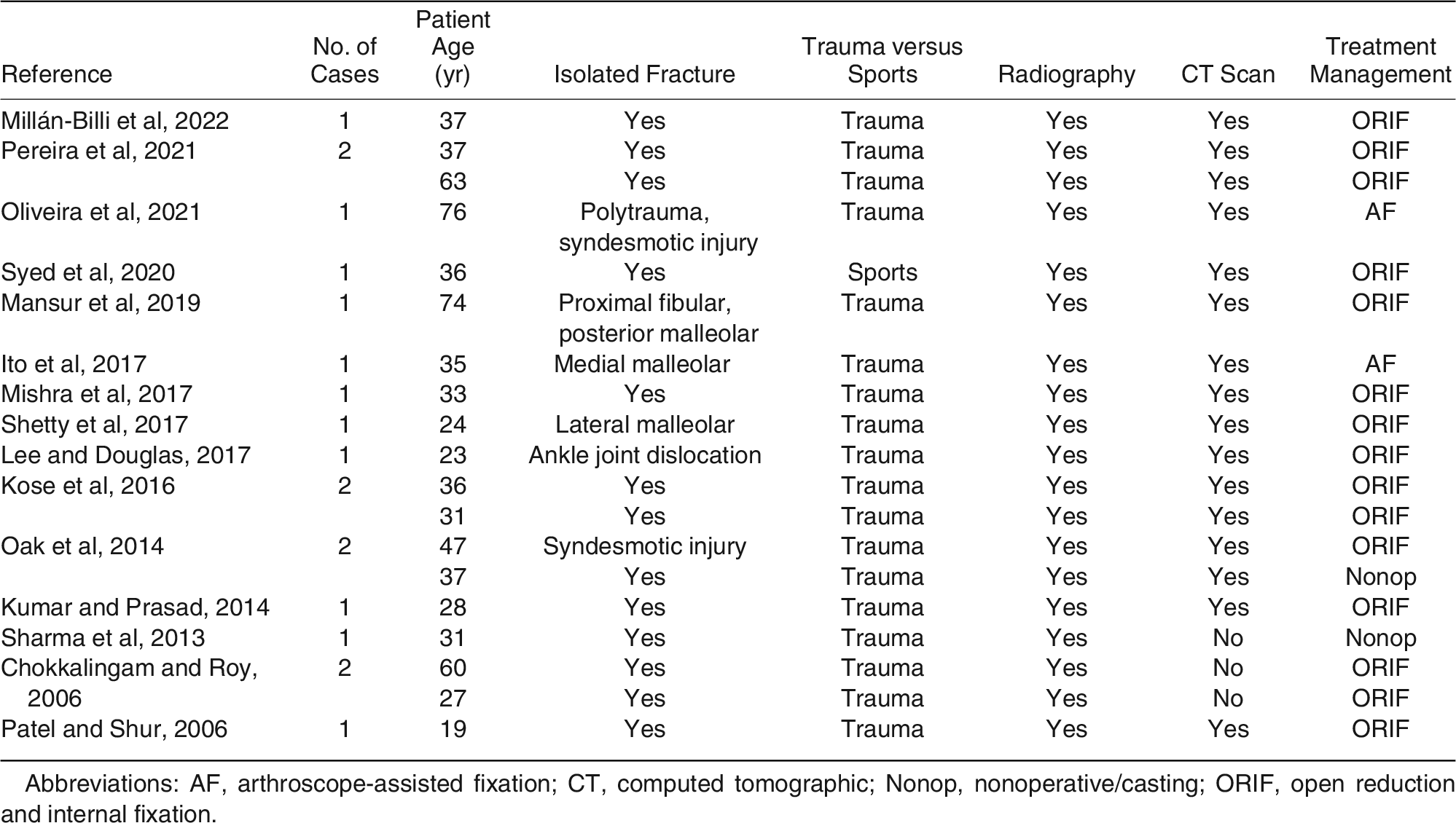
Table 1 summarizes the 15 literature review references that met the inclusion criteria containing a total of 20 cases. The mean age was 50 years (range, 19–76 years). Most case reports of the data revealed an adult isolated Tillaux fracture, whereas others included syndesmotic injury (n = 2), malleolar fractures (n = 3), and dislocation (n = 1). Most of the ankle fractures were sustained by traumatic injury and all case reports underwent initial plain film radiographic evaluation. Only three of the 20 case reports did not proceed with a CT scan. As for treatment, open reduction and internal fixation was the most common surgical treatment method (n = 11) seen in the data, followed by arthroscope-assisted fixation technique (n = 2) and nonoperative treatment (n = 2).
The rarity of this type of fracture in adults may cause it to be overlooked in traditional ankle films and may require a high index of suspicion for further evaluation. Whenever there is a high index of suspicion or concern, careful attention to the syndesmosis area in traumatic ankle injuries may assist in uncovering this diagnosis along with preventing potential future complications [2] In addition, a CT scan can help confirm the suspected diagnosis, rule out other injuries of the tibia and its articulations, and provide more information on the best course of action relating to surgical intervention. Because of the nature of being an intra-articular type of fracture, ensuring that the articulation is congruous and that any loose osseous fragments are reduced is of utmost importance to allow the normal anatomical function of the ankle [3]
Some authors have argued that a “true” isolated Tillaux fracture is one of the three most common occult fractures that may result from a typical ankle sprain or an inversion injury, and only approximately 12% of Tillaux-Chaput fractures are associated with malleolar fractures in adults. Radiographic films of the ankle may not completely reveal this fracture because of the rarity of isolated Tillaux fractures along with superimposition of the fibula and tibia where the fracture is located. Patients who present with “out-of-proportion” pain or return with no benefit from conservative treatment have been reported to have a CT scan that provided better visualization of the fracture. Therefore, the authors recommended that when there is a high suspicion of occult fracture or a decision for surgical intervention is made, a CT scan is recommended during the workup evaluation, to better visualize articular displacement and angulation of the avulsed fragment displacement and aid in perioperative decision making [4,5] Some reports, however, also detailed ankle operations for this injury without further CT scan advance imaging that went on to heal with uneventful results [6]
Associated injuries and concomitant fractures about the ankle have also been reported with Tillaux fractures. This report presented an ankle injury from a motor vehicle injury, where radiographs were taken that revealed a proximal fibular fracture and posterior malleolus fracture with no sign of Tillaux fracture. A CT scan was later obtained that revealed a fracture of the anterolateral aspect of the distal tibia. This case specifically shows a rare association between Tillaux and Volkmann high fibular fracture [7] Other reports described the identification of this Tillaux fracture during an irreducible ankle fracture-dislocation with soft-tissue interposition and others involving osseous obstructions that made closed reduction unsatisfactory. Because of minimal occurrences of Tillaux fractures in adults, there was relatively no suspicion of this preventing the reduction. Later, it was the understanding that educated assumptions were made that the external rotation injury caused the avulsion of the distal anterolateral tibia fragment, and the abduction force caused the widening of the syndesmosis, allowing the fragment to lodge in syndesmosis. This case provided another confirmation for always applying a more methodical approach to ankle fracture-dislocations. If closed reduction manipulation under anesthesia were to fail, proceeding with an oblique view radiograph may help rule out osseous obstruction. A CT scan would also allow better visualization and is more cost-effective than a magnetic resonance imaging scan and less time consuming [8] Another two cases reported on associated Tillaux fractures with a syndesmotic injury while falling down steps and bike rider collision. After radiographic films revealed a displaced intraarticular distal anterolateral tibial fracture, they were treated with closed reduction with internal rotation and dorsiflexion of the ankle and then placed in a long leg cast. Nondisplaced fractures can be treated with nonoperative management by internally rotating the foot in a long leg cast [9] One report argued also performing syndesmotic evaluation, even though mechanically a Tillaux fracture should present with no syndesmosis instability. It is therefore important to note that Tillaux fractures can be associated with syndesmotic injury; therefore, it is always best to be thorough before proceeding with conservative management [10]
Case Report
A 39-year-old patient presented to the emergency department at our Level I trauma center with an isolated, closed, displaced right ankle fracture he sustained from wrestling (Figure 1 and Figure 2). No other injuries were reported. Medical and surgical history were unremarkable, including social and family history. The ankle fracture dislocation required procedural sedation for reduction, which was performed by the emergency department staff on arrival. On physical examination, the patient remained neurovascularly intact after the closed reduction, and there were no open lesions or abrasions at the time of presentation. He was reduced and splinted and remained nonweightbearing until definitive surgical management, which was scheduled in the trauma room.
Figure 1.
Initial anteroposterior radiograph in the emergency department demonstrating displaced unstable ankle fracture.
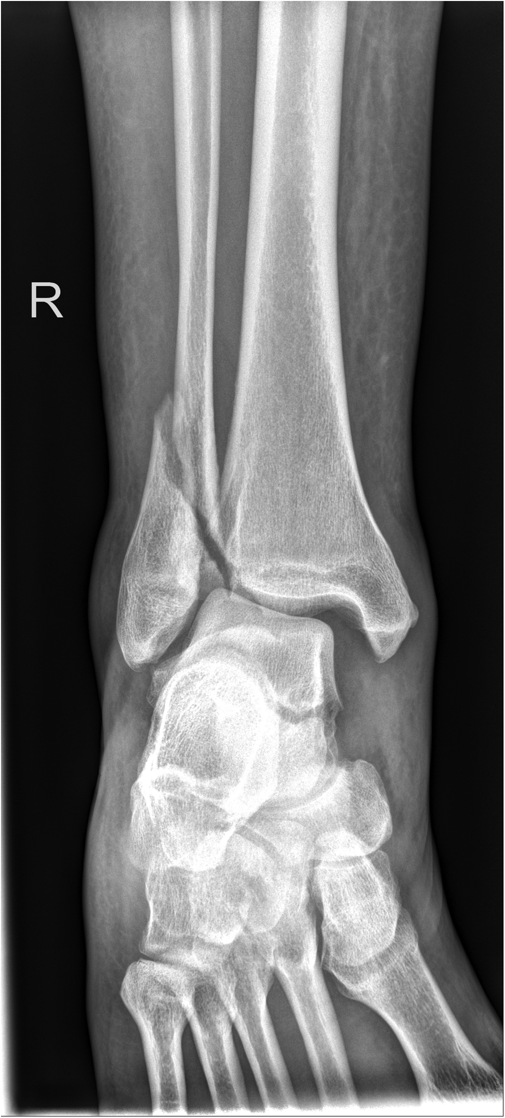
Figure 2.
Initial lateral radiograph in the emergency department demonstrating displaced unstable ankle fracture.
On the day of surgery, a popliteal and adductor canal regional anesthesia block was performed in the preoperative area. The patient was then brought into the operating room and induced under general anesthesia, and a thigh tourniquet was applied and set to 275 mm Hg. After proper standard preparation and draping were performed, including an appropriate timeout process, attention was then directed to the lateral ankle area. The fibula fracture was approached with a standard lateral incision, with care being taken to identify and retract the superficial peroneal nerve. The fracture was reduced, clamped, and fixated with a 3.5-mm cortical screw. A seven-hole one-third tubular plate was then positioned along the distal fibular to act as a neutralization plate, which was fixated to the fibula with nonlocking cortex screws and further fixated with 3.5-mm cortical screws proximal to the fracture and 4.0-mm cancellous screws distally.
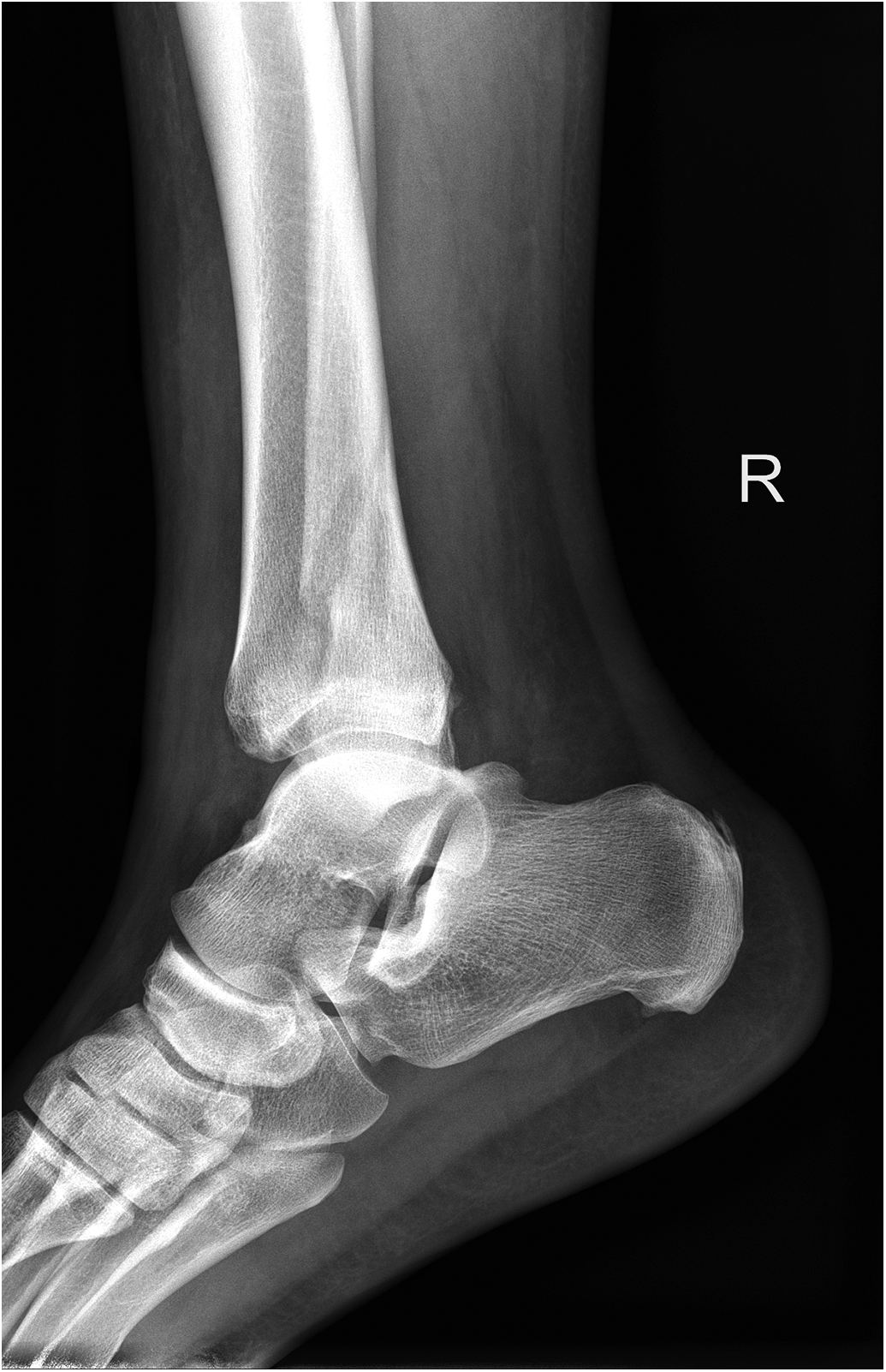
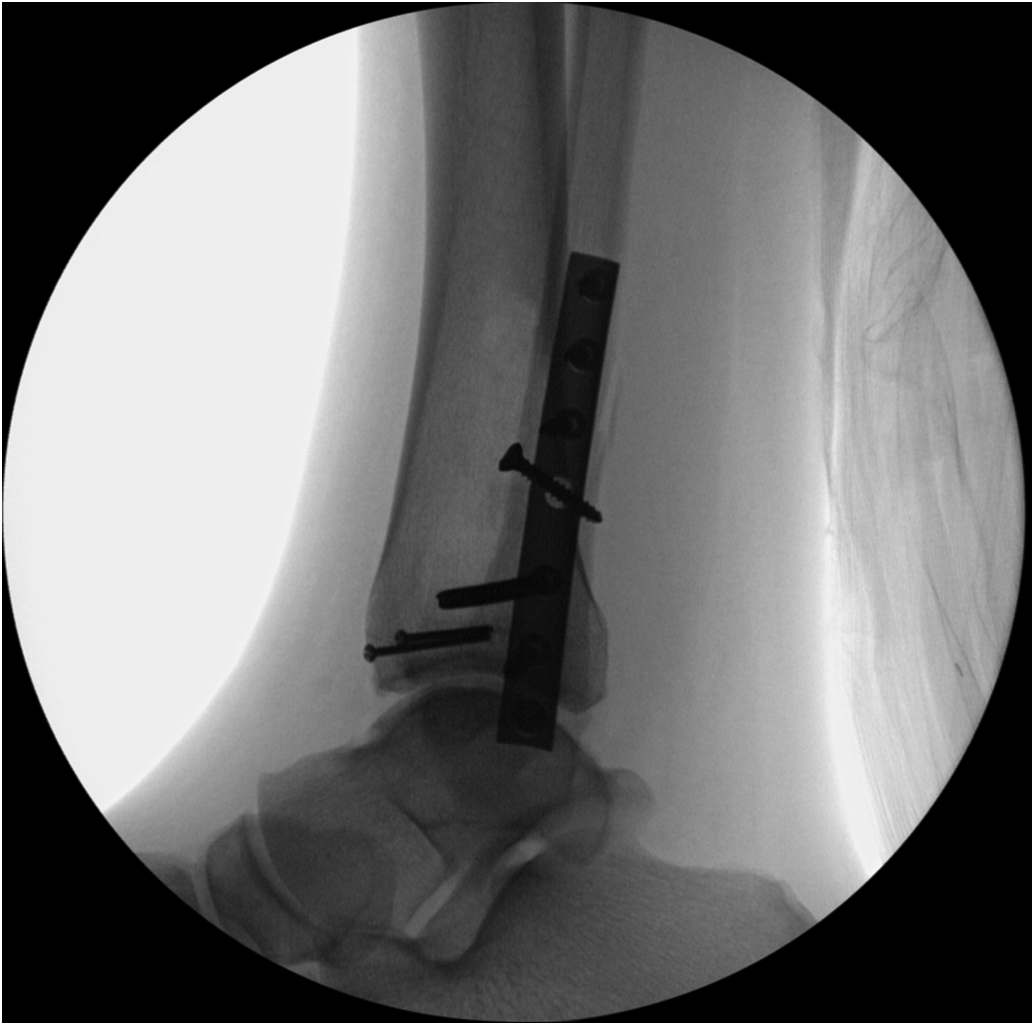
The Tillaux fracture fragment on the anterolateral distal tibia was then visualized with fluoroscopy, dissected, reduced, and provisionally pinned in place. Two 2.0-mm fully threaded screws were placed across the fracture site in lag fashion. The ankle joint was then stressed under live fluoroscopy and gapping at the syndesmosis was noted. A large manual clamp was used to reduce the syndesmosis, and a fully threaded 4.0-mm screw was placed across the syndesmosis through the third most distal hole in the plate. The ankle joint was then noted to be appropriately reduced in stable alignment (Figure 3 and Figure 4). The patient tolerated the anesthesia and procedure well and was taken to the recovery room in stable condition.
Figure 3.
Postoperative anteroposterior radiograph demonstrating restored alignment of ankle fracture, with stable internal fixation including fixation of the Tillaux fracture.
Figure 4.
Postoperative lateral radiograph demonstrating restored alignment of ankle fracture, with stable internal fixation including fixation of the Tillaux fracture.
Discussion
In adults, this fracture may not be readily considered as part of the differential fracture pattern diagnosis. As in our case report, it was not apparent on initial standard radiographic films. The common forces of a Tillaux fracture include high-energy external rotational motion at the level of the ankle joint that typically affects the anteroinferior tibiofibular ligament and its attachment to the distal lateral tibia. As common as a supination external rotation mechanism may be in ankle injuries, these fractures appear to occasionally occur in adolescents with open tibial physis and are more significantly rare in adults with closed tibial physis.
During a typical ankle fracture injury presentation, it may be easily unrecognized on typical ankle plain radiographs. Commonly performed radiographs of the anteroposterior and lateral films of the ankle may reveal minimal, if any, osseous changes, and thus this fracture may not be as apparent on these views as they would on an oblique view, especially if the observer reading the images is not particularly looking for this fracture fragment.
Sometimes, a high index of suspicion can be helpful during this phase of injury assessment. Irreducible ankle fracture-dislocations can be caused by soft-tissue interposition; however, some cases involve osseous obstructions that make closed reduction unsatisfactory. These intraarticular-type fractures typically require anatomical reduction and stabilization by open reduction and internal fixation to avoid long-term complications such as ankle instability, ankle pain, and posttraumatic osteoarthritis.
Operative treatment of this fracture has consisted of primary internal fixation along with reduction and fixation of other associated ankle fractures. Nonoperative treatment with minimal displacement of less than 2 mm was treated with a short leg cast in neutral position. Some reports of this type of fracture were also associated with injury to the medial malleolus or deltoid ligament. This article also argues that if displacement is larger than 2 mm to proceed with a CT scan to further evaluate displacement and then attempt closed reduction under anesthesia. If this reduction fails, proceed with surgical intervention for anatomical reduction [1] One case report noted that after a CT scan was obtained, a 3-mm diastasis was revealed, and operative management was performed. Contralateral films of the unaffected ankle were taken at the time of injury to ensure that the growth plate had closed, which it had. This case presents the potential causes of delayed ossification, such as hypothyroidism, hyperparathyroidism, and other genetic expressions that could affect closure. Therefore, these fractures that typically occur in pediatric patients and adolescents, and may occur more often in older patients who have delayed ossification of their growth plates, which may be also difficult to objectively assess with traditional ankle plain films [12] The most common fixation options reported were cannulated 4.5-mm lag screws, headless compression screws, and 3.5-mm screws [13–15] Another report included the use of arthroscopy as part of fixation-assisted technique for fixating a Tillaux fracture [13]
Conclusions
Tillaux fractures in adults are rare and they have been scarcely reported in our literature. With only a few reports noted in our literature, there are insufficient data to describe any classification association with this injury pattern. Considering the location of the fracture and ankle function, there is sufficient understanding of potential posttraumatic ankle arthritis without anatomical reduction. Traditional standard ankle plain film radiographs may not be sufficient to visualize this type of fracture. Adding an oblique view and possibly using a CT scan in the presence of high suspicion and physical findings have been reported to be key in the proper identification, assessment, and treatment of this type of injury.
Financial Disclosure: None reported.
Conflict of Interest: None reported.
References
- TillauxP. Recherches cliniques et expérimentales sur les fractures malléolaires. Bull Acad Med 1872, 21, 817. [Google Scholar]
- OliveiraN; PinhoP; BaptistaM; et al. Missed Tillaux fracture and syndesmosis injury in adult: arthroscopic assisted reduction and fixation. Rev Bras Ortop (Sao Paulo) 2021, 56, 399. [Google Scholar]
- KumarN; PrasadM. Tillaux fracture of the ankle in an adult: a rare injury. J Foot Ankle Surg 2014, 53, 757. [Google Scholar]
- KoseO; YukselHY; GulerF; et al. Isolated adult Tillaux fracture associated with Volkmann fracture—a unique combination of injuries: report of two cases and review of the literature. J Foot Ankle Surg 2016, 55, 1057. [Google Scholar]
- ItoK; TanakaY; KatsuiR; et al. Corrective osteotomy for malunion of a Tillaux-Chaput facture: a case report. Clin Med Case Rep 2017, 1, 103. [Google Scholar]
- ChokkalingamS; RoyS. Adult Tillaux fractures of ankle: case report. Internet J Orthop Surg 2006, 6, 1. [Google Scholar]
- MansurH; LimaT; GoncalvesC; et al. Adult Tillaux fracture in association with Volkmann and Maisonneuve fractures: a case report. JAPMA 2019, 109, 477. [Google Scholar]
- LeeC; DouglasTJ. Irreducible ankle fracture from an interposed Chaput fragment: a case report. JBJS Case Connect 2017, 7, e42. [Google Scholar]
- MishraPK; PatidarV; SinghSP. Chaput tubercle fracture in an adult—a rare case report. J Clin Diag Res 2017, 11, RD01. [Google Scholar]
- OakNR; SabbBJ; KadakiaAR; et al. Isolated adult Tillaux fracture: a report of two cases. J Foot Ankle Surg 2014, 53, 489. [Google Scholar]
- SharmaB; ReddyIS; MeanockC. The adult Tillaux fracture: one not to miss. BMJ Case Rep 2013, 2013, bcr2013200105. [Google Scholar]
- PatelA; ShurV. Juvenile Tillaux ankle fracture pattern in a skeletally mature adult. Foot 2006, 16, 54. [Google Scholar]
- ShettyS; SasidharanB; ShettyS; et al. The Tillaux fracture in adult. Orthopaed Surg Traumatol 2017, 1, 114. [Google Scholar]
- SyedT; StoreyP; RochaR; et al. Tillaux fracture in adult: a case report. Ortho J MPC 2020, 26, 95. [Google Scholar]
- PereiraEC; RosaJM; DinizSE; et al. The rare isolated adult Chaput-Tillaux fracture: two case reports and review of the literature. Int J Res Orthop 2020, 7, 134. [Google Scholar]
- Millán-BilliA; Fa-BinefaM; Gómez-MasdeuM; et al. Tillaux fracture in adults—case report and review of literature. J Clin Orthop Trauma 2022, 27, 10189. [Google Scholar]
© 2023 American Podiatric Medical Association