


Abstract
A 70-year-old patient with multiple medical problems presented to us with displaced closed very distal tibia and fibula fractures and a prior total knee replacement on the affected extremity. We treated the patient with an isolated fibula open reduction and internal fixation. At a 1-year follow-up, both the tibial and fibular fractures had healed, and the patient had an excellent outcome. “Fibula-only” fixation of very distal tibia-fibula fractures appears to be a viable option to manage these difficult fractures. The novelty of the case report lies in that this technique has not been described previously in the literature.
Distal tibia fracture with associated distal fibula fractures can be seen in high-velocity injuries or in elderly patients with ground level fall.[1] These fractures can be difficult to treat, and there is no clear consensus on the optimal surgical strategy for these injuries.[2] Given the limited soft-tissue cover, poor vascularity of the region, and proximity to the ankle joint, the surgical treatment option often needs to be tailored to each patient to prevent infection, nonunion, malunion, and posttraumatic ankle joint arthritis and stiffness.A variety of surgical treatment options are used: intramedullary (IM) nailing or open reduction and internal fixation (ORIF) with plate of tibia along with IM nailing or ORIF with plate of fibula.[3] A recent meta-analysis of 1,332 patients with distal tibia fractures treated with IM nails or plates demonstrated a high incidence of knee pain with the IM nailing and high rates of infection with plates.[3] Performing an IM nail fixation of very distal tibia fractures can be technically challenging.[4]
An ORIF of the tibia is also associated with complications. The distal tibia has poor vascularity and wound healing potential. Several studies have demonstrated wound-related complications and infections with distal tibia plating.[5,6,7]
Nonoperative management of complex unstable ankle fractures is reserved for medically unfit patients who cannot tolerate surgery. Nonoperative management of such fractures results in increased stiffness, shortening, and malunion, which affect long-term outcomes.[8] In addition, the prolonged immobilization needed might lead to ankle stiffness and is not well tolerated by elderly patients. We present a case of a 70-year-old woman with a displaced closed distal tibia fracture and a fibula fracture that we managed successfully with an isolated fibula ORIF. The patient was informed that data concerning the case would be submitted for publication and the patient agreed.
Case Report
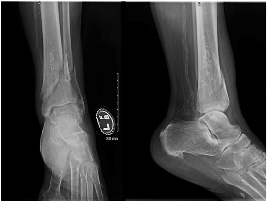
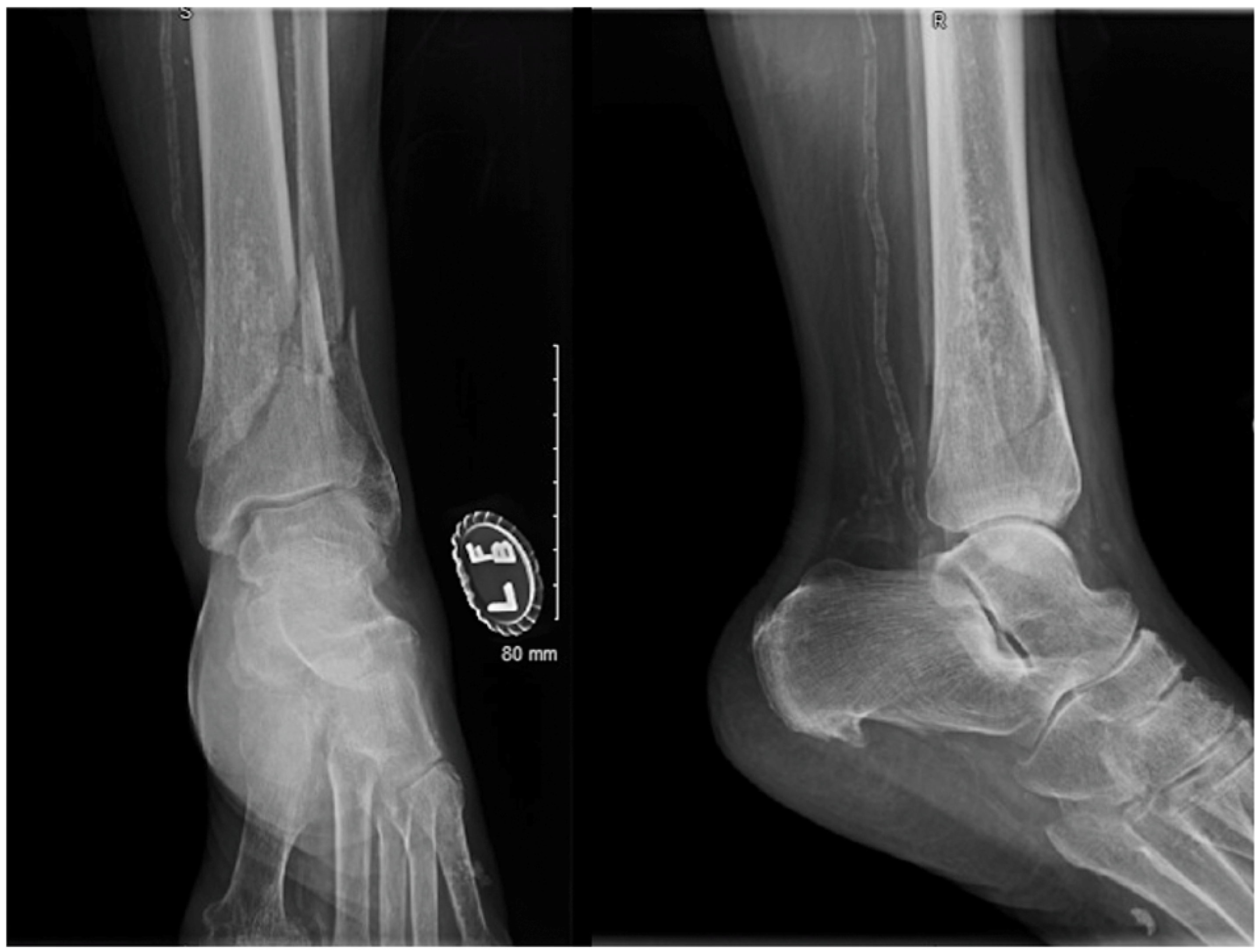
A 70-year-old female patient presented to our emergency department with a history of fall. She sustained closed left very distal tibia and fibula fractures (
Figure 1
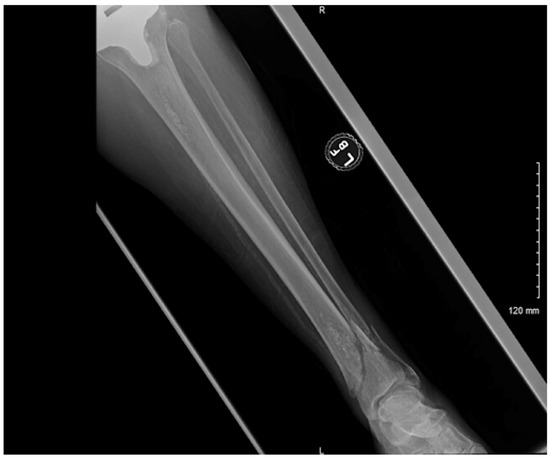
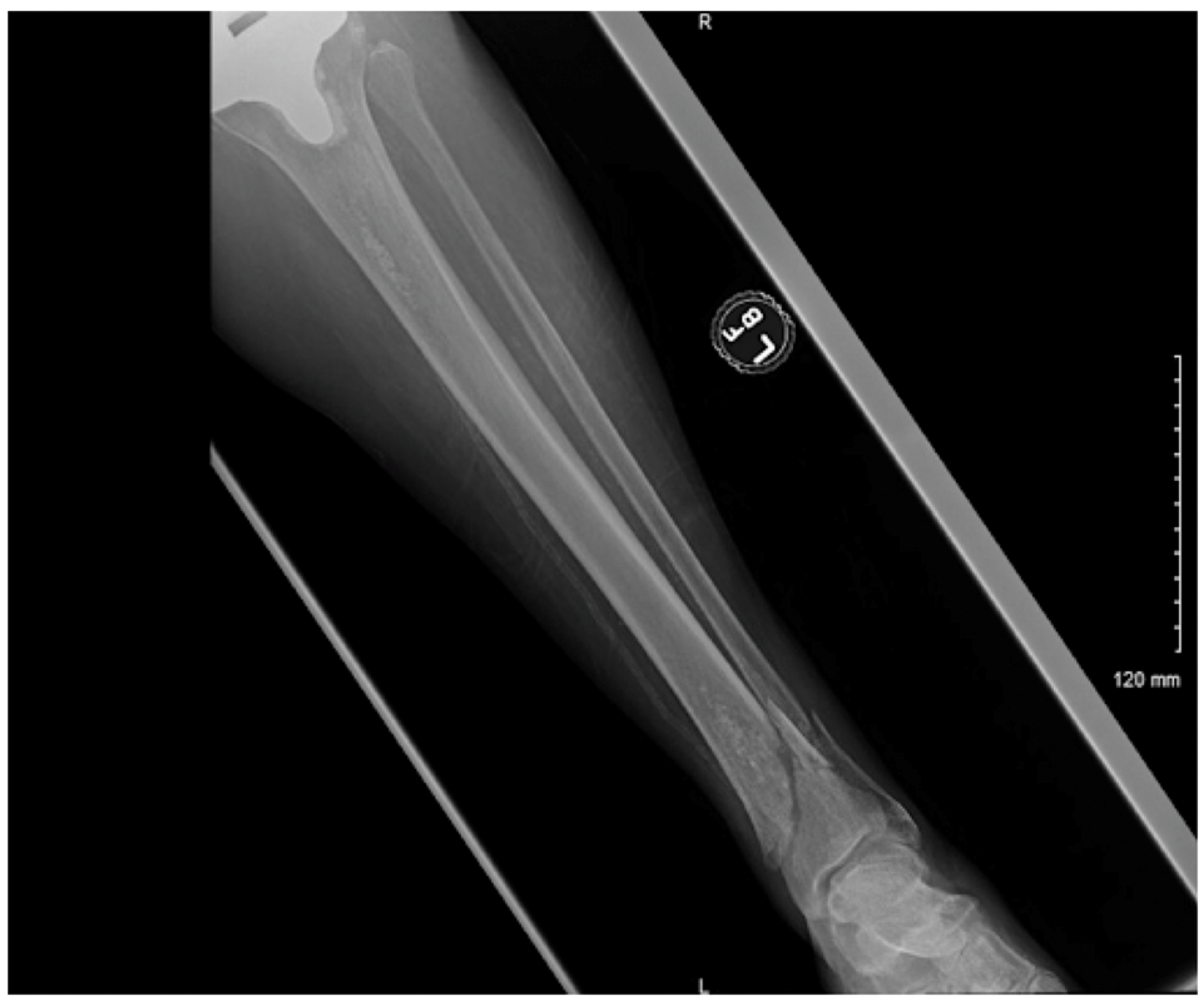
). The fracture was reduced and splinted in the emergency room. The patient was morbidly obese (body mass index, 43 kg/m2). Her other medical history included hypertension and peripheral vascular disease. She had a surgical history of a total knee replacement on the ipsilateral extremity (
Figure 2
). Before the accident, she was able to walk 3 blocks with a roller-walker.
Figure 1.
Initial radiographs at the time of injury.
Figure 2.
Ipsilateral total knee replacement on the injured extremity.
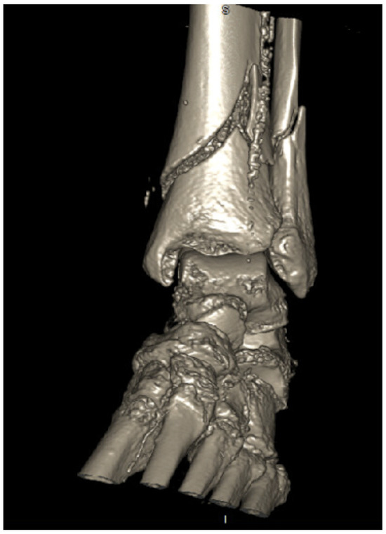
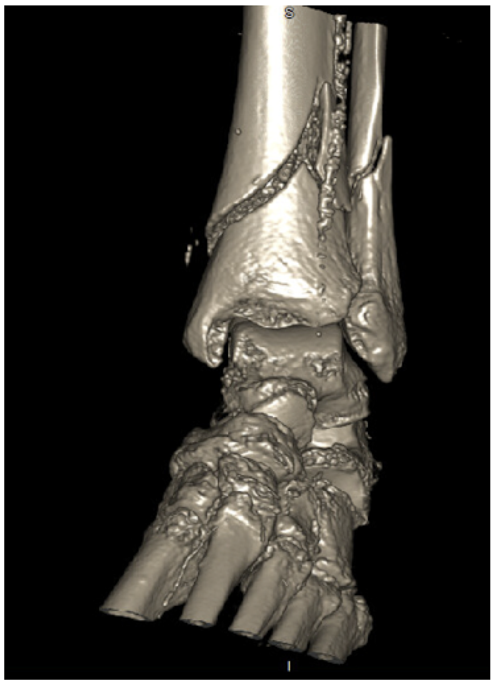
Radiographs and a computed tomographic scan with three-dimensional reconstruction were obtained. This revealed a very distal extra-articular tibia and fibula fracture (
Figure 3
).
Figure 3.
Computed tomographic scan with threedimensional reconstruction of the ankle.
As the fracture was unstable in the splint, nonoperative management was not suitable. All the described surgical treatment options for a fracture of this type in the orthopedic literature were reviewed. As the patient had a total knee implant on the same extremity, antegrade IM nailing of the tibial fracture would have been difficult to perform, as the entry point on the proximal tibia would have been difficult to identify. A retrograde tibia nailing would result in loss of ankle motion. An ORIF of the distal tibia was considered high risk for wound dehiscence and infection given the patient’s morbid obesity and peripheral vascular disease; therefore, a decision was made to perform an isolated fibula ORIF with plate to achieve anatomical alignment of both tibial and fibula fractures.
Operative Technique
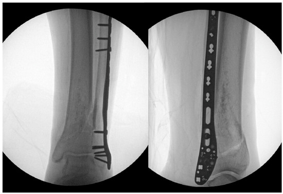
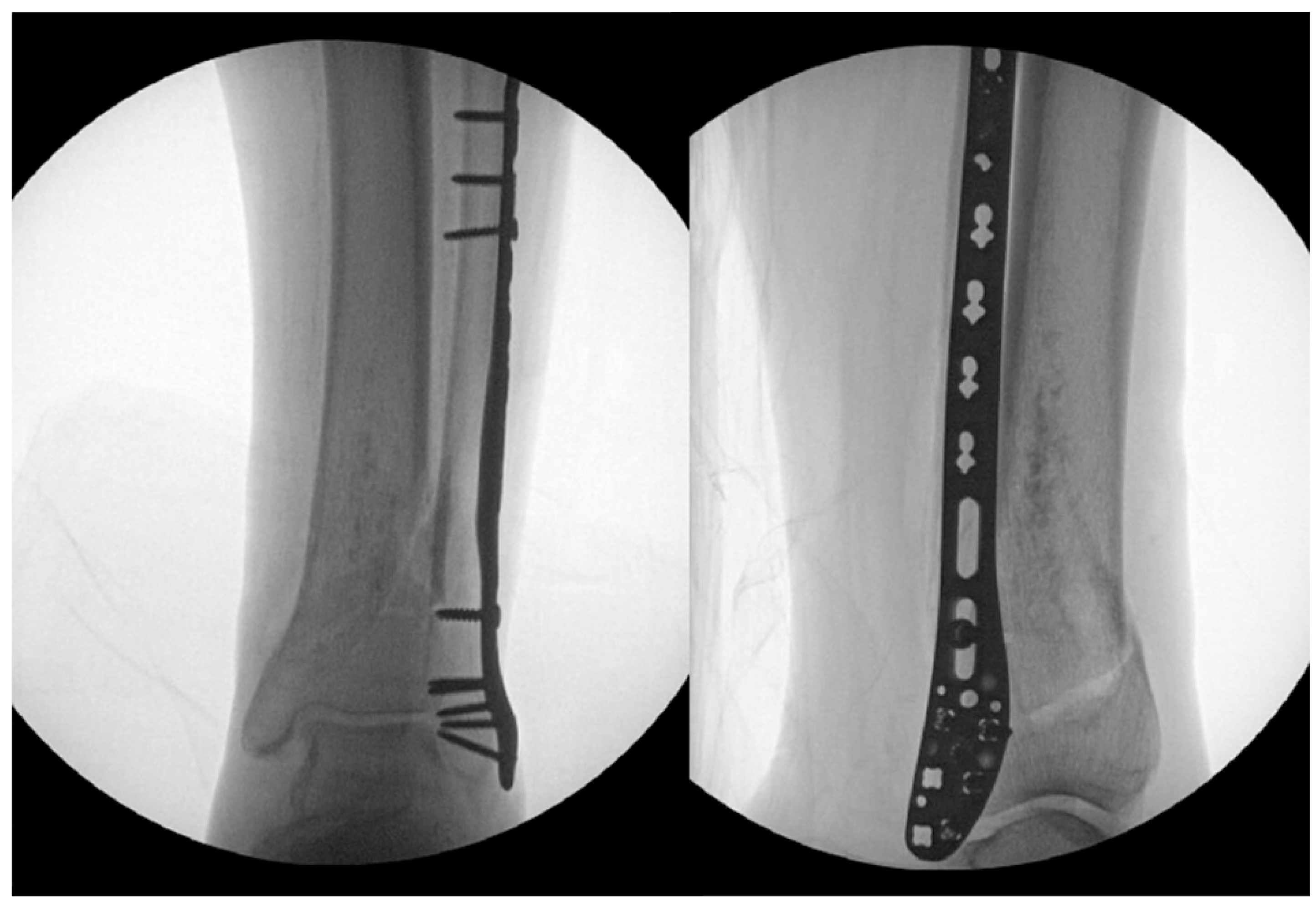
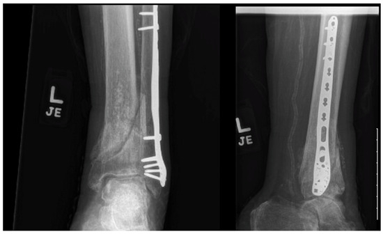
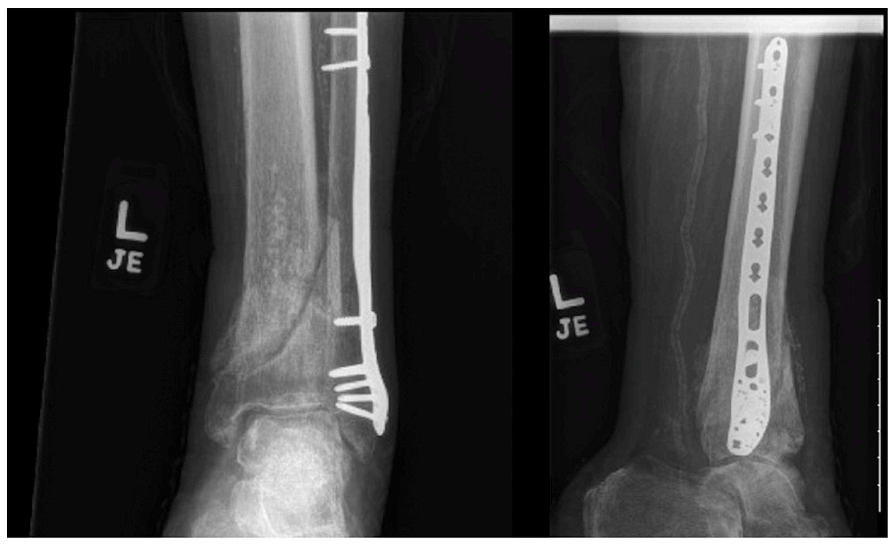
Surgery was performed under regional anesthesia with the patient in the supine position. A posterolateral skin incision was used to approach the fibula fracture. It was noted intraoperatively that the fracture was highly unstable. Once the fibula was reduced anatomically and fixed internally with plate and screws, it was noted that the tibia fracture was in fact stable, as demonstrated by live fluoroscopy films. Fluoroscopic images also demonstrated excellent restoration of the length and alignment of both the tibia and fibula. The syndesmosis was intact; thus, a decision was made not to use any syndesmotic screws (Figure 4).
Figure 4.
Fibula-only fixation with adequate restoration of the tibial length and alignment.
Postoperative Protocol
A posterior short leg splint was placed with the ankle in neutral position for 2 weeks. This was replaced by a controlled ankle movement boot at 2 weeks. At 4 weeks, the patient was allowed ankle range of motion (ROM) as tolerated. The left lower extremity was kept nonweightbearing for 2 months postoperatively, after which partial weightbearing was started.
At 3-month follow-up, the patient demonstrated full ROM of the ankle, reported no pain, and had returned to walking pain-free. A radiograph at 3 months demonstrated ongoing healing of distal tibia and fibula fractures (Figure 5).
Figure 5.
Three-month postoperative radiographs.
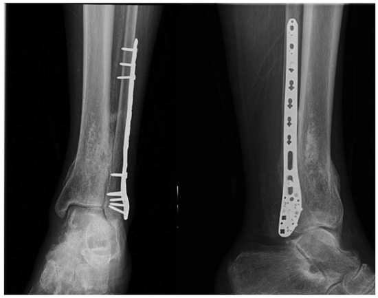
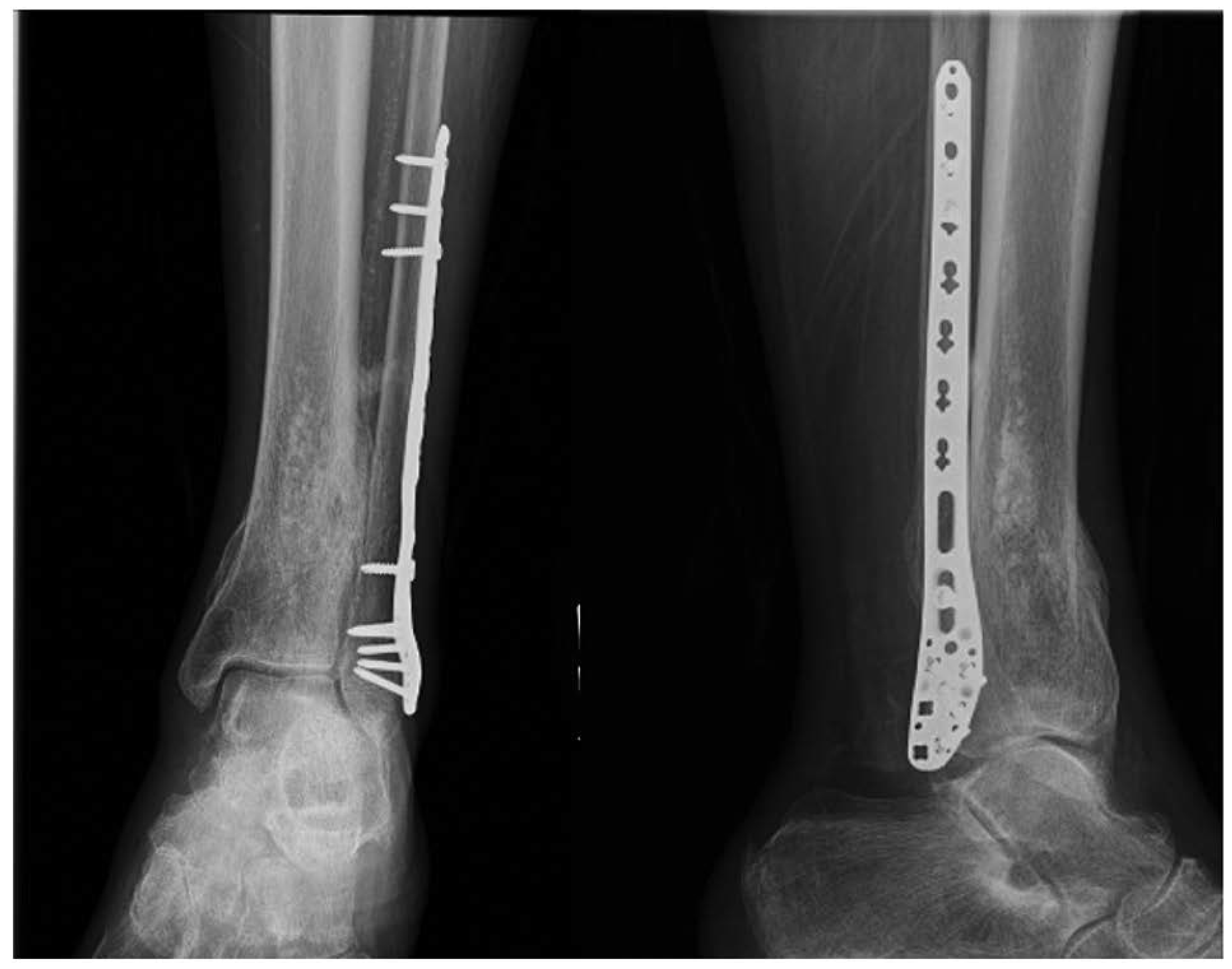
At the latest follow-up at 1 year, the patient returned to her previous level of ambulation with a roller walker. Radiographs demonstrated healed fractures. Her ankle was pain-free. Her ankle ROM was 10° of dorsiflexion and 30° of plantarflexion. Her American Orthopaedic Foot and Ankle Score was 91 (Figure 6).
Figure 6.
One-year postoperative radiographs.
Discussion
Unstable ankle fractures in geriatric patients are difficult to manage. Several treatment options have been described in the literature on management of these fractures.
Nonoperative treatment of distal tibia fractures has been described in the literature. However, this results in an increased incidence of stiffness and malalignment.[8] Nonoperative management requires prolonged use of a lower leg cast, which is not well tolerated by elderly patients. In our patient, we were able to transition to a controlled ankle movement boot early in the postoperative period and began early ROM of the ankle.
Extra-articular tibia fractures such as the one described in our case report can be managed with a closed IM nailing of the tibia with or without fibula fixation.[10,11] However, our patient had an ipsilateral total knee replacement in the same extremity. The entry point for an antegrade tibia nail is close to the articular surface of the knee joint, and a total knee implant would have precluded obtaining an ideal starting point for an antegrade nail. In addition, the surgical technique is difficult to perform. A recent meta-analysis showed a high incidence of malunion of more than 20% with this approach.[11]
Distal tibial plating with or without fibular plating has been described. However, several case reports and case series have shown increased soft-tissue and wound healing complications with the use of this approach.[12,13] The distal tibial skin has a limited soft-tissue envelope, and wounds in this site have poor healing capacity.[14] The patient presented in our report presented with a history of uncontrolled diabetes and peripheral vascular disease. Literature has shown higher rates of wound-related complications and infections with the use of large distal tibial incisions and the use of distal tibial plates in patients with diabetes and peripheral vascular disease.[14]
Closed retrograde tibial nailing[15] with or without preparing the ankle joint has been described for unstable complex ankle fractures in geriatric patients. The disadvantage of the procedure is the loss of ROM of the ankle.
A decision to perform an isolated fibula fixation was undertaken in our patient, as the risks of other available surgical treatment options outweighed potential benefits. This case report demonstrates a safer and simple technique of isolated fibula fixation of combined distal tibia and fibula fracture. We propose that in these fractures, anatomical fixation of the fibula would act as an “internal splint” and restore the length and alignment of the tibia. The syndesmosis was intact in our patient and there was no motion of the tibial fracture after the fibula was fixed in our patient. Syndesmotic screws and percutaneous compression screws on the tibia could be added to obtain additional stability of the tibia, if determined necessary.
The patient demonstrated excellent outcomes with healed tibia and fibula fracture. On careful review of the postoperative radiographs, an 8° sagittal malalignment was noted immediately after surgery. At final follow-up, radiographs demonstrated healed distal tibia, and fibula fractures with an 8° tibial malunion in the sagittal plane were observed. There was no malunion in the coronal plane. This suggests that the fixation construct was stable and did not result in any postoperative motion of the fracture sites.
There is no current literature found on isolated fibula ORIF for displaced, closed distal tibia and fibula fractures despite extensive search. Our case report is unique in this regard, and it offers a simple yet effective technique to manage these challenging injuries.
Conclusions
Our case report demonstrates that an isolated fibula ORIF of very distal tibia and fibula fractures can result in an excellent outcome. The surgical technique is simple to perform and avoids wound-related complications and infections associated with distal tibia ORIF and plating.
Financial Disclosure
None reported.
Conflicts of Interest
None reported.
References
- WANI IH, UL GANI N, YASEEN M, ET AL: Operative management of distal tibial extra-articular fractures—intramedullary nail versus minimally invasive percutaneous plate osteosynthesis. Ortop Traumatol Rehabil 2017, 19, 537. [CrossRef] [Scilit] [PubMed]
- CASSTEVENS C, LE T, ARCHDEACON MT, ET AL: Management of extra-articular fractures of the distal tibia: intramedullary nailing versus plate fixation. J Am Acad Orthop Surg 2012, 20, 675. [CrossRef] [Scilit]
- BLEEKER NJ, VAN DE WALL BJM, IJPMA FFA, ET AL: Plate vs. nail for extra-articular distal tibia fractures: how should we personalize surgical treatment? A meta-analysis of 1332 patients. Injury 2021, 52, 345. [CrossRef] [Scilit] [PubMed]
- EL ATTAL R, HANSEN M, ROSENBERGER R, ET AL: Intramedullary nailing of the distal tibia illustrated with the Expert tibia nail [in German]. Oper Orthop Traumatol 2011, 23, 397.
- JOVENIAUX P, OHL X, HARISBOURE A, ET AL: Distal tibia fractures: management and complications of 101 cases. Int Orthop 2010, 34, 583. [CrossRef] [Scilit] [PubMed]
- LAU TW, LEUNG F, CHAN CF, ET AL: Wound complication of minimally invasive plate osteosynthesis in distal tibia fractures. Int Orthop 2008, 32, 697. [CrossRef] [Scilit] [PubMed]
- SATHIYAKUMAR V, THAKORE RV, IHEJIRIKA RC, ET AL: Distal tibia fractures and medial plating: factors influencing reoperation. Int Orthop 2014, 38, 1483. [CrossRef] [Scilit] [PubMed]
- SARMIENTO A: Distal tibia fracture: opinion: nonoperative treatment. J Orthop Trauma 2006, 20, 75. [CrossRef] [Scilit]
- KRISTENSEN KD, KIAER T, BLICHER J: No arthrosis of the ankle 20 years after malaligned tibial-shaft fracture. Acta Orthop Scand 1989, 60, 208. [CrossRef] [Scilit] [PubMed]
- WASKY PR, BELTRAN MJ: Extreme nailing or less invasive plating of lower extremity periarticular fractures. Orthop Clin North Am 2021, 52, 111. [CrossRef] [Scilit] [PubMed]
- PENG J, LONG X, FAN J, ET AL:. Concomitant distal tibia-fibula fractures treated with intramedullary nailing, with or without fibular fixation: a meta-analysis. J Foot Ankle Surg 2021, 60, 109. [CrossRef] [Scilit] [PubMed]
- KARIYA A, JAIN P, PATOND K, ET AL: Outcome and complications of distal tibia fractures treated with intramedullary nails versus minimally invasive plate osteosynthesis and the role of fibula fixation. Eur J Orthop Surg Traumatol 2020, 30, 1487. [CrossRef] [Scilit] [PubMed]
- VIBERG B, KLEVEN S, HAMBORG-PETERSEN E, ET AL: Complications and functional outcome after fixation of distal tibia fractures with locking plate—a multicentre study. Injury 2016, 47, 1514. [CrossRef] [Scilit] [PubMed]
- MUZAFFAR N, BHAT R, YASIN M: Complications of minimally invasive percutaneous plating for distal tibial fractures. Trauma Mon 2016, 21, e22131.
- TAYLOR BC, HANSEN DC, HARRISON R, ET AL: Primary retrograde tibiotalocalcaneal nailing for fragility ankle fractures. Iowa Orthop J 2016, 36, 75.
© 2023 American Podiatric Medical Association