


Abstract
Dislocation of the proximal interphalangeal joint of the fifth toe is an uncommon injury. When it is diagnosed in the acute phase, closed reduction is commonly an adequate treatment option. We describe a rare case of a 7-year-old patient who presented with late-diagnosed isolated dislocation of the proximal interphalangeal joint of the fifth toe. Although there are a few reported cases of late-diagnosis combined fracture-dislocation of the toes in both adult and pediatric age groups in the literature, belatedly diagnosed dislocation of the fifth toe without accompanying fracture in the pediatric population, to our knowledge, has not yet been reported. This patient achieved good clinical outcomes after treatment via open reduction and internal fixation.
Isolated traumatic dislocation of the interphalangeal toe joint is an uncommon foot injury usually associated with other foot injuries [1]. Although the closed reduction is successful when diagnosed in the acute phase [2], chronic and irreducible dislocations of the toes may need to be treated with open reduction and internal fixation [3]. Chronic and recurrent dislocations of the fifth toe are relatively rare in the adult and pediatric age groups [4].
A few reported cases of late-diagnosis combined fracture-dislocation of the toes in both adult and pediatric age groups exist in the literature. However, to our knowledge, belated-diagnosis isolated dislocation of the fifth toe without accompanying fracture in the pediatric population has not yet been reported. In the present report, we describe a rare case of a 7-year-old patient who presented with late-diagnosed isolated dislocation of the proximal interphalangeal joint of the fifth toe. The patient and his family consented to publication of this study of his illness and treatment with relevant personal information deleted.
Case Report
A 7-year-old boy was admitted to the trauma center due to deformity in the fifth toe of his right foot. The trauma occurred 2 months before admission. Pain, swelling, and deformity occurred in the fifth toe of the foot after striking a football without wearing football shoes. The patient’s family was reluctant to immediately take him to the hospital for fear of the ongoing coronavirus disease 2019 pandemic. Instead, he was taken to a local bonesetter. A bonesetter is a practitioner of joint manipulation. Before the advent of chiropractors, osteopaths, and physical therapists, bonesetters were the main providers of this type of treatment. Traditionally, they practiced without any formal training in accepted medical procedures. Bonesetters would also reduce joint dislocations and “re-set” bone fractures. The practice of joint manipulation and treating fractures dates back to ancient times and has roots in most countries. The bonesetter applied an inappropriate treatment to the patient. After this treatment, the patient’s foot remained in bandages for approximately 1 month. The patient’s family determined that although the pain and edema in the fifth toe had improved, the deformity persisted.
The patient, who was then taken to several different hospitals by his family, was referred to our trauma center at the end of the second month after the initial trauma. In our first examination of the patient, there was no observed pain and swelling. The only pathologic finding of the patient was a valgus deformity of the fifth toe. The patient’s only complaint was that he could not wear shoes due to the deformed toe. Weightbearing comparative radiographs of both feet were taken. Complete dislocation of the proximal interphalangeal joint was detected without signs of fracture (Fig. 1). The situation at that time was discussed with the family in detail. The patient’s family was informed that although reduction might not be achieved owing to chronic dislocation, closed reduction under anesthesia would be attempted. If a successful closed reduction could not be achieved, surgical treatment would be performed. The patient was taken to the operating table under general anesthesia, and closed reduction was attempted using gentle maneuvers under fluoroscopy. This was not successful.
Figure 1.
Preoperative anteroposterior (A) and lateral (B) radiographs showing the isolated proximal interphalangeal joint dislocation.
Surgical Treatment
Surgery was performed under general anesthesia. The patient was in the supine position, with tourniquet pressure applied to the relevant extremity. A dorsal longitudinal skin incision centered over the fifth toe was made. By preserving the extensor tendon, the proximal interphalangeal joint was reached. It was observed that the joint was occupied by granulation tissue with entrapped joint capsule remnants. These tissues were completely removed without damaging the cartilage. The joint was reduced. One Kirschner wire was placed in a retrograde manner from the tip of the distal phalanx to maintain the satisfactory reduction and alignment (Fig. 2). The wound was irrigated, the tourniquet was deflated, the bleeding was controlled, and, finally, the wound edges were sutured accordingly.
Figure 2.
Postoperative oblique (A), anteroposterior (B), and lateral (C) radiographs showing reduction and fixation by Kirschner wire.
Postoperative Follow-up
Intravenous cefazolin sodium was administered postoperatively on the first day. The patient was immobilized with a nonweightbearing short-leg cast after surgery. The patient had no wound problems, such as drainage, erythema, or swelling. On the second postoperative day he was discharged from the hospital. The first follow-up after surgery was performed on postoperative day 10, and sutures were removed. Successive follow-up visits were scheduled once every 10 days. Anteroposterior and lateral foot radiographs were recorded at each visit (Fig. 3). The Kirschner wire and cast were removed 4 weeks after the surgery because follow-up radiographs revealed an acceptable alignment, and the patient started full weightbearing activity without a cast. There were no complications observed during the early postoperative period or up to the time of the last follow-up.
Figure 3.
First follow-up anteroposterior (A) and lateral (B) radiographs showing good alignment.
The patient had no concerns at the 8-month follow-up visit, and his fifth toe exhibited good alignment radiologically (Fig. 4) and clinically (Fig. 5). There was no limitation of range of motion or restriction of activity.
Figure 4.
Final follow-up anteroposterior radiograph showing good alignment.
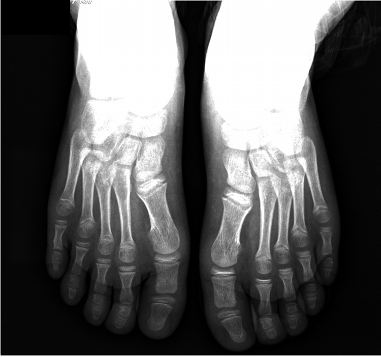
Figure 5.
Final follow-up anteroposterior (A) and lateral (B) photographs showing good alignment clinically.
Discussion
Chronic and recurrent dislocations of the fifth toe are relatively rare in both adult and pediatric age groups [4]. In a case report of chronic irreducible isolated dislocation of the proximal interphalangeal joint of the fifth toe, Vaseenon et al [1] performed an open reduction of the joint, and the corrected position of the proximal interphalangeal joint was maintained by inserting a longitudinal Kirschner wire. They reported good clinical and radiographic outcomes [1]. In this case, the patient was in an adult group, different from our case.
Ween and Schipper [3] reported a case of an irreducible fracture-dislocation of the proximal interphalangeal joint of the fifth toe in a 65-year-old man. In this report, after a dorsal approach, the interposition of the short flexor tendon and the volar plate was revealed in the interphalangeal joint. The interposition was corrected, and reduction was successfully accomplished. The distal fracture fragment was fixed with a Kirschner wire. They also reported no repeated dislocations, and satisfactory function of the toe was maintained. In this report, the patient was in the adult group and had a combined fractured-dislocation, unlike the present patient.
In pediatric patients, these conditions are extremely uncommon, and only a few case reports can be found in the literature. A fracture-dislocation of the fifth toe in a 2-year-old girl was reported by Kushare et al [4] as they performed open reduction after dissecting the soft tissues and clearing out scar tissue in the proximal interphalangeal joint space. Because of the chronic nature of the fracture-dislocation, the reduction was unstable, and they used Kirschner wire to maintain the reduction. After 18 months of follow-up, they reported good clinical alignment of the fifth toe with no residual pain. In this report, the patient was in the pediatric age group, but there was a combined fracture and dislocation.
In another case of a 10-year-old girl with a dislocation of the proximal interphalangeal joint of the fourth toe and fracture of the articular base of the middle phalanx, the authors performed an open reduction revealing an incarcerated volar plate at the fracture site and reported good outcomes in terms of clinical alignment and pain [5].
In conclusion, we present, to our knowledge, a previously unpublished clinical scenario of a belatedly diagnosed dislocation of the fifth toe without accompanying fracture in a 7-year-old boy. Note that such incidents may occur in children and that treatment should be arranged in a timely manner. Irreducible isolated proximal interphalangeal joint dislocation of the fifth toe after appropriately closed reduction attempts can safely be treated with open reduction and Kirschner wire fixation using a dorsal approach in the pediatric age group.
Financial Disclosure
None reported.
Conflicts of Interest
None reported.
References
- Vaseenon T, Cheewawattanachai C, Sirirungruangsarn Y: Chronic irreducible dislocation of the proximal interphalangeal joint of the fifth toe: a case report. BMC Res Notes 7: 76, 2014.
- Fugate DS, Thomson JD, Christensen KP: An irreducible fracture dislocation of a lesser toe: a case report. Foot Ankle 11: 317, 1991.
- Ween M, Schipper BI: Irreducible fracture of the proximal interphalangeal joint of the fifth toe J Emerg Med 44: e63, 2013.
- Kushare VI, Elabd A, Attia E: Missed fracture dislocation of the fifth toe in a two-year-old child: a case report. Foot Ankle Surg 60: 172, 2021.
- Neubauer T, Wagner M, Quell M: Interphalangeal dislocation of the fourth toe with avulsion-fracture in a child: report of a case. Foot Ankle Int 18: 175, 1997.
© 2023 American Podiatric Medical Association